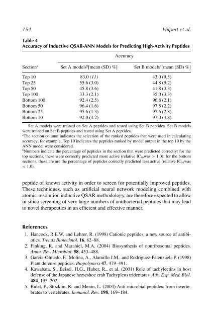
154 Hilpert et al. Table 4 Accuracy of Inductive QSAR-ANN Models for Predicting High-Activity <strong>Peptide</strong>s Accuracy Section a Set A models b [mean (SD) %] Set B models b [mean (SD) %] Top 10 83.0 (11) 43.0 (9.5) Top 25 55.6 (3.0) 44.8 (9.2) Top 50 45.8 (3.6) 41.8 (3.3) Top 100 33.3 (2.1) 35.0 (3.3) Bottom 100 92.4 (2.5) 96.8 (2.1) Bottom 50 96.4 (1.6) 97.8 (2.2) Bottom 25 95.6 (1.3) 97.6 (2.8) Bottom 10 92.0 (4.2) 97.0 (4.8) Set A models were trained on Set A peptides and tested using Set B peptides. Set B models were trained on Set B peptides and tested using Set A peptides. a The section column indicates the selection of the ranked peptides that were used in calculating accuracy; for example, Top 10 indicates the peptides ranked by model output in the top 10 by the ANN model were considered. b Numbers indicate the percentage of peptides in the section that were predicted correctly: for the top sections, these were correctly predicted more active (relative IC50was > 1.0); for the bottom sections, these are are the percentage of peptides correctly predicted less active (relative IC50was < 1.0). peptide of known activity in order to screen for potentially improved peptides. These techniques, such as artificial neural network modeling combined with atomic-resolution inductive QSAR methodology, are therefore expected to allow in silico screening of very large numbers of antibacterial peptides that may lead to novel therapeutics in an efficient and effective manner. References 1. Hancock, R.E.W. and Lehrer, R. (1998) Cationic peptides: a new source of antibiotics. Trends Biotechnol. 16, 82–88. 2. Finking, R. and Marahiel, M.A. (2004) Biosynthesis of nonribosomal peptides. Annu. Rev. Microbiol. 58, 453–488. 3. Garcia-Olmedo, F., Molina, A., Alamillo J.M., and Rodriguez-Palenzuela P. (1998) Plant defense peptides. Biopolymers 47, 479–491. 4. Kawabata, S., Beisel, H.G., Huber, R., et al. (2001) Role of tachylectins in host defense of the Japanese horseshoe crab Tachypleus tridentatus. Adv. Exp. Med. Biol. 484, 195–202. 5. Bulet, P., Stocklin, R. and Menin, L. (2004) Anti-microbial peptides: from invertebrates to vertebrates. Immunol. Rev. 198, 169–184.
Cationic Antimicrobial <strong>Peptide</strong>s 155 6. Iwanaga, S. and Kawabata, S. (1998) Evolution and phylogeny of defense molecules associated with innate immunity in horseshoe crab. Front. Biosci. 3, D973–D984. 7. Imler, J.L. and Bulet, P. (2005) Antimicrobial peptides in Drosophila: structures, activities and gene regulation. Chem. Immunol. Allergy 86, 1–21. 8. Cannon, J.P., Haire, R.N. and Litman G.W. (2002) Identification of diversified genes that contain immunoglobulin-like variable regions in a protochordate. Nat. Immunol. 3, 1200–1207. 9. Litman, G.W., Anderson, M.K. and Rast, J.P. (1999) Evolution of antigen binding receptors. Annu. Rev. Immunol. 17, 109–147. 10. Nochi, T. and Kiyono, H. (2006) Innate immunity in the mucosal immune system. Curr. Pharm. Des. 12, 4203–4213. 11. Yang, D., Biragyn, A., Hoover, D.M., Lubkowski, J. and Oppenheim, J.J. (2004) Multiple roles of antimicrobial defensins, cathelicidins, and eosinophil-derived neurotoxin in host defense. Annu. Rev. Immunol. 22, 181–215. 12. Li, J., Xu, X., Xu, C., et al. (2007) Anti-infection peptidomics of amphibian skin. Mol. Cell. Proteomics. Epub ahead of print, Jan 31, 2007. 13. Yang, D., Biragyn, A., Kwak, L.W. and Oppenheim, J.J. (2002) Mammalian defensins in immunity: more than just microbicidal. Trends Immunol. 23, 291–296. 14. Jenssen, H., Hamill, P. and Hancock, R.E.W. (2006) <strong>Peptide</strong> antimicrobial agents. Clin. Microbiol. Rev. 19, 491–511. 15. Papo, N. and Shai, Y. (2005) Host defense peptides as new weapons in cancer treatment. Cell. Mol. Life Sci. 62, 784–790. 16. Gallo, R.L., Ono, M., Povsic, T., et al. (1994) Syndecans, cell surface heparan sulfate proteoglycans, are induced by a proline-rich antimicrobial peptide from wounds. Proc. Natl. Acad. Sci. USA 91, 11035–11039. 17. Hancock, R.E.W. and Rozek, A. (2002) Role of membranes in the activities of antimicrobial cationic peptides. FEMS Microbiol. Lett. 206, 143–149. 18. Mani, R., Cady, S.D., Tang, M., Waring, A.J., Lehrer, R.I. and Hong, M. (2006) Membrane-dependent oligomeric structure and pore formation of a beta-hairpin antimicrobial peptide in lipid bilayers from solid-state NMR. Proc. Natl. Acad. Sci. USA 103, 16242–16247. 19. Hwang, P.M. and Vogel, H.J. (1998) Structure-function relationships of antimicrobial peptides. Biochem. Cell. Biol. 76, 235–246. 20. Gazit, E., Miller, I.R., Biggin, P.C., Sansom, M.S.P., and Shai, Y. (1996) Structure and orientation of the mammalian antibacterial peptide cecropin P1 within phospholipid membranes. J. Mol. Biol. 258, 860–870. 21. Brogden, K.A. (2005) Antimicrobial peptides: pore formers or metabolic inhibitors in bacteria? Nat. Rev. Microbiol. 3, 238–250. 22. Paschke, M. (2006) Phage display systems and their applications. Appl. Microbiol. Biotechnol. 70, 2–11. 23. Westerlund-Wikstrom, B. (2000) <strong>Peptide</strong> display on bacterial flagella: principles and applications. Int. J. Med. Microbiol. 290, 223–230. 24. Yan, X. and Xu, Z. (2006) Ribosome-display technology: applications for directed evolution of functional proteins. <strong>Drug</strong> Discov. Today 11, 911–916.
- Page 2 and 3:
Peptide-Based Drug Design
- Page 4 and 5:
METHODS IN MOLECULAR BIOLOGY TM Pep
- Page 6 and 7:
Preface Natural products chemistry
- Page 8 and 9:
Contents Preface...................
- Page 10 and 11:
Contributors Nikolinka Antcheva •
- Page 12 and 13:
Contributors xi Alessandro Tossi
- Page 14 and 15:
2 Otvos advance of computer power a
- Page 16 and 17:
4 Otvos see derivatives active in b
- Page 18 and 19:
6 Otvos was designed based on ligan
- Page 20 and 21:
8 Otvos 27. Borghouts, C., Kunz, C.
- Page 22 and 23:
10 Bulet Key Words: Invertebrate im
- Page 24 and 25:
12 Bulet 6. 5 �L or higher volume
- Page 26 and 27:
14 Bulet 2.5.2.1. MALDI-TOF-MS 1. M
- Page 28 and 29:
16 Bulet 7. Small-volume low-protei
- Page 30 and 31:
18 Bulet 3. Centrifuge between 8000
- Page 32 and 33:
20 Bulet internal diameter). Increa
- Page 34 and 35:
22 Bulet interest. As no instrument
- Page 36 and 37:
24 Bulet 3. Incubate the plates in
- Page 38 and 39:
26 Bulet bioactive peptides from th
- Page 40 and 41:
28 Bulet 21. Chernysh, S., Kim, S.I
- Page 42 and 43:
3 Sequence Analysis of Antimicrobia
- Page 44 and 45:
Sequence Analysis of Antimicrobial
- Page 46 and 47:
Sequence Analysis of Antimicrobial
- Page 48 and 49:
Sequence Analysis of Antimicrobial
- Page 50 and 51:
Sequence Analysis of Antimicrobial
- Page 52 and 53:
Sequence Analysis of Antimicrobial
- Page 54 and 55:
Sequence Analysis of Antimicrobial
- Page 56 and 57:
Sequence Analysis of Antimicrobial
- Page 58 and 59:
4 The Spot Technique: Synthesis and
- Page 60 and 61:
The Spot Technique 49 3. For amine
- Page 62 and 63:
The Spot Technique 51 2. Biotinylat
- Page 64 and 65:
The Spot Technique 53 the solutions
- Page 66 and 67:
The Spot Technique 55 solution. Ens
- Page 68 and 69:
The Spot Technique 57 (see Fig. 3)
- Page 70 and 71:
The Spot Technique 59 Fig. 5. Princ
- Page 72 and 73:
The Spot Technique 61 library, the
- Page 74 and 75:
The Spot Technique 63 7. Regenerati
- Page 76 and 77:
The Spot Technique 65 following rul
- Page 78 and 79:
The Spot Technique 67 20. Atherton,
- Page 80 and 81:
The Spot Technique 69 50. Bolger, G
- Page 82 and 83:
5 Analysis of A� Interactions Usi
- Page 84 and 85:
Analysis of Aβ Interactions 73 are
- Page 86 and 87:
Analysis of Aβ Interactions 75 Ana
- Page 88 and 89:
Analysis of Aβ Interactions 77 3.
- Page 90 and 91:
Analysis of Aβ Interactions 79 4.
- Page 92 and 93:
Analysis of Aβ Interactions 81 5.
- Page 94 and 95:
Analysis of Aβ Interactions 83 2.
- Page 96 and 97:
Analysis of Aβ Interactions 85 9.
- Page 98 and 99:
6 NMR in Peptide Drug Development J
- Page 100 and 101:
NMR of Peptides 89 usually require
- Page 102 and 103:
NMR of Peptides 91 limiting the siz
- Page 104 and 105:
NMR of Peptides 93 and STD NMR expe
- Page 106 and 107:
NMR of Peptides 95 The basis of the
- Page 108 and 109:
NMR of Peptides 97 Fig. 5. Overlay
- Page 110 and 111:
NMR of Peptides 99 Fig. 7. Schemati
- Page 112 and 113:
NMR of Peptides 101 4.4.2. Saturati
- Page 114 and 115: NMR of Peptides 103 4.6. Diffusion
- Page 116 and 117: NMR of Peptides 105 Fig. 13. Propos
- Page 118 and 119: NMR of Peptides 107 protein prevent
- Page 120 and 121: NMR of Peptides 109 6. Wuthrich, K.
- Page 122 and 123: NMR of Peptides 111 45. Morris, K.
- Page 124 and 125: NMR of Peptides 113 78. Aumailley,
- Page 126 and 127: 116 Copps et al. Fig. 1. Potential
- Page 128 and 129: 118 Copps et al. Fig. 2. Flowchart
- Page 130 and 131: 120 Copps et al. 10. In accordance
- Page 132 and 133: 122 Copps et al. Fig. 3. DSSP for g
- Page 134 and 135: 124 Copps et al. ingly with the sam
- Page 136 and 137: 126 Copps et al. 19. Essman, U., Pe
- Page 138 and 139: 128 Hilpert et al. Key Words: Scree
- Page 140 and 141: 130 Hilpert et al. Table 1 (Continu
- Page 142 and 143: 132 Hilpert et al. different natura
- Page 144 and 145: 134 Hilpert et al. wt A C D E F …
- Page 146 and 147: 136 Hilpert et al. methods primaril
- Page 148 and 149: 138 Hilpert et al. Table 2 (Continu
- Page 150 and 151: 140 Hilpert et al. Table 2 (Continu
- Page 152 and 153: 142 Hilpert et al. Table 2 (Continu
- Page 154 and 155: 144 Hilpert et al. Table 3 Summary
- Page 156 and 157: 146 Hilpert et al. Table 3 (Continu
- Page 158 and 159: 148 Hilpert et al. of substituting
- Page 160 and 161: 150 Hilpert et al. in models that c
- Page 162 and 163: 152 Hilpert et al. than control. Gi
- Page 166 and 167: 156 Hilpert et al. 25. Pini, A., Gi
- Page 168 and 169: 158 Hilpert et al. 55. Giacometti,
- Page 170 and 171: 9 Investigating the Mode of Action
- Page 172 and 173: Proline-Rich Antimicrobial Peptides
- Page 174 and 175: Proline-Rich Antimicrobial Peptides
- Page 176 and 177: Proline-Rich Antimicrobial Peptides
- Page 178 and 179: Proline-Rich Antimicrobial Peptides
- Page 180 and 181: Proline-Rich Antimicrobial Peptides
- Page 182 and 183: Proline-Rich Antimicrobial Peptides
- Page 184 and 185: Proline-Rich Antimicrobial Peptides
- Page 186 and 187: 10 Serum Stability of Peptides Håv
- Page 188 and 189: Serum Stability of Peptides 179 2.
- Page 190 and 191: Serum Stability of Peptides 181 9.
- Page 192 and 193: Serum Stability of Peptides 183 �
- Page 194 and 195: Serum Stability of Peptides 185 13.
- Page 196 and 197: 11 Preparation of Glycosylated Amin
- Page 198 and 199: Glycosylated Amino Acid Synthesis 1
- Page 200 and 201: Glycosylated Amino Acid Synthesis 1
- Page 202 and 203: Glycosylated Amino Acid Synthesis 1
- Page 204 and 205: Glycosylated Amino Acid Synthesis 1
- Page 206 and 207: Glycosylated Amino Acid Synthesis 1
- Page 208 and 209: Glycosylated Amino Acid Synthesis 1
- Page 210 and 211: Glycosylated Amino Acid Synthesis 2
- Page 212 and 213: Glycosylated Amino Acid Synthesis 2
- Page 214 and 215:
Glycosylated Amino Acid Synthesis 2
- Page 216 and 217:
Glycosylated Amino Acid Synthesis 2
- Page 218 and 219:
12 Synthesis of O-Phosphopeptides o
- Page 220 and 221:
Solid-Phase Synthesis of O-Phosphop
- Page 222 and 223:
Solid-Phase Synthesis of O-Phosphop
- Page 224 and 225:
Solid-Phase Synthesis of O-Phosphop
- Page 226 and 227:
Solid-Phase Synthesis of O-Phosphop
- Page 228 and 229:
Solid-Phase Synthesis of O-Phosphop
- Page 230 and 231:
Solid-Phase Synthesis of O-Phosphop
- Page 232 and 233:
13 Peptidomimetics: Fmoc Solid-Phas
- Page 234 and 235:
Peptidomimetics 225 O O R S N H Pep
- Page 236 and 237:
Peptidomimetics 227 not without its
- Page 238 and 239:
Peptidomimetics 229 Oxidation step:
- Page 240 and 241:
Peptidomimetics 231 of peptidosulfo
- Page 242 and 243:
Peptidomimetics 233 8. Allow the re
- Page 244 and 245:
Peptidomimetics 235 3.3.2. Solid-Ph
- Page 246 and 247:
Peptidomimetics 237 On the other ha
- Page 248 and 249:
Peptidomimetics 239 nitrogen (73).
- Page 250 and 251:
Peptidomimetics 241 3. Cover the re
- Page 252 and 253:
Peptidomimetics 243 efficient synth
- Page 254 and 255:
Peptidomimetics 245 55. Alsina, J.,
- Page 256 and 257:
14 Synthesis of Toll-Like Receptor-
- Page 258 and 259:
Lipopeptides 249 2. Materials 2.1.
- Page 260 and 261:
Lipopeptides 251 Fig. 1. Structural
- Page 262 and 263:
Lipopeptides 253 8. To enable lipid
- Page 264 and 265:
Lipopeptides 255 Representative res
- Page 266 and 267:
Lipopeptides 257 Fig. 2. Immunogeni
- Page 268 and 269:
Lipopeptides 259 8. A large plastic
- Page 270 and 271:
Lipopeptides 261 26. Nardin, E.H.,
- Page 272 and 273:
264 Otvos response, synthetic pepti
- Page 274 and 275:
266 Otvos Fig. 3. Schematic present
- Page 276 and 277:
268 Otvos 3. Methods The main goal
- Page 278 and 279:
270 Otvos 3.2.3. Purification and Q
- Page 280 and 281:
272 Otvos 6. Meyer, D., and Torres,
- Page 282 and 283:
16 Cysteine-Containing Fusion Tag f
- Page 284 and 285:
Targeted Therapeutic and Imaging Ag
- Page 286 and 287:
Targeted Therapeutic and Imaging Ag
- Page 288 and 289:
Targeted Therapeutic and Imaging Ag
- Page 290 and 291:
Targeted Therapeutic and Imaging Ag
- Page 292 and 293:
Targeted Therapeutic and Imaging Ag
- Page 294 and 295:
Targeted Therapeutic and Imaging Ag
- Page 296 and 297:
Targeted Therapeutic and Imaging Ag
- Page 298 and 299:
Targeted Therapeutic and Imaging Ag
- Page 300 and 301:
Targeted Therapeutic and Imaging Ag
- Page 302 and 303:
Index A A� peptides aggregation a
- Page 304 and 305:
Index 297 phosphopeptides influenci
- Page 306 and 307:
Index 299 for genetically synthetic
- Page 308 and 309:
Index 301 peptidosulfonamide, disad
- Page 310:
Index 303 solid phase, 180, 214 sul