reSOLUTION_Research_09_Neuroscience - Leica Microsystems
reSOLUTION_Research_09_Neuroscience - Leica Microsystems
reSOLUTION_Research_09_Neuroscience - Leica Microsystems

You also want an ePaper? Increase the reach of your titles
YUMPU automatically turns print PDFs into web optimized ePapers that Google loves.
confocal microscoPy<br />
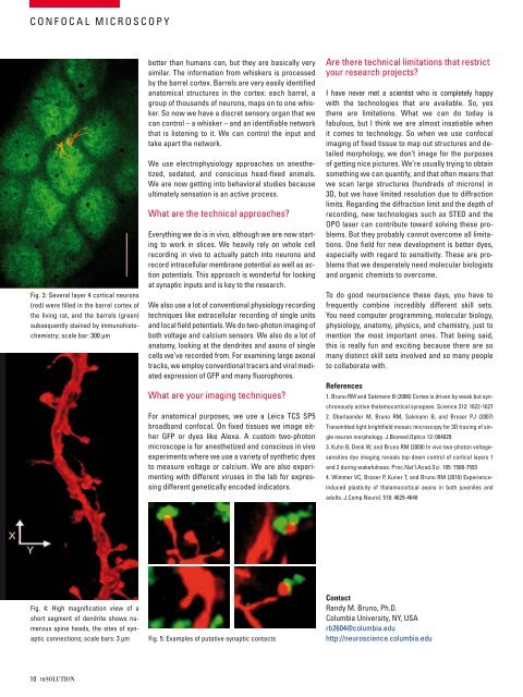
fig. 3: several layer 4 cortical neurons<br />
(red) were filled in the barrel cortex of<br />
the living rat, and the barrels (green)<br />
subsequently stained by immunohistochemistry;<br />
scale bar: 300 μm<br />
10 resolutioN<br />
better than humans can, but they are basically very<br />
similar. the information from whiskers is processed<br />
by the barrel cortex. barrels are very easily identified<br />
anatomical structures in the cortex: each barrel, a<br />
group of thousands of neurons, maps on to one whisker.<br />
so now we have a discret sensory organ that we<br />
can control – a whisker – and an identifiable network<br />
that is listening to it. We can control the input and<br />
take apart the network.<br />
We use electrophysiology approaches on anesthetized,<br />
sedated, and conscious head-fixed animals.<br />
We are now getting into behavioral studies because<br />
ultimately sensation is an active process.<br />
What are the technical approaches?<br />
everything we do is in vivo, although we are now starting<br />
to work in slices. We heavily rely on whole cell<br />
recording in vivo to actually patch into neurons and<br />
record intracellular membrane potential as well as action<br />
potentials. this approach is wonderful for looking<br />
at synaptic inputs and is key to the research.<br />
We also use a lot of conventional physiology recording<br />
techniques like extracellular recording of single units<br />
and local field potentials. We do two-photon imaging of<br />
both voltage and calcium sensors. We also do a lot of<br />
anatomy, looking at the dendrites and axons of single<br />
cells we’ve recorded from. for examining large axonal<br />
tracks, we employ conventional tracers and viral mediated<br />
expression of gfP and many fluorophores.<br />
What are your imaging techniques?<br />
for anatomical purposes, we use a leica tcs sP5<br />
broadband confocal. on fixed tissues we image either<br />
gfP or dyes like alexa. a custom two-photon<br />
microscope is for anesthetized and conscious in vivo<br />
experiments where we use a variety of synthetic dyes<br />
to measure voltage or calcium. We are also experimenting<br />
with different viruses in the lab for expressing<br />
different genetically encoded indicators.<br />
fig. 4: High magnification view of a<br />
short segment of dendrite shows numerous<br />
spine heads, the sites of synaptic<br />
connections; scale bars: 3 μm fig. 5: examples of putative synaptic contacts<br />
are there technical limitations that restrict<br />
your research projects?<br />
i have never met a scientist who is completely happy<br />
with the technologies that are available. so, yes<br />
there are limitations. What we can do today is<br />
fabulous, but i think we are almost insatiable when<br />
it comes to technology. so when we use confocal<br />
imaging of fixed tissue to map out structures and detailed<br />
morphology, we don’t image for the purposes<br />
of getting nice pictures. We’re usually trying to obtain<br />
something we can quantify, and that often means that<br />
we scan large structures (hundreds of microns) in<br />
3d, but we have limited resolution due to diffraction<br />
limits. regarding the diffraction limit and the depth of<br />
recording, new technologies such as sted and the<br />
oPo laser can contribute toward solving these problems.<br />
but they probably cannot overcome all limitations.<br />
one field for new development is better dyes,<br />
especially with regard to sensitivity. these are problems<br />
that we desperately need molecular biologists<br />
and organic chemists to overcome.<br />
to do good neuroscience these days, you have to<br />
frequently combine incredibly different skill sets.<br />
you need computer programming, molecular biology,<br />
physiology, anatomy, physics, and chemistry, just to<br />
mention the most important ones. that being said,<br />
this is really fun and exciting because there are so<br />
many distinct skill sets involved and so many people<br />
to collaborate with.<br />
References<br />
1. bruno rm and sakmann b (2006) cortex is driven by weak but synchronously<br />
active thalamocortical synapses. science 312: 1622-1627<br />
2. oberlaender m, bruno rm, sakmann b, and broser PJ (2007)<br />
transmitted light brightfield mosaic microscopy for 3d tracing of single<br />
neuron morphology. J.biomed.optics 12: 064029<br />
3. Kuhn b, denk W, and bruno rm (2008) in vivo two-photon voltagesensitive<br />
dye imaging reveals top-down control of cortical layers 1<br />
and 2 during wakefulness. Proc.nat’l.acad.sci. 105: 7588-7593<br />
4. Wimmer Vc, broser P, Kuner t, and bruno rm (2010) experienceinduced<br />
plasticity of thalamocortical axons in both juveniles and<br />
adults. J.comp.neurol. 518: 4629-4648<br />
Contact<br />
randy m. bruno, Ph.d.<br />
columbia university, ny, usa<br />
rb2604@columbia.edu<br />
http://neuroscience.columbia.edu