reSOLUTION_Research_09_Neuroscience - Leica Microsystems
reSOLUTION_Research_09_Neuroscience - Leica Microsystems
reSOLUTION_Research_09_Neuroscience - Leica Microsystems

Create successful ePaper yourself
Turn your PDF publications into a flip-book with our unique Google optimized e-Paper software.
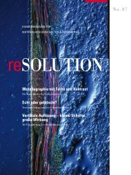
suPerresolution<br />
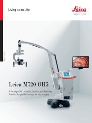
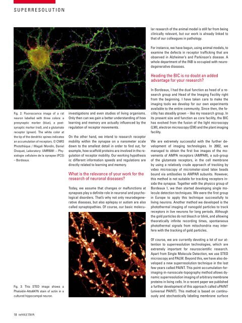
fig. 2: fluorescence image of a rat<br />
neuron labelled with three colors: a<br />
presynaptic marker (blue), a postsynaptic<br />
marker (red), and a glutamate<br />
receptor (green). the white color at<br />
the tip of the dendritic spines indicates<br />
an accumulation of receptors. © cnrs<br />
Photothèque / magali mondin, daniel<br />
choquet, laboratory: umr5<strong>09</strong>1 – Physiologie<br />
cellulaire de la synapse (Pcs)<br />
– bordeaux.<br />
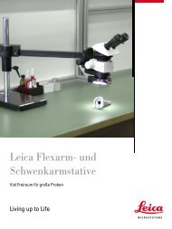
fig. 3: this sted image shows a<br />
Phaloidin-atto647n stain of actin in a<br />
cultured hippocampal neuron.<br />
18 resolutioN<br />
investigations and even studies of living organisms.<br />
only then can we gain a better understanding of how<br />
learning and memory are actually influenced by the<br />
regulation of receptor movements.<br />
on the other hand, we intend to research receptor<br />
mobility within the synapse on a nanometer scale<br />
down to the smallest detail in order to find out, for<br />
example, how scaffold proteins are involved in the regulation<br />
of receptor mobility. our working hypothesis<br />
is: different information speeds and regulations are<br />
directly related to learning and memory.<br />
What is the relevance of your work for the<br />
research of neuronal diseases?<br />
today, we assume that changes or malfunctions at<br />
synapses play a definite role in neuronal and psychological<br />
disorders. that’s why not only neurodegenerative<br />
diseases, but also epilepsy or autism are also<br />
called synaptopathies. of course, our basic molecu-<br />
lar research of the animal model is still far from being<br />
clinically relevant, but our work is already linked to<br />
that of our colleagues in pathology.<br />
for instance, we have begun, using animal models, to<br />
examine the defects in receptor trafficking that are<br />
observed in alzheimer’s and Parkinson’s disease. a<br />
whole department of the inb is occupied with neurodegenerative<br />
diseases.<br />
Heading the bic is no doubt an added<br />
advantage for your research?<br />
in bordeaux, i had the dual function as head of a research<br />
group and Head of the imaging facility right<br />
from the beginning. i have taken care to make the<br />
imaging tools we develop for our own experiments<br />
available to the entire community. since then, the facility<br />
has steadily grown – like my research group. in<br />
its present size and function as core facility, the bic<br />
has evolved from the fusion of the light microscopy<br />
(lm), electron microscopy (em) and the plant imaging<br />
facility.<br />
We are extremely successful with the further development<br />
of imaging technologies. in 2002, we<br />
managed to obtain the first live images of the movements<br />
of amPa receptors (amPar), a sub-group<br />
of the glutamate receptors, in the cell membrane<br />
by using a relatively crude approach of tracking by<br />
video microscopy of micrometer-sized latex beads<br />
bound via antibodies to amPar subunits. However,<br />
this method is not suitable for tracking receptors inside<br />
the synapse. together with the physics group of<br />
bordeaux 1, we then started developing single molecule<br />
detection techniques. We were the first group<br />
in europe to apply this technique successfully to<br />
living neurons. another method we developed is the<br />
photothermal imaging of nanogold particles to track<br />
receptors in live neurons for long periods. although<br />
the gold particles do not bleach or blink, and allowing<br />
theoretically infinite recording times, spontaneous<br />
photothermal signals from mitochondria may interfere<br />
with the tracking of gold particles.<br />
of course, we are currently devoting a lot of our attention<br />
to superresolution technologies, which are<br />
extremely important for neuroscientific research.<br />
apart from single molecule detection, we use sted<br />
microscopy and Palm. beyond this, we have also developed<br />
a new superresolution technique in the last<br />
few years called Paint. this point-accumulation-forimaging-in-nanoscale-topography<br />
method allows dynamic<br />
superresolution imaging of arbitrary membrane<br />
proteins in living cells. in a recent paper we published<br />
a further development of this approach called uPaint<br />
(universal Paint). this method is based on continuously<br />
and stochastically labeling membrane surface