reSOLUTION_Research_09_Neuroscience - Leica Microsystems
reSOLUTION_Research_09_Neuroscience - Leica Microsystems
reSOLUTION_Research_09_Neuroscience - Leica Microsystems

You also want an ePaper? Increase the reach of your titles
YUMPU automatically turns print PDFs into web optimized ePapers that Google loves.
HigH content screening<br />
CYTOO’s HCA Platform and <strong>Leica</strong> HCS A<br />
Normalize cells<br />
dr. constantin nelep and dr. Joanne young, cytoo cell architects<br />
the combination of cytoo’s Hca Platform with the leica Hcs a solution provides a highly reproducible high content analysis solution.<br />
conventional cell culture conditions on uniform adhesive supports lead to high morphological variability and uncontrolled cell migration.<br />
this situation is in sharp contrast to what is found in tissue, where cells respond to external spatial information (both from the extracellular<br />
matrix and neighboring cells) and adopt a reproducible polarized architecture, necessary for proper tissue function.<br />
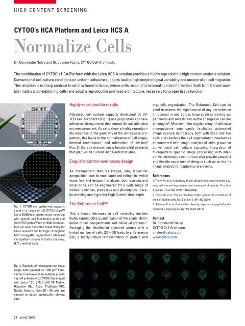
fig. 1: cytoo micropatterned supports<br />
come in a range of: (a) cytoochips<br />
(up to 20,000 micropatterns per coverslip<br />
with special cell localization grid) and<br />
(b) cytooplates (up to 5000 micropattern<br />
per well) dedicated respectively for<br />
basic research and for High-throughput<br />
microscopy/Hcs applications. standard<br />
micropattern shapes include crossbow,<br />
H, y, l and full disks.<br />
fig. 2: example of micropatterned Hela<br />
single cells (seeded on 1100 µm 2 fibronectin<br />
crossbow-shape patterns providing<br />
cell polarization). cytoochip imaged<br />
with leica tsc sPe / las af matrix.<br />
objective 63x. actin: Phalloidin-fitc;<br />
nuclei: Hoechst. only 20 – 50 cells are<br />
needed to obtain statistically relevant<br />
data.<br />
24 resolutioN<br />
a<br />
b<br />
Highly reproducible results<br />
advanced cell culture supports developed by cytoo<br />
cell architects (fig. 1) use proprietary concave<br />
adhesive micropatterns that control the cell adhesion<br />
microenvironment. as cells show a highly reproducible<br />
response to the geometry of the adhesive micropattern,<br />
this leads to the normalization of cell shape,<br />
internal architecture 1 and orientation of division 2<br />
(fig. 2) thereby overcoming a fundamental obstacle<br />
that plagues all current High content studies.<br />
exquisite control over assay design<br />
as micropattern features (shape, size, molecular<br />
composition) can be modulated and refined in myriad<br />
ways, live and endpoint analyses, both existing and<br />
novel ones, can be engineered for a wide range of<br />
cellular activities, processes and phenotypes, thereby<br />
enabling much greater High content data depth.<br />
the reference cell<br />
this dramatic decrease in cell variability enables<br />
highly reproducible quantification of the spatial distribution<br />
of cell compartments and individual proteins 1,3 .<br />
averaging the distribution observed across only a<br />
limited number of cells (20 – 50) leads to a reference<br />
cell, a highly robust representation of protein and<br />
organelle organization. the reference cell can be<br />
used to assess the significance of any perturbation<br />
introduced in and across large scale screening experiments<br />
and reveals any subtle changes in cellular<br />
phenotype 2 . moreover, the regular array of adhesive<br />
micropatterns significantly facilitates automated<br />
image capture microscopy both with fixed and live<br />
cells and resolves the cell segmentation headaches<br />
encountered with image analysis of cells grown on<br />
conventional cell culture supports. integration of<br />
micropattern specific image processing with interactive<br />
microscope control can also provide powerful<br />
and flexible experimental designs such as on-the-fly<br />
image analysis for capturing rare events.<br />
References<br />
1. thery, m. et al. anisotropy of cell adhesive microenvironment governs<br />
cell internal organization and orientation of polarity. Proc natl<br />
acad sci u s a 103, 19771-19776 (2006).<br />
2. thery, m. et al. the extracellular matrix guides the orientation of<br />
the cell division axis. nat cell biol 7, 947-953 (2005).<br />
3. schauer, K. et al. Probabilistic density maps to study global endomembrane<br />
organization. nat methods (2010).<br />
Contact<br />
dr. constantin nelep<br />
cytoo cell architects<br />
cnelep@cytoo.com<br />
www.cytoo.com