reSOLUTION_Research_09_Neuroscience - Leica Microsystems
reSOLUTION_Research_09_Neuroscience - Leica Microsystems
reSOLUTION_Research_09_Neuroscience - Leica Microsystems

You also want an ePaper? Increase the reach of your titles
YUMPU automatically turns print PDFs into web optimized ePapers that Google loves.
suPerresolution<br />
Discovering Cellular Morphology Beyond the Diffraction Limit<br />
confocal Nanoscopy Goes<br />
Multicolor<br />
dr. Jochen J. sieber, leica microsystems<br />
scientists strive to understand the architecture of life. they want to learn how biological structures are arranged in respect to one<br />
another. do they co-localize within or are they excluded from the same superstructure? does localization follow a special pattern<br />
and how does the overall arrangement reflect the biological function? multicolor superresolution imaging allows these fundamental<br />
questions to be addressed by far-field fluorescence microscopy in unprecedented detail.<br />
14 resolutioN<br />
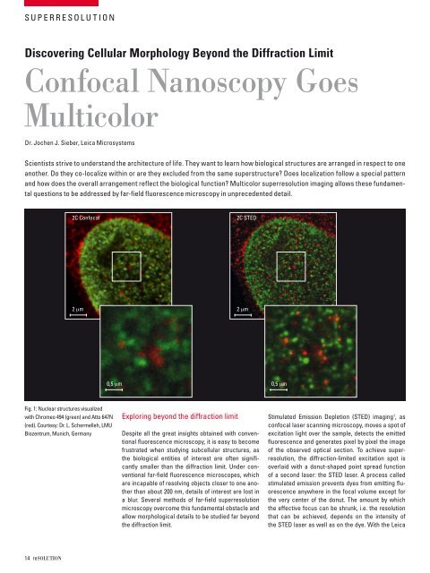
2c confocal<br />
2 mm<br />
0,5 mm<br />
fig. 1: nuclear structures visualized<br />
with chromeo 494 (green) and atto 647n<br />
(red). courtesy: dr. l. schermelleh, lmu<br />
biozentrum, munich, germany<br />
exploring beyond the diffraction limit<br />
2c sted<br />
2 mm<br />
despite all the great insights obtained with conventional<br />
fluorescence microscopy, it is easy to become<br />
frustrated when studying subcellular structures, as<br />
the biological entities of interest are often significantly<br />
smaller than the diffraction limit. under conventional<br />
far-field fluorescence microscopes, which<br />
are incapable of resolving objects closer to one another<br />
than about 200 nm, details of interest are lost in<br />
a blur. several methods of far-field superresolution<br />
microscopy overcome this fundamental obstacle and<br />
allow morphological details to be studied far beyond<br />
the diffraction limit.<br />
0,5 mm<br />
stimulated emission depletion (sted) imaging 1 , as<br />
confocal laser scanning microscopy, moves a spot of<br />
excitation light over the sample, detects the emitted<br />
fluorescence and generates pixel by pixel the image<br />
of the observed optical section. to achieve superresolution,<br />
the diffraction-limited excitation spot is<br />
overlaid with a donut-shaped point spread function<br />
of a second laser: the sted laser. a process called<br />
stimulated emission prevents dyes from emitting fluorescence<br />
anywhere in the focal volume except for<br />
the very center of the donut. the amount by which<br />
the effective focus can be shrunk, i.e. the resolution<br />
that can be achieved, depends on the intensity of<br />
the sted laser as well as on the dye. With the leica