reSOLUTION_Research_09_Neuroscience - Leica Microsystems
reSOLUTION_Research_09_Neuroscience - Leica Microsystems
reSOLUTION_Research_09_Neuroscience - Leica Microsystems

Create successful ePaper yourself
Turn your PDF publications into a flip-book with our unique Google optimized e-Paper software.
stereotaXic<br />
<strong>Leica</strong> Angle Two for Precise 3D Brain Access<br />
avoid confounding,<br />
improve accuracy<br />
charles W. scouten, Ph.d., leica microsystems<br />
Humans and larger animals have great variation in brain size, and even in the relative size of different structures within brains of a<br />
given size. fortunately for neuroscience research, rodent brains are made with more quality control; they are much more uniform<br />
animal to animal within a species and age range. this uniformity makes it possible to create coordinate maps or atlases of the brain,<br />
showing the three dimensional location of every structure in the brain relative to a zero point. for common age ranges among common<br />
laboratory species like rats, mice and hamsters, bregma, a point on the skull where the bone plates have grown together, is<br />
usually used as the zero point. together with a suitable manipulator and oriented head holder, such maps can be used to position<br />
probes at a preplanned site in the brains of several rodents. the instrument for moving a probe to a given coordinate in space – inside<br />
the brain – is a stereotaxic instrument.<br />
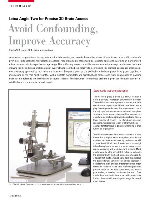
fig. 1: the leica angle two stereotaxic instrument improves accuracy in small animal brain surgery.<br />
32 resolutioN<br />
stereotaxic instrument function<br />
the reason to place a probe at a known location in<br />
brain is to study localization of function in the brain.<br />
the brain is a very heterogeneous structure, and different<br />
sites and regions have different functional roles to<br />
play. learning to understand that organization is one of<br />
the major goals of neuroscience, and requires regional<br />
studies of brain. Various stains and immune-markers<br />
can show regional chemical variation in brain. stereotaxic<br />
insertion of probes – for stimulation, injection,<br />
recording, microdialysis, lesion or other functions – is<br />
an important technique to gain understanding of brain<br />
functional organization.<br />
traditional stereotaxic instruments consist of a head<br />
holder that is aligned with a manipulator, with the manipulator<br />
movements measured by vernier scales with<br />
a resolution of 100 microns. a newer idea is to use digital<br />
scales in place of vernier, and obtain easier, less error<br />
prone reading and resolution to 10 microns. manipulators<br />
can be tilted and rotated, but doing so breaks<br />
the alignment with the head holder, and changes the<br />
distance that must be moved along each axis to reach<br />
the desired target. sometimes an angled approach is<br />
necessary, to avoid arteries, or other structures important<br />
to the research. in this case, the investigator must<br />
perform math on the atlas coordinates, and usually<br />
pilot studies, to develop coordinates that work. once<br />
that is done, the manipulator is locked in place, since<br />
further changes in tilt would again change the coordinates<br />
needed.