reSOLUTION_Research_09_Neuroscience - Leica Microsystems
reSOLUTION_Research_09_Neuroscience - Leica Microsystems
reSOLUTION_Research_09_Neuroscience - Leica Microsystems

You also want an ePaper? Increase the reach of your titles
YUMPU automatically turns print PDFs into web optimized ePapers that Google loves.
stereotaXic<br />
34 resolutioN<br />
hairs mark the target, and blue cross-hairs mark the tip<br />
location. the user can see on screen where the tip is<br />
in brain at any given time, and watch the graphics as<br />
the tip approaches the target. for angled approaches,<br />
the atlas will update plates to the one in which the tip is<br />
in or over. a circle, then a double circle, alerts the user<br />
that the target is reached to avoid overshoot.<br />
Virtual skull flat<br />
the head must be held in a standard position. usually,<br />
this is skull flat, as defined by george Paxinos. With<br />
conventional stereotaxic instruments, the user touches<br />
the probe down at bregma, takes a zero reading (or zeroes<br />
a digital display), then moves the probe caudally<br />
to the lambda point and touches the tip down again.<br />
Hopefully, both points will be at the same vertical reading.<br />
if not, a repetitive trial and error adjustment of a<br />
dovetail supporting the animal’s teeth begins, in order<br />
to adjust head tilt to the zero plane. eventually, the<br />
user decides it is close enough. in contrast, with the<br />
Virtual skull flat feature found only on the leica angle<br />
two, the user touches down at bregma and lambda,<br />
and the computer calculates how tilted the head is<br />
and corrects the linear axes distance to the target for<br />
where it is, given the head tilt. there is no need to ever<br />
physically adjust the degree of head tilt; the computer<br />
directs the user to the correct target even though the<br />
head is tilted.<br />
named target icons<br />
a user may save a set of target coordinates as an icon<br />
on the screen below the atlas. to reset those coordinates<br />
as the desired target, the user double clicks on<br />
the icon when in set target mode. this is very useful<br />
when there are multiple experiments in progress, and<br />
avoids mistakes in re-entering coordinates.<br />
bilateral surgeries<br />
after performing a surgery on one side of the brain, to<br />
position a probe in the symmetrical opposite side of the<br />
brain, the user clicks a minus sign after the ml (medial<br />
to lateral) coordinate to reset the target to the same<br />
point on the other side. there is no need to return to<br />
bregma or remeasure head tilt. the computer displays<br />
how to reach that point on the other side, even if the<br />
manipulator is rotated or tilted.<br />
final report<br />
after each surgery, the user clicks on the export position<br />
button to save the present coordinates and tilts to<br />
a line in a file. the animal id, date, and other information<br />
is typed in as needed. at the end of the experiment,<br />
the user can print a one page report with a line<br />
for every animal, and an accurate listing of the exact<br />
atlas coordinates, and the tilt and rotation, at which the<br />
manipulation was performed.<br />
References<br />
1. Jacob W. skovira, John H. mcdonough, and tsung-ming shih. Protection<br />
against sarin-induced seizures in rats by direct brain microinjection<br />
of scopolamine, midazolam or mK-801. Journal of molecular<br />
neuroscience, Volume 40, numbers 1-2 / January, 2010.<br />
2. y. sztainberg, y. Kuperman, m. tsoory, m. lebow, and a. chen . the<br />
anxiolytic effect of environmental enrichment is mediated via amygdalar<br />
crf receptor type 1. molecular Psychiatry (19 January 2010) |<br />
doi:10.1038/mp.20<strong>09</strong>.151.<br />
3. martha W. bagnall, brian zingg, alexandra sakatos, setareh H.<br />
moghadam, Hanns ulrich zeilhofer, and sascha du lac1. glycinergic<br />
Projection neurons of the cerebellum. the Journal of neuroscience,<br />
august 12, 20<strong>09</strong>, 29(32):10104-10110.<br />
4. thomas l. Kash, William P. nobis, robert t. matthews, and danny g.<br />
Winder. dopamine enhances fast excitatory synaptic transmission in<br />
the extended amygdala by a crf-r1-dependent Process. the Journal<br />
of neuroscience, december 17, 2008, 28(51):13856-13865.<br />
to see a video of the leica angle two demonstration,<br />
go to www.myneurolab.com<br />
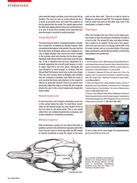
fig. 3: location of bregma and lambda as<br />
defined by dr. george Paxinos (george<br />
Paxinos and charles Watson: the rat<br />
brain in stereotaxic coordinates,<br />
academic Press, new york, 1998, p. 11).<br />
courtesy of academic Press.