reSOLUTION_Research_09_Neuroscience - Leica Microsystems
reSOLUTION_Research_09_Neuroscience - Leica Microsystems
reSOLUTION_Research_09_Neuroscience - Leica Microsystems
Create successful ePaper yourself
Turn your PDF publications into a flip-book with our unique Google optimized e-Paper software.
confocal microscoPy<br />
OPO IR Laser in Developmental Biology<br />
deep tissue imaging – From<br />
Visible to iR Wavelengths<br />
dr. andrea Pfeifer and dr. bernd sägmüller, leica microsystems<br />
embryonic development relies on genetic coding and non-coding informations. in particular, physical forces generated by blood<br />
flow are critical for proper development of the cardiovascular system. to gain new insight into the fundamental control of cell<br />
response to physical changes and to study the dynamics and roles of biological flow during the development of the zebrafish,<br />
dr. Julien Vermot established his lab last year at the institute of genetics and molecular and cellular biology (igbmc) in strasbourg,<br />
france. He belongs to the first lab to use the leica dm6000 cfs equipped with an oPo/ti:sa infrared source for deep tissue<br />
imaging and infared excitation wavelengths up to 1300 nm.<br />
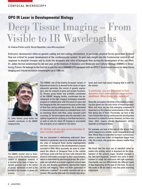
dr. Julien Vermot, group leader and<br />
scientific coordinator of the igbmc<br />
imaging facility<br />
the igbmc, located close to strasbourg,<br />
is one of the leading european<br />
centers of biomedical research. it<br />
is devoted to the study of higher eukaryotic<br />
genomes and to the control<br />
of genetic expression as well as the<br />
analysis of the function of genes and<br />
proteins.<br />
6 resolutioN<br />
the igbmc, one of the leading european centers of<br />
biomedical research, is devoted to the study of higher<br />
eukaryotic genomes, the control of genetic expression,<br />
and the analysis of gene and protein functions.<br />
dr. Vermot, group leader and scientific coordinator<br />
of the igbmc imaging facility, coordinates the development<br />
of the light imaging techniques scientific<br />
program in collaboration with the board of users and<br />
the imaging facility. His research focuses on the roles<br />
of fluid flow during embryogenesis. He is interested<br />
in characterizing fluid motion at a detailed level, such<br />
as watching blood cells flow, using resonant point<br />
scanning. dr. Vermot explains why the zebrafish is the<br />
optimal organism for studying in vivo fluid mechanics,<br />
as well as why he chose ir imaging to accomplish<br />
this and gives us an outlook for the future.<br />
dr. Vermot, can you give us an overview of<br />
your current research?<br />
We are interested in addressing embryonic development;<br />
in particular, we try to understand what are<br />
the roles of biological flows during organogenesis<br />
and their connections to the developmental program<br />
encoded in dna. more precisely, we want to understand<br />
the effects of biological flow at the cellular<br />
and tissue scale and find out how cells interpret the<br />
physical information provided by their environments,<br />
which is dominated by mechanical stress. We principally<br />
use zebrafish as a model organism and are keen<br />
to use and develop quantitative approaches based<br />
on live cell imaging. “How are flows generated in<br />
embryonic cavities?” is another question we try to<br />
answer. We usually deal with micrometer size struc-<br />
tures and need high-speed imaging that is safe for<br />
the animal.<br />
in particular, you are interested in fluid<br />
dynamics from embryonic development to<br />
adulthood. What drives your research?<br />
basically, we explore the limits of the models, proposing<br />
that genes are the only driver of morphogenesis.<br />
more and more, we see that emerging complexity is<br />
dependent on the physical environments of the cells,<br />
flow being one of them. Practically, we look at the<br />
role of blood flow during cardiovascular development<br />
because it is related to human diseases, but there are<br />
many other organs whose development strictly<br />
depends on biological flows.<br />
for example, we look at the role of cilia driven flow,<br />
which happens at a smaller scale compared to blood<br />
flow. blood flow is controlled by heart contractions,<br />
which is about two orders of magnitude bigger than<br />
cilia. as a result, cilia generate a slower, smaller flow<br />
profile.<br />
We found that the inner ear of zebrafish relies on<br />
motile cilia activity, which is important for the development<br />
of the sensory organ. another example is<br />
the ‘left-right-organizer,’ present very early in development<br />
to break the embryonic left-right symmetry.<br />
importantly, we can differentiate the different types<br />
of flow depending on the type of cilia beat. to see at<br />
this scale in 3d we need a very fast point scanning<br />
instrument.


