reSOLUTION_Research_09_Neuroscience - Leica Microsystems
reSOLUTION_Research_09_Neuroscience - Leica Microsystems
reSOLUTION_Research_09_Neuroscience - Leica Microsystems

Create successful ePaper yourself
Turn your PDF publications into a flip-book with our unique Google optimized e-Paper software.
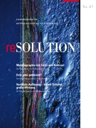
customer magazine for neuroscience<br />
and cell biology<br />
resolutioN<br />
CARS and Confocal – a Successful Affair<br />
<strong>Leica</strong> TCS CARS Opens New Ways for <strong>Research</strong><br />
Sniffing Out the Secrets of Social Behavior<br />
The New Understanding of Olfactory Neurosensorics<br />
Avoid Confounding, Improve Accuracy<br />
<strong>Leica</strong> Angle Two for Precise 3D Brain Access<br />
No. <strong>09</strong><br />
special editioN
editorial<br />
contents<br />
2 resolutioN<br />
dear readers,<br />
the cell – the elementary unit of all living creatures and the object of desire of biomedical research all<br />
around the globe. a human adult consists of a hundred thousand billion cells, and there are roughly 220<br />
different types of cell. about 100 billion nerve cells perform their task in the human brain. How the smallest<br />
sub-units and molecular complexes of a cell work, how it communicates with other cells via the cell membrane,<br />
how changes on molecular level are connected with pathogenesis and how neuronal networks are<br />
capable of fascinating achievements such as learning and memory – there is still a long road to travel before<br />
these mysteries are fully solved. on the way, scientists are encountering many exciting questions, whose<br />
answers they are putting together like the pieces of a puzzle.<br />
We have therefore devoted this special issue of resolution to neuroscience and cell biology. We introduce<br />
you to new technologies such as cars microcopy, the oPo and the 2c sted. users report on their<br />
research approaches and results. altogether, we want this issue to give you an idea of our spectrum of<br />
system solutions and products that are helping biologists and neuroscientists make new discoveries.<br />
We, leica microsystems, are playing our part by cooperating closely with scientists and users to provide<br />
scientists with technologies and methods in the form of user-friendly systems that deliver fast and reproducible<br />
results.<br />
Have fun reading!<br />
anja schué didier goor<br />
communications & corporate identity european marketing manager research<br />
confocal microscoPy<br />
CARS and Confocal – a Successful Affair 03<br />
leica tcs cars opens new Ways<br />
for research<br />
Deep Tissue Imaging – From Visible to<br />
IR Wavelengths 06<br />
oPo ir laser in developmental biology<br />
Exploring the Concert of Neuronal Activities <strong>09</strong><br />
leica tcs sP5 supports research of<br />
synapses and cortical circuits<br />
suPerresolution<br />
The Missing Link to the Nanocosm of Life 11<br />
superresolution opens up new Perspectives<br />
in neurobiology<br />
Confocal Nanoscopy Goes Multicolor 14<br />
discovering cellular morphology beyond<br />
the diffraction limit<br />
Restless Receptors 17<br />
new insights into the dynamic<br />
organization of synapses<br />
HigH content screening<br />
Amplify the Power of Imaging 20<br />
High content screening automation for<br />
confocal and Widefield microscopes<br />
Combined Forces Improve Image Analysis 23<br />
definiens developer Xd and leica Hcs a<br />
Normalize Cells 24<br />
cytoo’s Hca Platform and leica Hcs a<br />
Looking for Rare Cells or Cellular Events? 25<br />
Picovitro Plates and leica Hcs a<br />
Widefield microscoPy<br />
Sniffing Out the Secrets of Social Behavior 26<br />
the new understanding of olfactory<br />
neurosensorics<br />
The Morbus Parkinson Puzzle 29<br />
single-cell analysis after laser microdissection<br />
stereotaXic<br />
Avoid Confounding, Improve Accuracy 32<br />
leica angle two for Precise 3d brain access<br />
Impact Neurotrauma – From War to Sports 35<br />
new tool to study traumatic brain injury<br />
Product neWs<br />
Cutting Edge Precision 37<br />
leica Vibratome series<br />
Explore Life in All Dimensions 38<br />
leica dmi6000 b with adaptive focus control<br />
registration 16<br />
imPrint 37
<strong>Leica</strong> TCS CARS Opens New Ways for <strong>Research</strong><br />
caRs and confocal –<br />
a successful affair<br />
dr. stefanie landwehr and Vanessa lurquin, Ph.d., leica microsystems<br />
since the advent of confocal microscopy, scientists have gained many new insights that lead to a deeper understanding of how<br />
life works. the most important drawback of single-photon and multiphoton confocal microscopy is the need to label the specimen.<br />
cars (coherent anti-stokes raman spectroscopy) addresses this issue because it is non-toxic, non-destructive, and minimally<br />
invasive. With the implementation of cars into the leica tcs sP5 broadband confocal system, leica microsystems adds new innovation<br />
to its product portfolio for live cell applications and small animal studies.<br />
staining processes have constraints: dyes bleach<br />
with time, can be phototoxic, and can influence subsequent<br />
research methods and camouflage subsequent<br />
research information. Processes with stained<br />
specimens are not suitable for long term analysis:<br />
atmospheric conditions destroy dyes, and dyes alter<br />
when in contact with air or moisture – any change<br />
to environmental condition will influence the experimental<br />
result. also, the staining process is time<br />
consuming. some samples cannot be stained at all,<br />
because the attachment of a fluorophore or the presence<br />
of the dye changes the functionality of the molecule<br />
or the organism.<br />
coherent anti-stokes raman<br />
spectroscopy – the technique<br />
cars is a third-order, nonlinear process that involves<br />
a pump beam at a frequency of ωp and a stokes<br />
beam at a frequency of ωs. the specimen is stimulated<br />
through a wave-mixing process. the anti-stokes<br />
signal at ωas=2ωp–ωs is generated in the phase matching<br />
direction as vibrational contrast at the frequency<br />
difference ∆ν=vsp–vs between the pump beam<br />
Energy<br />
PUMP<br />
Virtual state 1<br />
E<br />
Vibrational } states<br />
G<br />
G<br />
Energy<br />
PUMP<br />
STOKES<br />
and the stokes beam. this equals the frequency of<br />
the vibrational energy of a particular chemical bond.<br />
live molecular profiling with cars<br />
the main applications of cars microscopy are<br />
found in biological, pharmaceutical, and dermatological<br />
research, biomedical imaging, food processing,<br />
and materials science. its potential has been<br />
demonstrated for various biomedical applications,<br />
such as the imaging of lipid transport, protein concentrations,<br />
dna, rna, tissue in a living organism,<br />
and order in liquid crystals. by integrating cars<br />
technology into the leica tcs sP5, leica micro-<br />
systems offers the latest technology combined with<br />
an easy-to-use confocal system.<br />
High resolution images<br />
confocal microscoPy<br />
the conventional scanner is optimized for morphological<br />
studies as in brain and skin, or for imaging subcellular<br />
features such as the cytoskeleton. it allows<br />
sampling up to 8196 x 8196 pixels per image, combining<br />
a large field of view with high resolution. also, fig. 1: cars energy diagram<br />
Virtual state 1<br />
E<br />
} Energy<br />
Vibrational<br />
energy<br />
PUMP<br />
STOKES<br />
PROBE<br />
Virtual state 2<br />
G<br />
E<br />
Energy<br />
PUMP<br />
STOKES<br />
PROBE<br />
CARS<br />
E<br />
Virtual state 2<br />
}<br />
G<br />
Vibrational energy<br />
which leads to an image<br />
neuroscience 3
confocal microscoPy<br />
fig. 2: skin of a mouse ear (maximum Projection),<br />
raman shift at 2849 cm -1 (which corresponds to cH 2<br />
stretching vibration)<br />
4 resolutioN<br />
the speed of the scanner can be adapted from 400 Hz<br />
to 2800 Hz in bidirectional mode.<br />
at the skin surface (fig. 2), long filaments corresponding<br />
to the hairs with chromospheres can be seen<br />
with a very strong signal because hairs are covered<br />
with oil. at the skin surface, a bright polygonal pattern<br />
outlines the corneocytes forming the top layer of<br />
the skin, the corneum. the signal arises from the intercellular<br />
space rich in lipids, cholesterol, and ceramids.<br />
in a second layer, bright structures surrounding<br />
the roots of hair are detected, the sebaceous glands.<br />
they are multicellular compartments packed with<br />
sebum reservoirs containing triglycerides and wax<br />
esters. at 70 – 80 µm of the surface, adipocytes, rich in<br />
fat, are found in the dermis. because the cars signal<br />
is generated only at the focal point, the 3d imaging<br />
capability is shown with the maximum projection.<br />
cars at video rate<br />
the resonant scanner on the leica tcs cars system<br />
provides the benefits of compact design and fast frame<br />
recording. the resonant scanner is based on the true<br />
confocal concept of point-illumination and point-observation.<br />
the resonant scanner allows a speed of 16000 Hz<br />
frequency in bidirectional mode. at a frame size of<br />
512 x 512 pixels, the system acquires 29 images per<br />
second. With lower sampling, the speed can be in-<br />
fig. 3: unstained skin of a mouse ear imaged with<br />
leica tcs cars. sebacouce glands are multicellular<br />
compartments, which contain triglycerides and<br />
wax ester. While non-lipid components appear as<br />
dark areas in the image, the fatty components are<br />
much brighter. due to their cH 2 bondings they can be<br />
perfectly imaged with cars and lead to sharp, crisp<br />
imaging results.<br />
creased up to 290 frames per second at a resolution<br />
of 512 x 32 pixels. dynamic processes with high time<br />
resolution can be imaged and measured or a linescan<br />
can be taken at full rate.<br />
an averaging function is available on the leica tcs<br />
cars system. applying averaging improves the signal-to-noise<br />
ratio, which is especially useful in the<br />
case of the resonant scanner. it is possible to find<br />
the right compromise between image quality and<br />
acquisition speed, depending on the imaging requirements.<br />
as with the leica tcs cars the amplitude<br />
of the confocal resonant scanner is tunable, which<br />
allows it to zoom in by applying smaller amplitudes.<br />
With this feature it is possible to focus into regions<br />
of interest while acquiring images at video-rate. the<br />
pan function – another helpful device to quickly move<br />
into interesting areas, which are not necessarily in<br />
the center of the microscopic field – is also available<br />
with the resonant scanner.<br />
subsequently cars microscopy opens new ways<br />
to visualize structures based on intrinsic vibrational<br />
properties without staining or labeling the specimen.<br />
the specimen does not suffer from perturbation by<br />
the dye or photo-bleaching. cars opens new methods<br />
of research, especially in cell biology, neurosciences,<br />
pharmacology, dermatology, and medical<br />
imaging.<br />
fig. 4: the cross-section of a mouse tail imaged labelfree<br />
with epi-cars (reflected light, red areas) and<br />
f-sHg (transmitted light, green areas) combining<br />
information about lipids and structural properities of<br />
the specimen.
Making CARS Microscopy Accessible<br />
“Many research areas, whether from a life science or materials science background,<br />
require fast, non-invasive imaging with high spatial resolution, high molecular<br />
specificity, and high sensitivity. As an optical method by which contrast is<br />
generated on the basis of spectroscopic properties intrinsic to the sample, CARS<br />
microscopy can offer all of this. In neuroscience, for example, CARS microscopy<br />
can have an impact comparable to two-photon microscopy, except that it does<br />
not rely on the introduction of fluorescent labels.<br />
The advent of commercial CARS microscopy systems is certainly a major step<br />
toward making the technique accessible to researchers interested in its application.<br />
The <strong>Leica</strong> TCS CARS is especially attractive, since it offers hands-off<br />
operation while allowing the full potential of CARS microscopy to be used. Its<br />
complete integration into the well-established <strong>Leica</strong> TCS SP5 environment<br />
allows every user familiar with confocal microscopy to rapidly take advantage of<br />
all new possibilities offered by CARS microscopy.”<br />
fig. 5: the deviation of fatty components in food can<br />
be shown with leica tcs cars. the maximum projection<br />
shows that lipid components (red) are located<br />
in all regions of a potato chip but not only at the<br />
surface. green parts in the chip provide structural<br />
information taken with only an multiphoton laser.<br />
fig. 6: deviation of lipid droplets in cream, overlay<br />
image. the green background shows the water (acquired<br />
at 3150 cm -1 ) while the red dots are fatty components<br />
(acquired at 2850 cm -1 ).<br />
confocal microscoPy<br />
Prof. andreas zumbusch, department<br />
of chemistry, university of Konstanz,<br />
germany, heads a research group for<br />
Physical chemistry. He focuses on<br />
single molecule fluorescence spectroscopy<br />
and microscopy, as well as<br />
on the development and application of<br />
non-linear optical microscopy.<br />
fig. 7: these cars pictures show lipid-rich adipocytes<br />
of the subcutaneous fat layer of mouse skin.<br />
the left image is taken at full speed of the resonant<br />
scanner, acquiring images at a rate of 29 images /<br />
second in bi-directional mode, i.e., at video rate.<br />
Within the pixel dwell time of 120 ns only a few cars<br />
photons are detected, resulting in a noisy image. the<br />
image on the right corresponds to averaging over 30<br />
images taken with the resonant scanner, i.e., corresponds<br />
to the image quality of the non-resonant<br />
scanner.<br />
neuroscience 5
confocal microscoPy<br />
OPO IR Laser in Developmental Biology<br />
deep tissue imaging – From<br />
Visible to iR Wavelengths<br />
dr. andrea Pfeifer and dr. bernd sägmüller, leica microsystems<br />
embryonic development relies on genetic coding and non-coding informations. in particular, physical forces generated by blood<br />
flow are critical for proper development of the cardiovascular system. to gain new insight into the fundamental control of cell<br />
response to physical changes and to study the dynamics and roles of biological flow during the development of the zebrafish,<br />
dr. Julien Vermot established his lab last year at the institute of genetics and molecular and cellular biology (igbmc) in strasbourg,<br />
france. He belongs to the first lab to use the leica dm6000 cfs equipped with an oPo/ti:sa infrared source for deep tissue<br />
imaging and infared excitation wavelengths up to 1300 nm.<br />
dr. Julien Vermot, group leader and<br />
scientific coordinator of the igbmc<br />
imaging facility<br />
the igbmc, located close to strasbourg,<br />
is one of the leading european<br />
centers of biomedical research. it<br />
is devoted to the study of higher eukaryotic<br />
genomes and to the control<br />
of genetic expression as well as the<br />
analysis of the function of genes and<br />
proteins.<br />
6 resolutioN<br />
the igbmc, one of the leading european centers of<br />
biomedical research, is devoted to the study of higher<br />
eukaryotic genomes, the control of genetic expression,<br />
and the analysis of gene and protein functions.<br />
dr. Vermot, group leader and scientific coordinator<br />
of the igbmc imaging facility, coordinates the development<br />
of the light imaging techniques scientific<br />
program in collaboration with the board of users and<br />
the imaging facility. His research focuses on the roles<br />
of fluid flow during embryogenesis. He is interested<br />
in characterizing fluid motion at a detailed level, such<br />
as watching blood cells flow, using resonant point<br />
scanning. dr. Vermot explains why the zebrafish is the<br />
optimal organism for studying in vivo fluid mechanics,<br />
as well as why he chose ir imaging to accomplish<br />
this and gives us an outlook for the future.<br />
dr. Vermot, can you give us an overview of<br />
your current research?<br />
We are interested in addressing embryonic development;<br />
in particular, we try to understand what are<br />
the roles of biological flows during organogenesis<br />
and their connections to the developmental program<br />
encoded in dna. more precisely, we want to understand<br />
the effects of biological flow at the cellular<br />
and tissue scale and find out how cells interpret the<br />
physical information provided by their environments,<br />
which is dominated by mechanical stress. We principally<br />
use zebrafish as a model organism and are keen<br />
to use and develop quantitative approaches based<br />
on live cell imaging. “How are flows generated in<br />
embryonic cavities?” is another question we try to<br />
answer. We usually deal with micrometer size struc-<br />
tures and need high-speed imaging that is safe for<br />
the animal.<br />
in particular, you are interested in fluid<br />
dynamics from embryonic development to<br />
adulthood. What drives your research?<br />
basically, we explore the limits of the models, proposing<br />
that genes are the only driver of morphogenesis.<br />
more and more, we see that emerging complexity is<br />
dependent on the physical environments of the cells,<br />
flow being one of them. Practically, we look at the<br />
role of blood flow during cardiovascular development<br />
because it is related to human diseases, but there are<br />
many other organs whose development strictly<br />
depends on biological flows.<br />
for example, we look at the role of cilia driven flow,<br />
which happens at a smaller scale compared to blood<br />
flow. blood flow is controlled by heart contractions,<br />
which is about two orders of magnitude bigger than<br />
cilia. as a result, cilia generate a slower, smaller flow<br />
profile.<br />
We found that the inner ear of zebrafish relies on<br />
motile cilia activity, which is important for the development<br />
of the sensory organ. another example is<br />
the ‘left-right-organizer,’ present very early in development<br />
to break the embryonic left-right symmetry.<br />
importantly, we can differentiate the different types<br />
of flow depending on the type of cilia beat. to see at<br />
this scale in 3d we need a very fast point scanning<br />
instrument.
How is the new leica system used for your<br />
research?<br />
zebrafish is a very imaging friendly animal. the larvae<br />
are transparent and easy to culture under a microscope.<br />
However, structures that generate flows are<br />
often localized in deep, light scattering tissues. in this<br />
case two-photon imaging is the modality of choice<br />
because it allows deep imaging with limited phototoxicity.<br />
the leica dm6000 cfs with an oPo helps<br />
to perform multicolor two-photon imaging using<br />
conventional fluorescent proteins, such as gfP and<br />
rfP. second Harmonic generation is possible. it also<br />
allows us to manipulate the tissue through femtosecond<br />
cell ablation where you can target single cells<br />
in the tissue and perform imaging. this technique is<br />
challenging, it may not work all the time but can give<br />
interesting results. two-photon microscopy is usually<br />
used to look deep into the sample. furthermore, as<br />
opposed to single-photon microscope techniques,<br />
two-photon imaging illuminates only the part of the<br />
sample you image, thereby it limits photobleaching<br />
and photodamage.<br />
today, many research projects are directed<br />
towards neurodegenerative,<br />
cancer, and lifestyle related diseases. How<br />
is your research connected with these<br />
diseases?<br />
most of us at igbmc work on basic research.<br />
However, many of our projects are linked to human<br />
diseases. most of the basic mechanisms in biology,<br />
when they go wrong, lead to such problems. We look<br />
at the origin of those diseases and do work that will<br />
lay the foundation for further and specific research to<br />
develop therapies. to do so, imaging is key and will be<br />
even more important in the future.<br />
Contact:<br />
dr. Julien Vermot<br />
igbmc imaging facility<br />
julien@igbmc.fr<br />
www.igbmc.fr<br />
confocal microscoPy<br />
00:00:00.105<br />
00:00:00.111<br />
00:00:00.117<br />
00:00:00.122<br />
00:00:00.128<br />
00:00:00.134<br />
zebrafish embryo expressing the dsred fluorescent protein under the control of a blood cell specific promoter. fast imaging with resonant<br />
scanner at 167 frames per second at 512 x 64 pixels. courtesy of Julien Vermot, igbmc imaging center, strasbourg, france.<br />
neuroscience 7
confocal microscoPy<br />
What is an OPO?<br />
imaging thick tissue sections as well as whole animals plays a growing role in life science research. obtaining spatial<br />
information in deep tissue areas is crucial to fully understanding biological processes. However, image quality decreases<br />
the deeper you image in the tissue, as light is scattered by biological specimens. current methods allow light to reach about<br />
one hundred microns deep with standard widefield or confocal fluorescence microscopy by using excitation sources in<br />
the visible range. unfortunately, it becomes impossible to penetrate hundreds of microns into the tissue while using visible<br />
light. because light scattering is dependent on the wavelength, better tissue penetration can be achieved by using longer<br />
excitation wavelengths. this is where excitation with infrared light, two-photon processes, and the oPo (optical parameter<br />
oscillator) can dramatically improve image quality.<br />
8 resolutioN<br />
How do we get longer excitation<br />
wavelengths?<br />
first, you need laser sources in the red and infrared.<br />
normally, these sources, called ti:sa lasers (titaniumsapphire),<br />
start with red wavelengths, e.g., 680 nm, and<br />
range into the infrared, e.g., 1080 nm. second, you need<br />
two photons to reach the fluorescent dye at approximately<br />
the same time. then the two photons of, for example,<br />
1000 nm, together equal the energy of an excitation<br />
wavelength of approximately 500 nm. this process is<br />
called multiphoton or two-photon imaging.<br />
When the maximum wavelength of the ir laser is at<br />
1080 nm, the longest reachable excitation in this twophoton<br />
process equals approximately 540 nm. However,<br />
many labels and dyes used in biological research need<br />
to be excited at longer wavelengths and cannot be used<br />
for two-photon imaging, unless an excitation wavelength<br />
longer than 1080 nm is used. With an optical parametric<br />
oscillator, or oPo, you can now use excitation<br />
wavelengths up to 1300 nm in the two-photon imaging<br />
process. this allows exciting dyes with an excitation<br />
maximum in standard one-photon microscopy of up to<br />
approximately 650 nm, which is a great improvement to<br />
confocal imaging. the more dyes that are possible and<br />
reachable with the two-photon process, the more information<br />
obtained from specimens with large imaging<br />
depths.<br />
What are the applications for oPo?<br />
if you look at the neurosciences, there is a field called<br />
connectomics, which is related to the connections between<br />
neurons, or between cells in general. to obtain<br />
a roadmap of connections between cells you need both<br />
a large overview and detailed resolution. the aim is to<br />
understand the function of the tissue – to look at how<br />
the circuits work. many other research areas can bene-<br />
tubulin stained with atto647n. two-photon excitation with oPo at 1200 nm.<br />
fit from the oPo. for example, in developmental biology<br />
it is crucial to protect tissue from photodamage during<br />
intravital embryo imaging as well as deep penetration<br />
of highly scattering tissues. Here, the longer excitation<br />
wavelengths generated by the oPo are optimal. additionally,<br />
the oPo is useful for using red and far red dyes<br />
for multiphoton imaging. even simultaneous excitation<br />
of two dyes at two different wavelengths is possible<br />
with the oPo.<br />
How does an oPo work?<br />
it is important to note that an oPo utilizes non-linear<br />
optics, which underlying physics are not easy to explain.<br />
However, think of single photons from a pump laser,<br />
which leave the ir source. in an optical resonator and a<br />
non-linear crystal, the pump photons overlap and<br />
produce a signal and an idler. those three waves, the<br />
pump, the signal and the idler, interact in the non-linear<br />
crystal. the signal – which is what you want – gains<br />
power with every round trip in the resonator. this is<br />
called parametric amplification of the signal, and the<br />
pump loses power accordingly. the signal is then<br />
coupled out and used for ir imaging.<br />
What does leica microsystems offer?<br />
We have fully integrated the control of the coherent<br />
compact oPo with our leica las af software, which<br />
greatly facilitates its operation. the leica tcs sP5<br />
mP and the new leica tcs mP, designed for infrared<br />
imaging with oPo, both extend the choice of excitation<br />
wavelengths from what was formerly up to 1080<br />
nm, to now up to 1300 nm. the user has the choice of<br />
three operation modes: single or sequential excitation<br />
with 1040 to 1300 nm with the oPo alone; 680 to 1040 nm<br />
with the ti:sa ir laser alone; or simultaneous excitation<br />
with both excitation sources, at 740 to 880 nm with the<br />
ti:sa laser, and 1030 to 1300 nm with the oPo.
confocal microscoPy<br />
<strong>Leica</strong> TCS SP5 Supports <strong>Research</strong> of Synapses and Cortical Circuits<br />
exploring the concert of<br />
Neuronal activities<br />
myriam gastard, Ph.d., leica microsystems<br />
How do real neural networks, composed of numerous different types of neurons, interconnected by complex arrangements of synapses,<br />
process information? randy m. bruno, Ph.d., assistant Professor at the department of neuroscience, columbia university,<br />
ny, usa is pursuing this question using the rodent whisker-barrel system. Here, anatomically and functionally distinct networks –<br />
barrels and barrel columns – are clearly identifiable, and the sensory transducers that provide input are directly controllable. With<br />
a variety of paired-recording techniques he investigates the mechanism for propagating information between thalamus and cortex,<br />
to study receptive field generation in excitatory and inhibitory neurons, and to demonstrate micro-organization of inputs to cortical<br />
columns. using imaging techniques such as confocal and two-photon microscopy, Prof. bruno visualizes neuronal dendritic arborization<br />
of neurons and their synaptic interconnections.<br />
Prof. bruno, can you describe your<br />
research interests?<br />
We want to understand cortical circuitry – to know<br />
how this one circuit, iterated over the entire neocortex,<br />
solves tactical, visual, and cognitive problems.<br />
the outcome of many laboratories’ research is that<br />
you have the same cell types, arranged in the same<br />
laminar structures, and having the same general connectivity<br />
with each other and with other areas of the<br />
brain. it is as if nature reiterated this one circuit for<br />
many different tasks. our goal is to reverse engineer<br />
that circuit.<br />
since i am a physiologist, we routinely record activity<br />
from individual neurons or groups of neurons to assess<br />
what the neuronal population is doing. but we<br />
all become anatomists in the process of doing this<br />
because we need to know how the neurons are connected,<br />
too.<br />
We can use conventional tracers or newer methodologies<br />
like viral expression of fluorescence<br />
protein, label large groups of anatomical connections.<br />
and, in the course of doing the single<br />
cell recordings, we can label single axons. as we<br />
start to look at pairs of neurons, we’re trying to figure<br />
out the connectivity between individual cell<br />
types, or two particular cells, and get back to what<br />
the real circuit is.<br />
Which model organisms do you use to<br />
investigate these different connectivities?<br />
to study the barrel cortex we work with rat and mouse.<br />
these two common laboratory species rely heavily<br />
on their senses of touch and smell because they are<br />
nocturnally active. rats have this very stereotypical<br />
pattern of whiskers on their faces, which they use<br />
for tactile sensation the way humans use finger tips.<br />
they swipe their whiskers back and forth over objects<br />
and textures as they explore their environments,<br />
and they do it with the same frequency of palpation<br />
that humans use when we stroke our finger tips<br />
across something. they have similar psycho-physical<br />
thresholds, so they can discriminate surfaces a little<br />
fig. 1: randy m. bruno, Ph.d., assistant<br />
Professor at the department of neuroscience,<br />
columbia university, ny, usa<br />
fig. 2: confocal microscopy mosaic of<br />
a layer 4 neuron filled in the barrel cortex<br />
of a living rat.<br />
neuroscience 9
confocal microscoPy<br />
fig. 3: several layer 4 cortical neurons<br />
(red) were filled in the barrel cortex of<br />
the living rat, and the barrels (green)<br />
subsequently stained by immunohistochemistry;<br />
scale bar: 300 μm<br />
10 resolutioN<br />
better than humans can, but they are basically very<br />
similar. the information from whiskers is processed<br />
by the barrel cortex. barrels are very easily identified<br />
anatomical structures in the cortex: each barrel, a<br />
group of thousands of neurons, maps on to one whisker.<br />
so now we have a discret sensory organ that we<br />
can control – a whisker – and an identifiable network<br />
that is listening to it. We can control the input and<br />
take apart the network.<br />
We use electrophysiology approaches on anesthetized,<br />
sedated, and conscious head-fixed animals.<br />
We are now getting into behavioral studies because<br />
ultimately sensation is an active process.<br />
What are the technical approaches?<br />
everything we do is in vivo, although we are now starting<br />
to work in slices. We heavily rely on whole cell<br />
recording in vivo to actually patch into neurons and<br />
record intracellular membrane potential as well as action<br />
potentials. this approach is wonderful for looking<br />
at synaptic inputs and is key to the research.<br />
We also use a lot of conventional physiology recording<br />
techniques like extracellular recording of single units<br />
and local field potentials. We do two-photon imaging of<br />
both voltage and calcium sensors. We also do a lot of<br />
anatomy, looking at the dendrites and axons of single<br />
cells we’ve recorded from. for examining large axonal<br />
tracks, we employ conventional tracers and viral mediated<br />
expression of gfP and many fluorophores.<br />
What are your imaging techniques?<br />
for anatomical purposes, we use a leica tcs sP5<br />
broadband confocal. on fixed tissues we image either<br />
gfP or dyes like alexa. a custom two-photon<br />
microscope is for anesthetized and conscious in vivo<br />
experiments where we use a variety of synthetic dyes<br />
to measure voltage or calcium. We are also experimenting<br />
with different viruses in the lab for expressing<br />
different genetically encoded indicators.<br />
fig. 4: High magnification view of a<br />
short segment of dendrite shows numerous<br />
spine heads, the sites of synaptic<br />
connections; scale bars: 3 μm fig. 5: examples of putative synaptic contacts<br />
are there technical limitations that restrict<br />
your research projects?<br />
i have never met a scientist who is completely happy<br />
with the technologies that are available. so, yes<br />
there are limitations. What we can do today is<br />
fabulous, but i think we are almost insatiable when<br />
it comes to technology. so when we use confocal<br />
imaging of fixed tissue to map out structures and detailed<br />
morphology, we don’t image for the purposes<br />
of getting nice pictures. We’re usually trying to obtain<br />
something we can quantify, and that often means that<br />
we scan large structures (hundreds of microns) in<br />
3d, but we have limited resolution due to diffraction<br />
limits. regarding the diffraction limit and the depth of<br />
recording, new technologies such as sted and the<br />
oPo laser can contribute toward solving these problems.<br />
but they probably cannot overcome all limitations.<br />
one field for new development is better dyes,<br />
especially with regard to sensitivity. these are problems<br />
that we desperately need molecular biologists<br />
and organic chemists to overcome.<br />
to do good neuroscience these days, you have to<br />
frequently combine incredibly different skill sets.<br />
you need computer programming, molecular biology,<br />
physiology, anatomy, physics, and chemistry, just to<br />
mention the most important ones. that being said,<br />
this is really fun and exciting because there are so<br />
many distinct skill sets involved and so many people<br />
to collaborate with.<br />
References<br />
1. bruno rm and sakmann b (2006) cortex is driven by weak but synchronously<br />
active thalamocortical synapses. science 312: 1622-1627<br />
2. oberlaender m, bruno rm, sakmann b, and broser PJ (2007)<br />
transmitted light brightfield mosaic microscopy for 3d tracing of single<br />
neuron morphology. J.biomed.optics 12: 064029<br />
3. Kuhn b, denk W, and bruno rm (2008) in vivo two-photon voltagesensitive<br />
dye imaging reveals top-down control of cortical layers 1<br />
and 2 during wakefulness. Proc.nat’l.acad.sci. 105: 7588-7593<br />
4. Wimmer Vc, broser P, Kuner t, and bruno rm (2010) experienceinduced<br />
plasticity of thalamocortical axons in both juveniles and<br />
adults. J.comp.neurol. 518: 4629-4648<br />
Contact<br />
randy m. bruno, Ph.d.<br />
columbia university, ny, usa<br />
rb2604@columbia.edu<br />
http://neuroscience.columbia.edu
Superresolution Opens up New Perspectives in Neurobiology<br />
the Missing link to<br />
the Nanocosm of life<br />
Wernher fouquet, Ph.d., neurocure berlin<br />
suPerresolution<br />
fully understanding the functionality and complexity of the human central nervous system remains one of the major open questions<br />
in modern science. in order to address this, understanding the structural architecture of the chemical synapse, a cellular specialization<br />
responsible for proper signal transduction and communication between neurons, is of crucial importance. considering the<br />
size of a synapse, e. g., drosophila nmJ of roughly 500 nm in diameter, it appears logical that, in order to visualize its spatial assembly,<br />
the resolution of image acquisition methods needs to be accordingly high. owing to their diffraction limited resolution, confocal<br />
and widefield fluorescence microscopes cannot properly display subsynaptic organization. stimulated emission depletion microscopy<br />
(sted) can be the method to overcome this barrier.<br />
in the past decades, there have been many significant<br />
scientific leaps to understand the nature of the human<br />
central nervous system. the most prominent of these<br />
findings involved the characterization of the chemical<br />
synapse, which describes a highly specialized<br />
compartment in neurons. Here, electrical impulses<br />
translate into a chemical signal to transmit information<br />
within a neuronal circuit onto a specific target cell.<br />
through this signal translation, information can rapidly<br />
be modulated and processed by either strengthening<br />
or weakening the transmission efficiency.<br />
therefore, synapses and their corresponding neuronal<br />
networks are thought to coordinate adequate<br />
responses to an environmental stimulus, learn and<br />
store information from past experiences, and finally<br />
form the basis for such immensely intricate processes<br />
as behavior and cognition. the answer to<br />
how this is possible lies deeply buried beneath the<br />
complexity of neuronal wiring and the multitude of<br />
synaptic proteins with their numerous functions and<br />
interactions.<br />
revealing biological nanostructures<br />
by dissecting the molecular composition of the synapse<br />
and its architecture, one can gather valuable<br />
information concerning the machinery of signal transduction<br />
and modulation. it is not surprising that the<br />
more that is known about the synapse, the higher the<br />
demand for visualizing small nuances that impact the<br />
structure‘s functionality. most synapses, though, are<br />
tiny cellular specializations. in order to explore the<br />
synaptic architecture using simple light microscopy,<br />
an image resolution that displays structures down to<br />
the molecular level cannot be achieved.<br />
one widely used method for reaching higher resolution<br />
is to use an electron microscope (em), which<br />
achieves higher resolution by irradiating the probes<br />
with considerably smaller wavelengths than used<br />
in light microscopy. through em and in combination<br />
with tomographic image processing, synaptic structures<br />
can be visualized with a resolution of only a<br />
few nanometers, which is many times higher than<br />
any light microscopy technique. a drawback is that<br />
fig. 1: Presynaptic t-bar structure depicted<br />
by different imaging techniques.<br />
(a) nmJ labeled with two antibodies<br />
for brP, either for the c-term (nc82)<br />
or n-term. because of its spherical<br />
structure of single subdivisions of the<br />
drosophila nmJ (boutons), single synapses<br />
(see inset) show a segregation<br />
of the brPn-term label toward the outside<br />
of the bouton. as in these regions<br />
the synaptic membrane lies perpendicular<br />
to the focal plane, a polarized<br />
orientation of brP from the membrane<br />
to cytoplasm may be implicated.<br />
(b) same structures now imaged with<br />
the leica tcs sted (green) and a confocal<br />
reference (red). in these images<br />
an architectural arrangement was<br />
described for brP that depicts striking<br />
similarities to the t-bar observed in<br />
electron micrographs (c).<br />
neuroscience 11
suPerresolution<br />
fig. 2: single synapses of different<br />
sizes show considerable structural<br />
rearrangement. sted images of gfP<br />
show dliprin-αgfP as discrete dots<br />
arranged around the synapse core labeled<br />
by brPnc82 (magenta), ranging<br />
from one to two dots at small, freshly<br />
assembled synapses and from four<br />
to five dots at mature ones. these architectural<br />
features weren‘t stipulated<br />
in em pictures nor in confocal images,<br />
demonstrating the importance of sted<br />
analysis for structural characterization<br />
at the drosophila neuromuscular junction.<br />
scale bar: 250 nm.<br />
12 resolutioN<br />
em often involves quite elaborate dehydration and<br />
contrasting procedures. even though very small<br />
structures are nicely displayed, attributing the visualized<br />
structures to the localization of one or more<br />
proteins via immuno-labeling remains tricky. furthermore,<br />
time-consuming difficulties arise when high<br />
resolution images are needed from bigger or thicker<br />
samples, and several em slices need to be merged or<br />
reconstructed.<br />
recent advances in light microscopy, such as the development<br />
of sted microscopy 1 , greatly contribute to<br />
this issue by offering a revolutionary simple method<br />
of fluorescence visualization with image resolution<br />
ranging down to 30 nm, and creating the fully new<br />
concept of nanobiophotonics.<br />
contributions of sted to neurobiology<br />
conventional fluorescent microscopy is perfectly suited<br />
for analysis of a biological specimen, since the localization<br />
of fluorescent dyes is easily assessed in both<br />
fixed and living specimens. sted goes one step further<br />
by enabling the detailed discrimination of even<br />
smaller cellular organelles and sub-compartments. in<br />
neurobiology many considerable achievements have<br />
been made, as described in a few examples below:<br />
synaptic vesicles (50 – 80 nm) are transport units used<br />
by the cell to harvest neurotransmitters, which on<br />
demand, are fused with the presynaptic membrane and<br />
release their content into the synaptic cleft. understanding<br />
the process of how such vesicles are formed,<br />
transported, and docked to the proper release<br />
site and how the endo/exocytosis vesicle recycling<br />
works is hugely important to the scientific community.<br />
recently, video rate sted imaging of live specimens<br />
was used to describe vesicle mobility along<br />
axons 2 . With the help of sted, the transport of vesicles<br />
was described more precisely, detecting even small<br />
changes in speed and direction otherwise unrecognizable<br />
in conventional image acquisition. in other<br />
experiments 3 the localization of a synaptic vesicle’s<br />
associated protein (synaptotagmin) was characterized<br />
upon vesicle fusion. their findings contributed<br />
to an overall understanding of how vesicle-specific<br />
proteins may be retrieved from the plasma membrane<br />
during endocytosis.<br />
temporal aspects of how single components of the<br />
synapse are incorporated into the protein matrix<br />
throughout synapse maturation, e.g. via synaptic precursor<br />
vesicles, are not yet fully understood.<br />
studies on the drosophila nmJ were performed<br />
to analyze the synapse structure and assembly 4,5,6 .<br />
Presynaptic electron dense structures named “tbars”<br />
(owing to their characteristic shape in electron<br />
micrographs) were shown to comprise bruchpilot<br />
(brP). brP is thought to play a role in signal transduction<br />
by acting as a presynaptic scaffolding protein.<br />
through the application of sted technology, in<br />
a synergistic combination with established imaging<br />
techniques, valuable information concerning the architecture<br />
of the roughly 250 nm size t-bar and adjacent<br />
structures was obtained (fig. 1). similar studies<br />
as in drosophila were performed on murine retina<br />
cells, where the composition of presynaptic proteins<br />
associated to precursor vesicles, which are thought<br />
to promote synaptogenesis, was described 7 .<br />
in the examples above, sted microscopy revealed<br />
a very precise distribution of fluorescently-tagged<br />
synaptic proteins, which until then were unrecognizable<br />
via conventional confocal imaging. When compared<br />
to data from electron micrographs, a whole<br />
new set of information was retrieved. but unlike em,<br />
sted, due to its simple methodology, allowed image<br />
acquisition not only on an uncomplicated and quick<br />
fashion, but also in a larger scale, thereby assisting in
a more extensive understanding of the synaptic structure<br />
and its impact on the signal transduction (fig. 2).<br />
thus, sted can be generally described as a “missing<br />
link” between confocal and electron microscopy.<br />
the sted findings concerning the characterization<br />
of the synaptic architecture broaden our understanding<br />
of the synapse function, which contributes to the<br />
general picture of how the central nervous system<br />
works and how complex processes such as learning<br />
and memory are accomplished.<br />
superresolution in live cell imaging<br />
live cell imaging, though, is where sted microscopy<br />
shows its most considerable strengths. understanding<br />
small structural changes, protein localization,<br />
turn-over rate or redistribution in live cells, especially<br />
during neuronal activity, is crucial for the characterization<br />
of synapses. this not only holds true for neurobiological<br />
research, but concerns many fields in<br />
biological and medical sciences. developments in<br />
sted technology, which were first limited to‚ aboveaverage<br />
bright and stable fluorescent dyes, such as<br />
atto ® 594 and atto ® 647n, more recently allowed the<br />
visualization of fluorescent proteins in live specimens<br />
with both recently developed far-red fluorescent proteins<br />
8 and commonly used markers such as egfP<br />
and eyfP 2,9,10,11 .<br />
With these improvements, the gain of resolution previously<br />
limited to fixed tissue, can now be achieved<br />
in live specimens. also, sted is the most straightforward<br />
technique to visualize dynamic protein reorganization,<br />
since it can penetrate tissue considerably<br />
(15 – 20 µm is typical), allows fast image acquisition,<br />
and doesn‘t depend on stochastic post-processing<br />
and reconstruction. sted, therefore, opens new<br />
possibilities of data acquisition including time-lapse<br />
and fraP experiments. With the application of this<br />
method a whole new range of questions regarding<br />
dynamic aspects may be addressed, enabling superresolution<br />
for live cell imaging (fig. 3).<br />
References<br />
1. Hell, s.W., dyba, m. & Jakobs, s. concepts for nanoscale resolution<br />
in fluorescence microscopy. current opinions in neurobiology<br />
14, 599-6<strong>09</strong>(2004).<br />
2. Westphal, V. et al. Video-rate far-field optical nanoscopy dissects<br />
synaptic Vesicle movement. science 320, 246-9(2008).<br />
3. Willig, K.i. et al. sted microscopy reveals that synaptotagmin<br />
remains clustered after synaptic Vesicle exocytosis. nature 440,<br />
935-939(2006).<br />
4. Kittel, r.J. et al. bruchpilot Promotes active zone assembly,<br />
ca2+ channel clustering, and Vesicle release. science 312, 1051-<br />
1054(2006).<br />
5. fouquet, W. et al. maturation of active zone assembly by drosophila<br />
bruchpilot. the Journal of cell biology 186, 129-145(20<strong>09</strong>).<br />
6. owald, d. et al. a syd-1 Homologue regulates Pre- and Postsynaptic<br />
maturation in drosophila. the Journal of cell biology 188,<br />
565-79(2010).<br />
7. regus-leidig, H. et al. early steps in the assembly of Photoreceptor<br />
ribbon synapses in the mouse retina: the involvement of Precursor<br />
spheres. the Journal of comparative neurology 512, 814-24(20<strong>09</strong>).<br />
8. morozova, K.s. et al. far-red fluorescent Protein excitable with<br />
red lasers for flow cytometry and superresolution sted nanoscopy.<br />
biophysical Journal 99, l13-5(2010).<br />
9. Hein, b., Willig, K.i. & Hell, s.W. stimulated emission depletion<br />
(sted) nanoscopy of a fluorescent Protein-labeled organelle inside<br />
a living cell. Proceedings of the national academy of sciences of<br />
the united states of america 105, 14271-14276(2008).<br />
10. nagerl, u.V. et al. live-cell imaging of dendritic spines by sted<br />
microscopy. Proceedings of the national academy of sciences of<br />
the united states of america 105, 18982-18987(2008).<br />
11. nägerl, u.V. & bonhoeffer, t. imaging living synapses at the nanoscale<br />
by sted microscopy. the Journal of neuroscience : the official<br />
journal of the society for neuroscience 30, 9341-6(2010).<br />
Contact<br />
Wernher fouquet, Ph.d.<br />
neurocure, ag sigrist<br />
wernher.fouquet@charite.de<br />
http://genetik.bcp.fu-berlin.de<br />
new e-mail from december 1, 2010<br />
wernher.fouquet@leica-microsystems.com<br />
suPerresolution<br />
fig. 3: live cell imaging with leica<br />
tcs sted cW. consecutive sted<br />
images (single confocal slices) of<br />
citrinedliprin-α in intact live drosophila<br />
larvae demonstrate, in principle,<br />
the possibilities for time lapse and<br />
fraP experiments.<br />
neuroscience 13
suPerresolution<br />
Discovering Cellular Morphology Beyond the Diffraction Limit<br />
confocal Nanoscopy Goes<br />
Multicolor<br />
dr. Jochen J. sieber, leica microsystems<br />
scientists strive to understand the architecture of life. they want to learn how biological structures are arranged in respect to one<br />
another. do they co-localize within or are they excluded from the same superstructure? does localization follow a special pattern<br />
and how does the overall arrangement reflect the biological function? multicolor superresolution imaging allows these fundamental<br />
questions to be addressed by far-field fluorescence microscopy in unprecedented detail.<br />
14 resolutioN<br />
2c confocal<br />
2 mm<br />
0,5 mm<br />
fig. 1: nuclear structures visualized<br />
with chromeo 494 (green) and atto 647n<br />
(red). courtesy: dr. l. schermelleh, lmu<br />
biozentrum, munich, germany<br />
exploring beyond the diffraction limit<br />
2c sted<br />
2 mm<br />
despite all the great insights obtained with conventional<br />
fluorescence microscopy, it is easy to become<br />
frustrated when studying subcellular structures, as<br />
the biological entities of interest are often significantly<br />
smaller than the diffraction limit. under conventional<br />
far-field fluorescence microscopes, which<br />
are incapable of resolving objects closer to one another<br />
than about 200 nm, details of interest are lost in<br />
a blur. several methods of far-field superresolution<br />
microscopy overcome this fundamental obstacle and<br />
allow morphological details to be studied far beyond<br />
the diffraction limit.<br />
0,5 mm<br />
stimulated emission depletion (sted) imaging 1 , as<br />
confocal laser scanning microscopy, moves a spot of<br />
excitation light over the sample, detects the emitted<br />
fluorescence and generates pixel by pixel the image<br />
of the observed optical section. to achieve superresolution,<br />
the diffraction-limited excitation spot is<br />
overlaid with a donut-shaped point spread function<br />
of a second laser: the sted laser. a process called<br />
stimulated emission prevents dyes from emitting fluorescence<br />
anywhere in the focal volume except for<br />
the very center of the donut. the amount by which<br />
the effective focus can be shrunk, i.e. the resolution<br />
that can be achieved, depends on the intensity of<br />
the sted laser as well as on the dye. With the leica
signal<br />
distance<br />
tcs sted, sub 60 nm lateral resolution is routinely<br />
achieved with e.g., atto 647n. confocal and sted<br />
microscopy are a perfect match and can be readily<br />
implemented in the same setup. With the available<br />
commercial realizations such as the leica tcs sted<br />
cW the user can toggle between confocal and sted<br />
resolution by a single mouse click.<br />
achieving 2c confocal superresolution<br />
sted microscopy allows fast and unbiased information<br />
to be obtained on the structure and organization<br />
of a biological entity of interest. the next step is to<br />
put this information into context. much can already<br />
be learned by exploring one structure with a resolution<br />
of e.g., 60 nm and imaging several confocal counterstains<br />
at the same time. nevertheless, seeing how<br />
two structures are organized in relation to one another<br />
in subdiffraction-limited detail opens the door<br />
to a completely new world of co-localization studies<br />
(fig. 2).<br />
two approaches for achieving two color (2c) sted<br />
images have been reported. the first is technically<br />
demanding and uses two separate sets of excitation<br />
and sted wavelengths for two spectrally separated<br />
dyes 2 . the second solution (the ‘one donut approach’)<br />
uses a standard fluorophore (stokes shift 10 – 30 nm)<br />
in combination with a large stokes shift dye (e.g., >100<br />
signal<br />
distance<br />
nm for chromeo 494) of partially overlapping emission<br />
spectra 3 . this allows the use of only one sted laser.<br />
the differences in the excitation spectra are exploited<br />
to distinguish the two dyes. to generate a 2c<br />
image, two frames are recorded with different excitation<br />
laser lines active in each frame. With the right<br />
dye combination and a balanced staining, brilliant 2c<br />
sted images are collected following this approach,<br />
even when the same band is detected for both channels.<br />
of course, the differences in emission spectra<br />
can also be exploited to help distinguish the dyes.<br />
the advantage of applying the same sted donut for<br />
both superresolution channels not only reduces the<br />
overall cost and complexity of the system, but also<br />
avoids chromatic aberration problems, which other<br />
implementations of superresolution microscopy need<br />
to compensate.<br />
Providing standard tools for co-localization<br />
studies at the nanoscale<br />
leica microsystems implemented 2c sted based on<br />
the ‘one donut approach’. both confocal superresolution<br />
systems, the leica tcs sted and the leica tcs<br />
sted cW, were enabled to perform co-localization<br />
studies in the sub 100 nm realm. to this end, a<br />
second excitation laser at 531 nm was introduced in<br />
addition to the 640 nm line for the leica tcs sted<br />
which realizes confocal nanoscopy with pulsed la-<br />
suPerresolution<br />
fig. 2: new quality of co-localization<br />
studies. confocal (left) and sted<br />
image (right) of the same structures.<br />
indicated intensity profiles of 860 nm<br />
shown below. dyes: chromeo 494<br />
(green); atto 647n (red).<br />
neuroscience 15
suPerresolution<br />
16 resolutioN<br />
sers in the deep red range. also, a special filter cube<br />
for the highly sensitive avalanche photodiode detectors<br />
(aPds) was designed for the recommended dye<br />
pair chromeo 494/atto 647n. this assures optimal dye<br />
separation without the need for post processing (fig.<br />
1 and 2).<br />
the leica tcs sted cW uses continuous wave lasers<br />
for sted imaging in the visible range. it facilitates<br />
confocal nanoscopy with green standard dyes,<br />
e.g., alexa 488, oregon green 488, fitc, chromeo 488,<br />
and also autofluorescent proteins (fPs) e.g. eyfP,<br />
citrin and Venus. a newly designed objective – the<br />
HcX Pl aPo 100x 1.40na oil sted orange – allows<br />
the tcs sted cW user to freely choose any argon<br />
line (458, 476, 488, 496, 514 nm) for excitation together<br />
with the 592 nm laser for sted. so a lot of dye and or<br />
fluorescent protein combinations have become applicable<br />
to 2c confocal nanoscopy.<br />
excellent 2c images are obtained e.g. from samples<br />
stained with bd Horizon V500 and oregon green 488<br />
without crosstalk between channels. fluorophore<br />
combinations showing a bigger spectral overlap also<br />
yielded good results after dye separation with the appropriate<br />
software package in the leica las af software.<br />
With the multitude of available dyes and fPs,<br />
especially in the visible range, more and more fluorophore<br />
combinations will prove useful for multicolor<br />
confocal nanoscopy.<br />
conclusions<br />
the optical sectioning capability of confocal microscopy<br />
has boosted co-localization studies to the next level.<br />
nevertheless, diffraction-limited resolution often<br />
masks important subcellular details in the recorded<br />
z-stacks. fully embedded in state-of-the-art confocal<br />
instruments, multicolor sted microcopy allows fast,<br />
unbiased and easy access to hidden morphologies<br />
within optical sections of cells, tissues or even organisms.<br />
the multicolor confocal nanoscopy systems<br />
leica tcs sted and leica tcs sted cW enable life<br />
science researchers to explore cellular architecture<br />
beyond the diffraction limit.<br />
References<br />
1. Hell s.W. and Wichmann J.: opt. lett. 19, 780–782 (1994)<br />
2. donnert g., et al.: biophys J. 92(8):l67-9 (2007)<br />
3. schmidt r, et al.: nat. methods 5(6):539-44 (2008)<br />
this article was published in:<br />
g.i.t. imaging & microscopy 4/2010, git Verlag gmbH & co Kg<br />
contact: dr. martin friedrich, martin.friedrich@wiley.com,<br />
www.imaging-git.com<br />
Register for upcoming resolutioN issues<br />
Would you like to read future issues of resolution, too? the following link will take you straight to the registration form:<br />
www.leica-microsystems.com/registration.01<br />
choose between different resolution editions and we would also be glad to<br />
hear your opinion of our magazine and any suggestions for topics.<br />
you can find this issue and all past issues of resolution on our website at:<br />
www.leica-microsystems.com/magazines.01<br />
CUSTOMER MAGAZINE FOR NEUROSCIENCE<br />
EUROPEAN EDITION<br />
<strong>reSOLUTION</strong><br />
Confocal Fixed Stage System<br />
Stereotaxic Atlases for <strong>Neuroscience</strong><br />
Immunohistochemistry in <strong>Research</strong> and Diagnosis<br />
No.<br />
07<br />
SPECIAL EDITION<br />
CUSTOMER MAGAZINE FOR LIFE SCIENCE RESEARCH<br />
EUROPEAN EDITION<br />
<strong>reSOLUTION</strong><br />
One of Our Last Horizons<br />
Deep Sea <strong>Research</strong> of the Senckenberg Society<br />
A New Challenge for<br />
Fluorescence Microscopy<br />
Stem Cell Biology in Cancer <strong>Research</strong><br />
The Fast Track to Superresolution<br />
<strong>Leica</strong> TCS STED CW – Continuous Wave STED<br />
No. 08
New Insights Into the Dynamic Organization of Synapses<br />
Restless Receptors<br />
anja schué, leica microsystems<br />
synapses are the switch-points in our brain for information transmission, learning and memory. and there is evidence to suggest that<br />
changes and malfunctions in synapses are partly responsible for a number of neurological and psychological disorders. neuroscientists<br />
already know a lot about how signals are transmitted from neuron to neuron. yet many synaptic processes are still not fully understood.<br />
dr. daniel choquet, research director at the institute for interdisciplinary neurosciences (iins) of the cnrs and bordeaux 2 university<br />
and Head of the bordeaux imaging center (bic) is researching signal transmission in the postsynaptic membrane. His studies and his<br />
developments of imaging techniques have provided new insights into the dynamics of glutamate receptors, which are involved in synaptic<br />
transmission in 80 % of excitatory cerebral nerve cells and play a key role in synaptic plasticity. the use of superresolution technologies<br />
is making an essential contribution to this research.<br />
dr. choquet, how has the understanding<br />
of the role of postsynaptic receptors<br />
changed?<br />
When i started working in bordeaux neuroscience<br />
institute (inb) in 1997, receptors for neurotransmitters<br />
were believed to be stable and rather immobile<br />
molecules whose activity and regulation were purely<br />
based on phosphorylation and structural modification.<br />
However, my earlier experience of cell biology<br />
made me wonder why the dynamics of neuronal<br />
receptors should be any less complex than those of<br />
other cell components.<br />
Various studies then proved that receptors are not<br />
firmly anchored in the membrane, but move in permanent<br />
exchange processes by endo- and exocytosis.<br />
some years later, we were able to show that the<br />
receptors also move in the plane of the cell membrane<br />
by lateral diffusion and travel relatively long<br />
distances within the synapse. in the last few years<br />
our laboratory ‘cellular Physiology of the synapse‘<br />
has been in close cooperation with the physics group<br />
of brahim lounis at the university of bordeaux 1 to<br />
start characterizing this mobility and examining the<br />
way it is regulated. in doing so, we made an amazing<br />
discovery for the knowledge of the time: the movements<br />
of the receptors are regulated by the neuron<br />
activity which, in turn, is directly connected with<br />
learning and memory.<br />
today, we know that receptors move very rapidly and<br />
that this mobility plays an essential role in the signal<br />
transmission between neurons. in fact the mobility of<br />
receptors controls the reliability of neuronal transmission.<br />
We have revealed that a minor modification<br />
of the mobility has a major impact on high frequency<br />
transmission. in addition this mobility enables the replacement<br />
of desensitized receptors by naïve receptors<br />
within a few milliseconds. this reduces synaptic<br />
depression and allows the neuron to transmit information<br />
at a higher frequency.<br />
these results have radically changed our understanding<br />
of neuronal physiology. besides the electrophysiological<br />
techniques, the light microscopic<br />
techniques have played a decisive role. they have<br />
literally shed new light on signal transmission functionality<br />
and synaptic plasticity.<br />
What issues are you working on at the<br />
moment?<br />
We are consolidating this knowledge by pursuing<br />
two different lines of research. We are extending our<br />
experiments, which have concentrated up to now on<br />
cell cultures and brain sections, to include ex vivo<br />
suPerresolution<br />
fig. 1: dr. daniel choquet (front row,<br />
right) with his team of the bordeaux<br />
imaging center<br />
neuroscience 17
suPerresolution<br />
fig. 2: fluorescence image of a rat<br />
neuron labelled with three colors: a<br />
presynaptic marker (blue), a postsynaptic<br />
marker (red), and a glutamate<br />
receptor (green). the white color at<br />
the tip of the dendritic spines indicates<br />
an accumulation of receptors. © cnrs<br />
Photothèque / magali mondin, daniel<br />
choquet, laboratory: umr5<strong>09</strong>1 – Physiologie<br />
cellulaire de la synapse (Pcs)<br />
– bordeaux.<br />
fig. 3: this sted image shows a<br />
Phaloidin-atto647n stain of actin in a<br />
cultured hippocampal neuron.<br />
18 resolutioN<br />
investigations and even studies of living organisms.<br />
only then can we gain a better understanding of how<br />
learning and memory are actually influenced by the<br />
regulation of receptor movements.<br />
on the other hand, we intend to research receptor<br />
mobility within the synapse on a nanometer scale<br />
down to the smallest detail in order to find out, for<br />
example, how scaffold proteins are involved in the regulation<br />
of receptor mobility. our working hypothesis<br />
is: different information speeds and regulations are<br />
directly related to learning and memory.<br />
What is the relevance of your work for the<br />
research of neuronal diseases?<br />
today, we assume that changes or malfunctions at<br />
synapses play a definite role in neuronal and psychological<br />
disorders. that’s why not only neurodegenerative<br />
diseases, but also epilepsy or autism are also<br />
called synaptopathies. of course, our basic molecu-<br />
lar research of the animal model is still far from being<br />
clinically relevant, but our work is already linked to<br />
that of our colleagues in pathology.<br />
for instance, we have begun, using animal models, to<br />
examine the defects in receptor trafficking that are<br />
observed in alzheimer’s and Parkinson’s disease. a<br />
whole department of the inb is occupied with neurodegenerative<br />
diseases.<br />
Heading the bic is no doubt an added<br />
advantage for your research?<br />
in bordeaux, i had the dual function as head of a research<br />
group and Head of the imaging facility right<br />
from the beginning. i have taken care to make the<br />
imaging tools we develop for our own experiments<br />
available to the entire community. since then, the facility<br />
has steadily grown – like my research group. in<br />
its present size and function as core facility, the bic<br />
has evolved from the fusion of the light microscopy<br />
(lm), electron microscopy (em) and the plant imaging<br />
facility.<br />
We are extremely successful with the further development<br />
of imaging technologies. in 2002, we<br />
managed to obtain the first live images of the movements<br />
of amPa receptors (amPar), a sub-group<br />
of the glutamate receptors, in the cell membrane<br />
by using a relatively crude approach of tracking by<br />
video microscopy of micrometer-sized latex beads<br />
bound via antibodies to amPar subunits. However,<br />
this method is not suitable for tracking receptors inside<br />
the synapse. together with the physics group of<br />
bordeaux 1, we then started developing single molecule<br />
detection techniques. We were the first group<br />
in europe to apply this technique successfully to<br />
living neurons. another method we developed is the<br />
photothermal imaging of nanogold particles to track<br />
receptors in live neurons for long periods. although<br />
the gold particles do not bleach or blink, and allowing<br />
theoretically infinite recording times, spontaneous<br />
photothermal signals from mitochondria may interfere<br />
with the tracking of gold particles.<br />
of course, we are currently devoting a lot of our attention<br />
to superresolution technologies, which are<br />
extremely important for neuroscientific research.<br />
apart from single molecule detection, we use sted<br />
microscopy and Palm. beyond this, we have also developed<br />
a new superresolution technique in the last<br />
few years called Paint. this point-accumulation-forimaging-in-nanoscale-topography<br />
method allows dynamic<br />
superresolution imaging of arbitrary membrane<br />
proteins in living cells. in a recent paper we published<br />
a further development of this approach called uPaint<br />
(universal Paint). this method is based on continuously<br />
and stochastically labeling membrane surface
fig. 4: sted image of a cultured hippocampal neuron<br />
biomolecules with fluorescent ligands in solution<br />
while imaging the samples with oblique illumination.<br />
another avenue we are able to explore thanks to<br />
our lm and em facilities is correlative light electron<br />
microscopy (clem). this enables em images<br />
of three-dimensional cellular nanostructures to be<br />
linked to fluorescence microscopic information on<br />
receptor localization, movement and interaction. our<br />
aim here is the application of superresolution Photonic<br />
imaging and em on one sample – i.e. super clem.<br />
What other projects are you supervising<br />
apart from your research work?<br />
the inb is one of europe’s leading neuroscientific<br />
communities and the umbrella organization for various<br />
groups and laboratories of the cnrs, inserm<br />
and inra. the institute has grown tremendously in<br />
the last few years – today, about 600 people from a<br />
wide variety of disciplines are working on neuroscientific<br />
projects. a new building with roughly 12,000<br />
square meters of lab space will be built within the<br />
next three years.<br />
one of the many activities with which the inb has<br />
made an international name for itself is the neurocampus<br />
project, which is receiving a 65 million euro<br />
grant from the aquitaine region. We are also, for<br />
example, project partners in the era-net neuron<br />
(network of european funding for neuroscience<br />
research). We are also strongly involved in the european<br />
esfri infrastructure project euro-bioimaging,<br />
whose french counterpart is france-bioimaging that<br />
unifies the most advanced french imaging core facilities<br />
in a comprehensive network. besides this, i<br />
am involved in fostering the participation of the inb<br />
in the national excellence initiative. and that’s not all<br />
by any means – it rarely gets boring here. in spite of<br />
all the work, it is extremely exciting to play a part in<br />
developing the achievement potential and the scientific<br />
reputation of the inb.<br />
References<br />
1. choquet, d. fast amPar trafficking for a high-frequency synaptic<br />
transmission. european J. neuroscience 32(2), 250-260 (2010)<br />
2. giannone, g., Hosy e., levet, f., constals, a., schulze, K., sobolevsky,<br />
a., rosconi, m., gouaux, e., tampé, r., choquet, d. cognet, l.<br />
dynamic superresolution imaging of endogenous Proteins on living<br />
cells at ultra-High density. biophysical J. 99(4), 1300–1310 (2010)<br />
3. borgdorff, a.J., and choquet, d. regulation of amPa receptor lateral<br />
movements. nature 417, 649-653. (2002)<br />
4. Heine, m., groc, l., frischknecht, r., beique, J.c., lounis, b., rumbaugh,<br />
g., Huganir, r.l., cognet, l., and choquet, d. surface mobility<br />
of postsynaptic amPars tunes synaptic transmission. science 320,<br />
201-205. (2008)<br />
Contact<br />
dr. daniel choquet<br />
institute for interdisciplinary neuro sciences<br />
bordeaux 2 university<br />
daniel.choquet@u-bordeaux2.fr<br />
www.inb.u-bordeaux2.fr<br />
suPerresolution<br />
fig. 5: superposed dic and fluorescence image of a cultured hippocampal neuron expressing gfP<br />
neuroscience 19
HigH content screening<br />
High Content Screening Automation for Confocal and Widefield Microscopes<br />
amplify the power<br />
of imaging<br />
Peter sendrowski, leica microsystems<br />
High content screening is a rapidly growing approach in life science research to answer complex questions in a shorter amount of<br />
time. as the paradigm shifts from descriptive imaging to quantitative analysis, researchers benefit from non-biased results using<br />
automated imaging systems to efficiently discover relationships between cells and organisms.<br />
fig. 1: High content screening with interactive system control for fully automated mitosis acquisition: microtubule secondary screen: scrambled srna tubulin (green), H2b (red).<br />
leica tcs sP5. objective: 63x oil (pre-scan); zoomed, maximum projection: 30 x 0.4 μm slices, 2 channels (high resolution). courtesy of christian conrad, embl, Heidelberg, germany.<br />
(1) mitocheck Project: www.mitocheck.org.<br />
20 resolutioN
leica microsystems introduces a new automated<br />
concept to obtain imaging data: leica Hcs a High<br />
content screening automation. the combination of<br />
a high resolution confocal point scanner or widefield<br />
system with las af matrix m3 software offers valuable<br />
benefits for biomedical research:<br />
• the highest resolution provides maximum information<br />
and is the basis for precise analysis.<br />
• sophisticated automation of the imaging system<br />
enables far more experiments to be run in a shorter<br />
amount of time in a standardized way. statistically<br />
relevant and unbiased datasets are generated.<br />
• leica Hcs a provides flexibility and facilitates easy<br />
adaptation to the experiments of today and tomorrow.<br />
by adding las af matrix m3, the value of an imaging<br />
system becomes more than the sum of its parts. leica<br />
Hcs a speeds up experimental throughput and amplifies<br />
laboratory capacity.<br />
intelligent microscopy<br />
leica microsystems provides the technology to perfectly<br />
match the needs of researchers. from basic<br />
mosaic up to 3d or 4d time resolved multiwell analysis,<br />
extensive automation capability converts confocal<br />
and widefield systems into fully featured high<br />
content screening devices.<br />
five new autofocus routines plus z-drift compensation<br />
ensure that the target specimen remains in focus.<br />
thus, high quality data is generated even during long<br />
term measurements. automated tracking functions<br />
move even cells that escape during observ-ation<br />
back into the central imaging field. a variety of multiple<br />
scan jobs can be freely combined at multiple<br />
positions, which offers maximum freedom for new<br />
experiment designs. With this, leica Hcs a offers<br />
the flexibility to easily combine low resolution prescreens<br />
with high resolution secondary scans.<br />
HigH content screening<br />
fig. 2: leica Hcs a includes re-engineered<br />
mosaic algorithms for excellent results<br />
at the push of a button – to visualize the<br />
finest details as well as information overviews:<br />
mouse diaphragm muscle stained<br />
against neuro filament 150. mosaic: xyz:<br />
5 x 5 x 101 images. (green: secondary<br />
antibody coupled to alexa fluor 488) and<br />
acetylcholine receptors (red: alphabungarotoxin<br />
coupled to alexafluor 647).<br />
courtesy of dr. rüdiger rudolf, cellular<br />
signaling in skeletal muscle, Karlsruhe<br />
institute of technology, germany.<br />
fig. 3: leica Hcs a supports frequently<br />
used multiwell plate formats to automatically<br />
study multi-dimensional<br />
experiments: time resolved or concentration<br />
dependent tests unveil true biological<br />
context. zebrafish, daniorerio,<br />
neurogenin - gfP. H2a courtesy of J.<br />
legradi, dr. u. liebel, Kit Karlsruhe<br />
institute of technology, germany.<br />
neuroscience 21
HigH content screening<br />
fig. 4: autofocus procedure: z-stack<br />
images are acquired at freely selectable<br />
positions. the focus positions<br />
are determined and stored in a color<br />
coded focus map.<br />
22 resolutioN<br />
based on las af matrix m3 software, the leica Hcs a<br />
package also offers top-level flexibility for defining<br />
screening patterns. it can be used for microtiter<br />
plates, chambered coverslips, spotted arrays, tissue<br />
micro arrays, petri dishes, and lab-on-a-chip applications.<br />
the single positions can combine with different<br />
recording parameters. all parameter settings can<br />
also be transferred by lan for use by all other leica<br />
Hcs a screening systems. the software platform offers<br />
a number of modules and functions that greatly<br />
enhance the efficiency of the imaging system.<br />
for system customization, leica microsystems created<br />
the computer aided microscopy (cam) interface,<br />
which allows remote control of the confocal or widefield<br />
system. the interface can be addressed by all<br />
modern programming languages including matlab<br />
or labview. While data streams to local storage devices<br />
on the fly, the laboratory’s image analysis runs<br />
in parallel. as soon as a rare event is detected during<br />
a pre-screen, the microscope, via the cam interface,<br />
can start a secondary scan at high resolution. the<br />
cells of interest are immediately analyzed, exactly<br />
when the rare events happen or hits are identified.<br />
fast acquisition, on-the-fly analysis, and feedback<br />
system control add new dimensions to imaging automation.<br />
Perfect integration<br />
the open architecture of leica Hcs a matches existing<br />
data structures. the open microscopy environment<br />
data structure ome.tiff is compatible with<br />
most existing image analysis solutions like imageJ 1,<br />
metamorph ® , leica mm af or definiens ® and<br />
ensures full compatibility with modern analysis<br />
platforms. researchers benefit from applying existing<br />
algorithms or can create new ones to automatically<br />
analyze data in 2, 3 or 4 dimensions. all information<br />
is present as the data exporter generates ome.tiff<br />
plus the Xml metadata structure on the fly.<br />
leica Hcs a imaging formats are platform independent<br />
and can be shared on apple mac tm osX, microsoft<br />
Windows ® or linuX2 platforms. the scalable<br />
data model offers easy collaboration between local<br />
teams, and fast data and information exchange.<br />
the leica Hcs a package is available for the leica<br />
tcs sP5 broadband confocal, leica tcs sPe personal<br />
confocal, leica tsc lsi macro confocal, and<br />
leica af7000, af6500, and af6000 widefield systems.<br />
a step ahead<br />
leica microsystems develops high-end technologies<br />
for a wide range of image acquisition techniques. based<br />
on this core competence, a joint partnership was<br />
developed to provide even more powerful customer<br />
solutions. definiens ag established an entirely new<br />
perspective in image analysis with its definiens cognition<br />
network technology ® to identify and measure biological<br />
specimens in 2d, 3d or 4d time lapse. automated<br />
tracking of cellular movements up to the quantification<br />
of developmental processes in whole organisms is<br />
now possible.<br />
leica Hcs a High content screening automation<br />
empowers researchers to gain scientific advantage<br />
by obtaining more results in a shorter time – efficiently<br />
and statistically robust.<br />
Annotations<br />
mac os X is a registered trademark of apple ® inc. Windows ® is<br />
a registered trademark of the microsoft ® corporation. (1) linux is a<br />
free unix-type operating system originally created by l. torvalds with<br />
the assistance of developers around the world.<br />
definiens ® is a registered trademark of definiens ag. (2) imageJ is<br />
a public domain Java image processing program inspired by national<br />
institutes of Health, niH image for Windows ® , mac os, mac<br />
os X and linux. metamorph ® is a registered trademark of mds<br />
analytical technologies. (3) open microscopy environment (ome) is<br />
a multi-site collaborative effort among academic laboratories and a<br />
number of commercial entities. ome is developed as a joint project<br />
between research-active laboratories at the dundee, nia baltimore,<br />
and Harvard medical school and loci. labVieW is a registered<br />
trademark of ni national instruments inc. matlab is a registered<br />
trademark of the mathWorks, inc., inc. c++ is a programming language<br />
standardized by iso. c# is a programming language developed<br />
by microsoft, inc.
Definiens Developer XD and <strong>Leica</strong> HCS A<br />
combined Forces<br />
improve image analysis<br />
dr. dorothee Kloos und eva tietz, definiens<br />
HigH content screening<br />
one of the greatest challenges in high content screening (Hcs) is the availability of effective technologies for image data analysis.<br />
Hcs creates significant information management challenges as a wealth of data needs to be reliably, reproducibly and efficiently<br />
analyzed. current image analysis tools are often unable to track the data from screens that contain more than a couple hundred<br />
genes. in a recent partnership, leica microsystems combined its Hcs automation package with powerful definiens software to<br />
offer more robust, efficient image analysis.<br />
definiens cognition network technology ®<br />
When nobel laureate Prof. gerd binnig founded<br />
the company definiens he made a radical departure<br />
from traditional, pixel-based image analysis<br />
approaches. definiens cognition network technology ®<br />
does not simply identify the ‘objects of interest’ but all of<br />
the intermediate objects together with their interrelationships<br />
(context). in effect, a model is built that is represented<br />
by definiens’ unique cognition network.<br />
it stores all of the objects, sub-objects and their semantic<br />
relationships in a clear hierarchy. the difference in<br />
approach is profound. it is the contextual information<br />
contained in the cognition network that enables the<br />
automated extraction of information – in exactly the<br />
same way as a human being makes sense of an image.<br />
fig. 1: four channel confocal image of cell culture (original data):<br />
matrix m3, interactive plate view, ome.tif format, approx. 7 mpx/ stack, 4 channels,<br />
1 stack per well<br />
2d, 3d, and time lapse applications<br />
definiens’ software suite is based on this unique<br />
technology. definiens developer Xd is an integrated<br />
development environment designed for researchers,<br />
scientists, and image analysis experts<br />
to create and test solutions for the full range of<br />
biomedical image analysis. it can handle any imaging<br />
modality, 2d, 3d and time lapse applications.<br />
cases that were previously only addressed by a human<br />
observer applying tedious manual segmentation<br />
can now be automated. this capability facilitates<br />
new experimental approaches and workflows, such<br />
as assay development. definiens developer Xd provides<br />
researchers with 2d, 3d and time-based tools<br />
for tracking cellular processes and generates useful<br />
data regarding cell morphology and structure, including<br />
correlating cell speed changes and persistence<br />
of movement.<br />
Contact<br />
dr. dorothee Kloos, definiens ag<br />
dkloos@definiens.com<br />
www.definiens.com<br />
fig. 2: development of fully automated 3d analysis of primary rat brain cells in<br />
definiens developer Xd<br />
neuroscience 23
HigH content screening<br />
CYTOO’s HCA Platform and <strong>Leica</strong> HCS A<br />
Normalize cells<br />
dr. constantin nelep and dr. Joanne young, cytoo cell architects<br />
the combination of cytoo’s Hca Platform with the leica Hcs a solution provides a highly reproducible high content analysis solution.<br />
conventional cell culture conditions on uniform adhesive supports lead to high morphological variability and uncontrolled cell migration.<br />
this situation is in sharp contrast to what is found in tissue, where cells respond to external spatial information (both from the extracellular<br />
matrix and neighboring cells) and adopt a reproducible polarized architecture, necessary for proper tissue function.<br />
fig. 1: cytoo micropatterned supports<br />
come in a range of: (a) cytoochips<br />
(up to 20,000 micropatterns per coverslip<br />
with special cell localization grid) and<br />
(b) cytooplates (up to 5000 micropattern<br />
per well) dedicated respectively for<br />
basic research and for High-throughput<br />
microscopy/Hcs applications. standard<br />
micropattern shapes include crossbow,<br />
H, y, l and full disks.<br />
fig. 2: example of micropatterned Hela<br />
single cells (seeded on 1100 µm 2 fibronectin<br />
crossbow-shape patterns providing<br />
cell polarization). cytoochip imaged<br />
with leica tsc sPe / las af matrix.<br />
objective 63x. actin: Phalloidin-fitc;<br />
nuclei: Hoechst. only 20 – 50 cells are<br />
needed to obtain statistically relevant<br />
data.<br />
24 resolutioN<br />
a<br />
b<br />
Highly reproducible results<br />
advanced cell culture supports developed by cytoo<br />
cell architects (fig. 1) use proprietary concave<br />
adhesive micropatterns that control the cell adhesion<br />
microenvironment. as cells show a highly reproducible<br />
response to the geometry of the adhesive micropattern,<br />
this leads to the normalization of cell shape,<br />
internal architecture 1 and orientation of division 2<br />
(fig. 2) thereby overcoming a fundamental obstacle<br />
that plagues all current High content studies.<br />
exquisite control over assay design<br />
as micropattern features (shape, size, molecular<br />
composition) can be modulated and refined in myriad<br />
ways, live and endpoint analyses, both existing and<br />
novel ones, can be engineered for a wide range of<br />
cellular activities, processes and phenotypes, thereby<br />
enabling much greater High content data depth.<br />
the reference cell<br />
this dramatic decrease in cell variability enables<br />
highly reproducible quantification of the spatial distribution<br />
of cell compartments and individual proteins 1,3 .<br />
averaging the distribution observed across only a<br />
limited number of cells (20 – 50) leads to a reference<br />
cell, a highly robust representation of protein and<br />
organelle organization. the reference cell can be<br />
used to assess the significance of any perturbation<br />
introduced in and across large scale screening experiments<br />
and reveals any subtle changes in cellular<br />
phenotype 2 . moreover, the regular array of adhesive<br />
micropatterns significantly facilitates automated<br />
image capture microscopy both with fixed and live<br />
cells and resolves the cell segmentation headaches<br />
encountered with image analysis of cells grown on<br />
conventional cell culture supports. integration of<br />
micropattern specific image processing with interactive<br />
microscope control can also provide powerful<br />
and flexible experimental designs such as on-the-fly<br />
image analysis for capturing rare events.<br />
References<br />
1. thery, m. et al. anisotropy of cell adhesive microenvironment governs<br />
cell internal organization and orientation of polarity. Proc natl<br />
acad sci u s a 103, 19771-19776 (2006).<br />
2. thery, m. et al. the extracellular matrix guides the orientation of<br />
the cell division axis. nat cell biol 7, 947-953 (2005).<br />
3. schauer, K. et al. Probabilistic density maps to study global endomembrane<br />
organization. nat methods (2010).<br />
Contact<br />
dr. constantin nelep<br />
cytoo cell architects<br />
cnelep@cytoo.com<br />
www.cytoo.com
Picovitro Plates and <strong>Leica</strong> HCS A<br />
looking for Rare cells<br />
or cellular events?<br />
fredrik Hellborg, Ph.d., Picovitro<br />
HigH content screening<br />
in many cell biology studies it is essential to identify and analyze a specific event or cell in order to understand the behavior of the<br />
cellular network or the whole organism. unfortunately, these events or cells are usually rare. today, the combination of Picovitro<br />
plates with the leica Hcs a solution enables researchers to grow and analyze large numbers of single physically isolated cells or<br />
clones of cells and overcome the obstacle of identifying and analyzing rare events or cells.<br />
Performing individual experiments<br />
in parallel<br />
the Picovitro plate is a small cell culture plate with<br />
many wells and a glass bottom. each well is 0.5 μl in<br />
volume, which is small enough for many cell types to<br />
survive as single cells but large enough to allow the<br />
cells to expand into clones of a few hundred cells if<br />
required. in this way it is possible to perform many<br />
individual experiments in parallel in a small format,<br />
and to find and study rare events of interest. each<br />
Picovitro plate has 672 wells and is the same size as<br />
a standard microscope slide. the glass bottom makes<br />
the plate excellent for analysis by most types of<br />
microscopy and/or other imaging techniques.<br />
identifying events of interest<br />
While Picovitro provides the platform for performing<br />
the experiments, leica microsystems provides the<br />
tools to analyze the experiments to find what is interesting.<br />
the automated high content screening<br />
solution from leica microsystems provides fully automated<br />
screens of Picovitro plates. the image analysis<br />
software identifies wells that contain events of<br />
interest such as cell division or chromosome condensation.<br />
these cells can be retrieved from the well or<br />
analyzed in more detail by, for example, imaging the<br />
particular well with higher magnification.<br />
by combining the Picovitro plates with automated<br />
high content screening, research and biotechnology<br />
labs are able to perform new cellular experiments<br />
which have not been possible with commonly available<br />
resources until now. this new solution leads to<br />
new ways of investigating cell behavior and destinies<br />
– and studying cell cultures at a new level of detail.<br />
Design & dimensions of Picovitro plates<br />
Picovitro plates are constructed of three layers: a glass bottom, a silicon microgrid that<br />
forms the wells, and a semipermeable membrane to facilitate gas exchange and avoid<br />
evaporation.<br />
outer measures [mm] 75 x 25<br />
no of wells 672<br />
Well pattern 14 x 48<br />
Well volume [nl] 500<br />
bottom plate [mm] 0.17<br />
complete assembly [mm] 0.87<br />
fig. 1: at the size of a conventional slide, 672<br />
micowells are etched in silica body, optimized<br />
for single cell culturing.<br />
Contact<br />
fredrik Hellborg, Ph.d.<br />
Picovitro ab<br />
fredrik.hellborg@picovitro.com<br />
www.picovitro.com<br />
fig. 2: semi-permeable silicone membrane<br />
prohibits evaporation, facilitates gas exchange,<br />
and avoids contamination<br />
neuroscience 25
Widefield microscoPy<br />
The New Understanding of Olfactory Neurosensorics<br />
sniffing out the secrets of<br />
social Behavior<br />
anja schué, leica microsystems<br />
in his novel “Perfume – the story of a murderer”, Patrick süskind put the power of odors into words better than anyone before him.<br />
it may be a fascinating idea, but no one will ever be able to create the perfect fragrance that makes a person irresistibly attractive.<br />
in the animal world, on the other hand, olfactory signals are an essential element of social communication. yet we are only just<br />
beginning to understand the complexities and functional differences of the sense of smell in mammals. Prof. marc spehr, head of<br />
the department of chemosensation at rWtH aachen university since 20<strong>09</strong>, is researching the neuronal mechanisms of olfactory<br />
perception and signal processing using the mouse model. He and his team are trying to find out how substances for social interaction<br />
are perceived and how this perception generates a specific type of behavior.<br />
fig. 1: Prof. marc spehr tested the new leica dm6000 fs fixed-stage microscope before the market launch.<br />
“We are highly satisfied with the microscope’s optics and stability. We find the large working distance and the<br />
space around the specimen impressive, too. the accessibility to the specimen area is important to neurophysiologists,<br />
as we need a lot of peripheral equipment.”<br />
26 resolutioN<br />
Prof. spehr, why are scientists becoming<br />
more and more interested in the nose?<br />
you could say this was triggered by the discovery<br />
of the multigene family of olfactory receptors by<br />
linda b. buck und richard axel in 1991, for which<br />
both were awarded the nobel Prize in 2004. since<br />
then, the nose, in particular the subject of molecular<br />
chemosensation, is definitely ”en vogue”. We have<br />
only recently gained new and exciting information<br />
that puts some of our previous knowledge of the<br />
mammalian sense of smell in jeopardy. We used to<br />
think the olfactory system consists of only two anatomically<br />
and functionally separate components:<br />
the nasal membrane or main olfactory system that<br />
actually smells, and the vomeronasal organ (Vno)<br />
or accessory olfactory system that detects socially<br />
relevant chemosignals, often referred to as pheromones.<br />
today, two additional olfactory subsystems, the<br />
septal organ and the grüneberg ganglion, have been<br />
discovered or rediscovered. and not only that – we<br />
now know that the functional structure of the sense<br />
of smell is far more complex and differentiated. for<br />
instance, some social signals are recognized via the<br />
main olfactory epithelium, just as some ‘conventional’<br />
odors are detected via the Vno. Within the subsystems,<br />
different neuron families are specialized<br />
for specific sensory stimuli and exhibit characteristic<br />
axonal projection patterns. in the last five years<br />
in particular, groundbreaking discoveries about<br />
such specialized nerve cells in the main olfactory
mucosa have revealed a totally different repertoire<br />
of signaling proteins than the canonical olfactory<br />
cells with which we are familiar. recent publications<br />
have shown, for example, that sensors of the<br />
grüneberg ganglion probably act as both smell and<br />
cold sensors.<br />
What have you discovered about olfactory<br />
receptors?<br />
Working together with Prof. ivan rodriguez from the<br />
university of geneva, we showed in 20<strong>09</strong> that the<br />
Vno not only comprises the two already known V1r<br />
and V2r receptor families, but also a third group,<br />
the formyl peptide receptors (fPr). two other representatives<br />
of this receptor type are known from<br />
immunology, where they play a key role in the chemotactic<br />
reaction of immune cells toward the site of a<br />
bacterial infection or inflammation. our results suggest<br />
that the fPrs can provide a mechanistic explanation<br />
for an already known sensory phenomenon<br />
in rodents: mice can smell whether other mice are<br />
healthy or sick.<br />
We still need to find out a lot more about the molecular<br />
processes of the sense of smell and the coding<br />
logic of social chemical signals. However, our new<br />
understanding of the olfactory system also leads to<br />
new and exciting issues. if we can decipher the different<br />
receptor types and their signal transmission<br />
strategies within neuronal networks, we will better<br />
understand the neurophysiological basis of social<br />
behavior in mammals.<br />
What issues are you working on<br />
at present?<br />
one of our current research projects focuses on<br />
what happens in a vomeronasal nerve cell when<br />
an fPr has bound its ligand, i.e. how this chemical<br />
binding signal translates into action potentials by<br />
the neurons. the biochemical translation of olfactory<br />
stimulation into the language of the brain is a<br />
key focus of our experiments. another topic we are<br />
currently working on is the processing of signals in<br />
the accessory olfactory bulb, the part of the olfactory<br />
brain that receives information from the Vno.<br />
We are trying to understand how excitatory and inhibitory<br />
neurons interact, and how the information<br />
originates that ultimately triggers changes in hormone<br />
levels and behavioral reactions.<br />
could this chemosensory knowledge be<br />
clinically relevant in future?<br />
at the rWtH aachen, we are in close contact with<br />
our colleagues in the department of Psychiatry. one<br />
of the symptoms of neurodegenerative diseases<br />
such as Parkinson’s or alzheimer’s is a damaged<br />
sense of smell. the exact link is not clear yet, but<br />
we know that this symptom is noticeable at an early<br />
stage of the disease. it would therefore be conceivable<br />
that this could play a role in early diagnostics.<br />
another extremely futuristic idea, though maybe<br />
not entirely absurd, is to develop an artificial Vno,<br />
with fPrs acting as a biosensor for diagnosis of<br />
medical disorders.<br />
can the findings from the animal models be<br />
transferred to humans, too?<br />
only to a limited extent, as the sense of smell is even<br />
more important for many animal species than for<br />
humans. this can already be seen from the number<br />
of relevant genes. a mouse has around 1200 genes<br />
exclusively encoding odorant receptors. only one<br />
third of these genes are still functional in humans.<br />
Humans no longer show a monomodality that is<br />
sometimes observed in animals. We simultaneously<br />
process different sensory stimuli, going primarily by<br />
visual impressions. although olfactory cues obviously<br />
trigger specific types of behavior in humans<br />
as well, we are yet to identify a human pheromone<br />
on the molecular level.<br />
Widefield microscoPy<br />
fig. 2: diagram of the olfactory subsystems of a mouse. the overall view shows four anatomically separate types<br />
of peripheral sensory tissue: the main olfactory epithelium (oe), the septal organ of masera (som), the grüneberg<br />
ganglion (gg), and the vomeronasal organ (Vno). the axons of sensory neurons of the oe, som, and gg project<br />
onto glomerular structures in the olfactory bulb (ob), whereas vomeronasal neurons form synaptic contacts with<br />
dendrites of mitral cells in the accessory olfactory bulb (aob).<br />
neuroscience 27
Widefield microscoPy<br />
fig. 3: tissue architecture and signal transduction pathways in the mouse Vno.<br />
a: confocal microscopic image of a coronal Vno cryosection. differentiated vomeronasal neurons express a<br />
green fluorescing protein (gfP). se sensory epithelium, l lumen, bV blood vessel. b: diagram of the vomeronasal<br />
system, coronal view. the sensory neuroepithelium can be divided into an apical (al) and a basal layer (bl). c:<br />
in the microvilli of the dendritic ends of the Vno neurons, the receptor-ligand interaction induces a complex biochemical<br />
signaling cascade to transform the chemical binding signal into electric pulses. Volatile low-molecular<br />
substances likely activate V1r receptors of the apical epithelium layer, whereas V2r receptors of the basal layer<br />
are probably stimulated by non-volatile macromolecules. fPr receptors are activated by pathogen- or inflammation-associated<br />
ligands. (Published in neuroforum 1/2001, spektrum akademischer Verlag)<br />
New <strong>Leica</strong> DM6000 FS Fixed-Stage Microscope<br />
• combines dic and epifluorescence with the recording or synchronization of<br />
electric signals<br />
• eliminates all mechanical vibrations and electric interference for a substantially<br />
improved stability of the experiments<br />
• Provides more clearance around the specimens due to the new HcX iraPo l<br />
25x/0.95 W objective for near infrared dic with the best possible access angle of<br />
41° and largest free working distance of 2.5 mm<br />
• enables penetration into the thickest samples, with unrivalled clear contrast and<br />
high resolution due to special dic prisms<br />
• ensures contact-free switchover and gentle submersion of the objectives into the<br />
aqueous nutrient medium with precise automatic refocusing due to the motorized<br />
two-position objective changer<br />
• offers extremely long working distance and generous space for peripherals<br />
28 resolutioN<br />
aggressive, territorial or sexual behavior is directly<br />
linked to olfactory perception in many mammals. in<br />
mice, we can control some behavioral phenotypes<br />
via specific chemical signals. i can’t imagine anything<br />
similar in humans. besides, social communication<br />
on the basis of chemical signals mainly works<br />
conspecifically. so when we find something in a<br />
mouse, we get new ideas on how and where to look<br />
for something potentially similar in humans.<br />
you use a wide variety of techniques for<br />
your experiments. How important is the<br />
microscope for your research?<br />
We use electrophysiological recordings and activity<br />
measurements, different imaging methods, molecular-biological,<br />
biochemical, and behavior analysis<br />
techniques. using a microscope, particularly for live<br />
cell imaging, is one of the central methods in our<br />
research. We mainly work with live tissue sections<br />
from the nose, the Vno, and the olfactory bulb. to<br />
correlate electrophysiological measurements with<br />
imaging data, we need a good fixed stage microscope<br />
equipped with the relevant optical techniques<br />
such as dic (differential interference contrast) and<br />
plenty of room for manipulation and perfusion systems,<br />
and patch clamp pre-amplifiers. for this type<br />
of upright microscopy, special water immersion<br />
objectives that show high transmission even in the<br />
near infrared and uV light range and fast, high-resolution<br />
cameras are essential. apart from the optics<br />
and accessibility to the area around the specimen,<br />
another main criterion is absolute microscope stability.<br />
We also use inverted microscopes and confocal<br />
systems for our different imaging approaches.<br />
Contact<br />
marc spehr, Ph.d.<br />
lichtenberg-Professor for chemosensation<br />
rWtH aachen university, germany<br />
m.spehr@sensorik.rwth-aachen.de<br />
www.sensorik.rwth-aachen.de
Single-cell Analysis After Laser Microdissection<br />
the Morbus<br />
parkinson puzzle<br />
dr. olaf spörkel, labscience communications<br />
Widefield microscoPy<br />
after morbus (m.) alzheimer, m. Parkinson is the second most common progressive neurodegenerative disease. before the first<br />
symptoms manifest themselves, up to 70 percent of dopamine-releasing neurons in the mid-brain have already died. dr. biol. hum.<br />
falk schlaudraff from the molecular neurophysiology team at the institute of applied Physiology of the university of ulm used<br />
modern laser microdissection methods to isolate and analyze cells from post mortem tissue specimens taken from m. Parkinson<br />
patients in order to gain molecular insight into the disease.<br />
dr. schlaudraff, what is the current state<br />
of m. Parkinson research?<br />
a characteristic sign of m. Parkinson is the deterioration<br />
of dopaminergic neurons in the mid-brain, specifically<br />
in the substantia nigra (sn, black substance).<br />
different causes and forms of this disease have been<br />
identified. in the case of the genetic familial form, for<br />
example, it has been possible to identify various genes<br />
that have a causal influence for m. Parkinson. However,<br />
we still don’t know whether all the relevant genes<br />
have been identified, or exactly how they contribute to<br />
pathogenesis. there are several theories on how the<br />
disease originates. m. Parkinson can be compared to<br />
a puzzle. We have already found many of the pieces<br />
and can put some of them together, but we don’t know<br />
what the whole picture looks like. We haven’t worked<br />
out the significance of some of the pieces yet, neither<br />
do we know how many pieces of the puzzle we still<br />
have to find before we can present a full picture of m.<br />
Parkinson and actually understand the disease.<br />
What was the focus of your research?<br />
i concentrated on gene expression analysis of individual<br />
dopaminergic midbrain neurons of the substantia nigra.<br />
these cells selectively degenerate as the m. Parkinson<br />
disease progresses. once a patient notices the cardinal<br />
symptoms of m. Parkinson, such as the resting<br />
tremor that is a characteristic of this disease, more<br />
than 70 percent of dopaminergic sn neurons have already<br />
died. one of the aims of my research was to develop<br />
optimized qPcr (quantitative polymerase chain<br />
reaction) assays. these enable valid comparison of<br />
the gene expression of promising gene candidates in<br />
individual neurons from human post mortem tissue of<br />
m. Parkinson patients with the gene expression of the<br />
same neurons from healthy control subjects. We developed<br />
a qPcr-based platform that could be used to isolate<br />
individual cells from native tissue and obtain highly<br />
comparable gene expression analyses. our analysis,<br />
for example the rna quality of the specimens, showed<br />
that the quality of the results is not influenced by the<br />
staining process and laser microdissection.<br />
Why are dopaminergic neurons in<br />
substantia nigra particularly affected?<br />
analyses of the different post mortem specimens are<br />
like snapshots in a specific stage of m. Parkinson. i can<br />
compare these snapshots with each other. but i can’t<br />
use these specimens to detect whether a changed gene<br />
expression is the result or the cause of the disease. We<br />
fig. 1: Working as a doctoral candidate<br />
under the supervision of Prof. dr. birgit<br />
liss in the molecular neurophysiology<br />
team at the institute of applied<br />
Physiology, m25, university of ulm, dr.<br />
biol. hum. falk schlaudraff researched<br />
morbus Parkinson at the molecular level.<br />
as part of his Ph.d. he analyzed the gene<br />
expression of human dopaminergic neurons,<br />
which he isolated from post mortem<br />
midbrain tissue of morbus Parkinson<br />
patients and control subjects using a<br />
laser microdissection system from leica<br />
microsystems.<br />
neuroscience 29
Widefield microscoPy<br />
30 resolutioN<br />
need to do further research on this subject.<br />
fig. 2: flow-chart representing the experimental procedure of the protocol for uV-lmd<br />
and quantitative rt–Pcr gene expression analysis of individual substantia nigra (sn)<br />
neurons from human postmortem Pd brains and controls. Published in: nucleic acids<br />
research, 2008, 1–16 doi:10.1<strong>09</strong>3/nar/gkn084, © oxford university Press.<br />
What other results did you obtain from<br />
your research?<br />
i detected an orchestrated change of gene expression<br />
in the selective dopaminergic neurons of m. Parkinson<br />
patients. this change affects genes that are involved<br />
in the regulation of the dopamine metabolism as well<br />
as genes that code for ion channels. We showed, for<br />
example, a higher expression of several genes involved<br />
in the synthesis and provision of dopamine in the<br />
surviving dopaminergic neurons. We also looked at<br />
various gene expression patterns of ion channels that<br />
regulate the activity of the dopaminergic neurons.<br />
Here too, we noticed a change in the expression of<br />
some of the examined genes of m. Parkinson patients.<br />
What do you use laser microdissection for?<br />
in the last few years there have been many studies<br />
comparing complete tissue specimens of substantia<br />
nigra of m. Parkinson patients with healthy tissue.<br />
However, this comparison is misleading, as in these<br />
patients, 70 percent of the neurons that are obviously<br />
involved in the disease are already degenerated at its<br />
onset. also, the composition and the sectioning of the<br />
examined brain tissue are extremely heterogeneous.<br />
so the “tissue” approach we have adopted up to now<br />
is like comparing apples with pears.<br />
We wanted to selectively view the midbrain dopaminergic<br />
neurons that are involved in the pathogenic<br />
process and used laser microdissection for validated<br />
comparison at the single-cell level. this technique<br />
makes it possible to accurately cut individual dopaminergic<br />
neurons out of complex tissue, without contact<br />
or contamination, and analyze the gene expression<br />
in individual cells. the most prevalent type of tissue<br />
in the brain is supporting tissue: glial cells are ten to<br />
50 times more common than the neurons we are interested<br />
in. Without laser microdissection, it would be<br />
almost impossible to clearly characterize the relatively<br />
rare nerve cells on a molecular level; they would not be<br />
distinguishable from background noise.<br />
the analysis of single cells frequently leads to different<br />
results from those obtained from a complete<br />
tissue examination. studies have shown that the<br />
expression of certain micrornas is changed in the<br />
tissue of m. Parkinson patients. We followed up these<br />
statements and at first we were able to confirm the<br />
results for the whole tissue. However, we also examined<br />
microdissected cells in parallel. Here we found<br />
that the microrna expression is not changed on a<br />
single cell level. this tissue artifact was detected<br />
with the aid of laser microdissection.<br />
What are the benefits of the laser<br />
microdissection system you use?<br />
We chose the leica microsystems lmd system,<br />
which enables contact-free dissection of single cells<br />
or, if necessary, larger areas of tissue. the dissected<br />
material is captured in the cap of a tube and can be<br />
processed immediately. leica microsystems also<br />
gave us excellent support. their technical service<br />
provided swift, comprehensive, and reliable answers<br />
to all sorts of questions.<br />
What developments do you expect?<br />
at present, there are no tests for diagnosing m. Parkinson<br />
at an early stage, and there are many variants<br />
of the disease and diseases with similar symptoms. a<br />
certain percentage of m. Parkinson cases are therefore<br />
diagnosed wrongly or not diagnosed at all. the<br />
prerequisite for successful treatment of m. Parkinson<br />
would be effective early diagnosis. if the disease<br />
could be diagnosed at an initial stage in which the
neurons are just beginning to degenerate, it might be<br />
possible to prevent progressive neuron degeneration<br />
so that the disease does not break out at all. the<br />
identification of biomarkers in blood or cerebral fluid<br />
is currently a major research focus. there are genes<br />
that are not only expressed in the brain, but ubiquitously<br />
– in all cells. if the expression of these genes<br />
were changed in the dopaminergic neurons of an m.<br />
Parkinson case, it would be possible to examine more<br />
easily accessible tissue for diagnostic purposes. the<br />
first steps have been taken, but there’s still a long way<br />
to go before such a test can be implemented, and further<br />
long-term studies need to be conducted.<br />
What projects are you working on at the<br />
moment?<br />
after obtaining my Ph.d. in July 2010, i started working<br />
in an r&d department of an industrial company,<br />
where i develop systems for analyzing nucleic acid.<br />
References<br />
1. schlaudraff, falk: Quantitative genexpressionsanalysen humaner<br />
dopaminerger neuron nach lasermikrodissektion aus post-mortem<br />
mittelhirngewebe von morbus Parkinson Patienten und Kontrollen,<br />
dissertation, universität ulm, medizinische fakultät.<br />
2. Kurz a, double Kl, lastres-becker i, tozzi a, tantucci m, bockhart<br />
V, bonin m, garcía-arencibia m, nuber s, schlaudraff f, liss<br />
b, fernández-ruiz J, gerlach m, Wüllner u, lüddens H, calabresi P,<br />
auburger g and gispert s (2010): a53t-alpha-synuclein overexpression<br />
impairs dopamine signaling and striatal synaptic Plasticity in<br />
old mice. Plos one.<br />
3. begus-nahrmann y, lechel a, obenauf ac, nalapareddy K, Peit e,<br />
Hoffmann e, schlaudraff f, liss b, schirmacher P, Kestler H, danenberg<br />
e, barker n, clevers H, speicher mr, rudolph Kl (20<strong>09</strong>): p53<br />
deletion impairs clearance of chromosomal-instable stem cells in<br />
aging telomere-dysfunctional mice. nature genetics.<br />
4. gründemann J, schlaudraff f, Haeckel o and liss b (2008): elevated<br />
alpha-synuclein mrna levels in individual uV-laser-microdissected<br />
dopaminergic substantia nigra neurons in idiopathic Parkinson‘s disease.<br />
nucleic acids research.<br />
5. aguado c, colon J, ciruela f, schlaudraff f, cabanero mJ, Watanabe<br />
m, liss b, Wickman K, lujan r (2007): cell type specific subunit<br />
composition of g-protein-gated potassium channels in the cerebellum.<br />
Journal of neurochemistry.<br />
Contact<br />
dr. biol. hum. falk schlaudraff<br />
falk.schlaudraff@gmx.de<br />
Widefield microscoPy<br />
fig. 3: lmd and mrna-expression analysis of individual substantia nigra da neurons from<br />
human Pd and control postmortem brains. (a and b) Pools of neuromelanin-positive [nm(+)]<br />
neurons were isolated via lmd of cresylviolet-stained horizontal midbrain cryosections<br />
from Pd (a) and control brains (b). upper panel: Pd (a) and control (b) cryosections after<br />
lmd of small pools of nm+ neurons from snpc. lower panels: representative Pd (a) and<br />
control (b) snpc nm(+) neurons before (left) and after dissection (right). insert: cap control<br />
after uV-lmd. scale bars: 250 mm, 20 mm, respectively. (c) scatter plot of a-synuclein<br />
gene-expression-levels in Pd and control brains. a-synuclein gene expression of each pool<br />
of 15 nm(+) and tH(+) snpc neurons is given as pg-equivalents of total cdna derived from<br />
human sn-tissue per cell (standard curve quantification), determined via quantitative realtime<br />
Pcr. bars represent mean a-synuclein expression for snpc pools of each brain±sem.<br />
(d) Plot of the mean a-synuclein cdna levels (±sem) against the rna integrity number for<br />
each brain. brain bank codes are indicated next to each dot. (e) linear regression between<br />
mean a-synuclein expression and rna integrity number for all individual analyzed control<br />
and Pd brains showed no positive correlation between higher rna quality of the tissue and<br />
detected a-synuclein expression levels. (controls: black dotted line, r 2 =0.0506; Pd brains:<br />
red dotted line, r 2 =0.9950; all analyzed brains combined: black line, r 2 =0.4369). Please note<br />
that Pd brains showed a strong inverse correlation between rna integrity and detected<br />
a-synuclein expression levels (red dotted line, r 2 =0.9950). (f) mean expression levels of<br />
a-syn, tH and eno2 were significantly higher in individual nm(+) sn da neurons from Pd<br />
brains compared to controls. Published in: nucleic acids research, 2008, 1–16 doi:10.1<strong>09</strong>3/<br />
nar/gkn084, © oxford university Press.<br />
neuroscience 31
stereotaXic<br />
<strong>Leica</strong> Angle Two for Precise 3D Brain Access<br />
avoid confounding,<br />
improve accuracy<br />
charles W. scouten, Ph.d., leica microsystems<br />
Humans and larger animals have great variation in brain size, and even in the relative size of different structures within brains of a<br />
given size. fortunately for neuroscience research, rodent brains are made with more quality control; they are much more uniform<br />
animal to animal within a species and age range. this uniformity makes it possible to create coordinate maps or atlases of the brain,<br />
showing the three dimensional location of every structure in the brain relative to a zero point. for common age ranges among common<br />
laboratory species like rats, mice and hamsters, bregma, a point on the skull where the bone plates have grown together, is<br />
usually used as the zero point. together with a suitable manipulator and oriented head holder, such maps can be used to position<br />
probes at a preplanned site in the brains of several rodents. the instrument for moving a probe to a given coordinate in space – inside<br />
the brain – is a stereotaxic instrument.<br />
fig. 1: the leica angle two stereotaxic instrument improves accuracy in small animal brain surgery.<br />
32 resolutioN<br />
stereotaxic instrument function<br />
the reason to place a probe at a known location in<br />
brain is to study localization of function in the brain.<br />
the brain is a very heterogeneous structure, and different<br />
sites and regions have different functional roles to<br />
play. learning to understand that organization is one of<br />
the major goals of neuroscience, and requires regional<br />
studies of brain. Various stains and immune-markers<br />
can show regional chemical variation in brain. stereotaxic<br />
insertion of probes – for stimulation, injection,<br />
recording, microdialysis, lesion or other functions – is<br />
an important technique to gain understanding of brain<br />
functional organization.<br />
traditional stereotaxic instruments consist of a head<br />
holder that is aligned with a manipulator, with the manipulator<br />
movements measured by vernier scales with<br />
a resolution of 100 microns. a newer idea is to use digital<br />
scales in place of vernier, and obtain easier, less error<br />
prone reading and resolution to 10 microns. manipulators<br />
can be tilted and rotated, but doing so breaks<br />
the alignment with the head holder, and changes the<br />
distance that must be moved along each axis to reach<br />
the desired target. sometimes an angled approach is<br />
necessary, to avoid arteries, or other structures important<br />
to the research. in this case, the investigator must<br />
perform math on the atlas coordinates, and usually<br />
pilot studies, to develop coordinates that work. once<br />
that is done, the manipulator is locked in place, since<br />
further changes in tilt would again change the coordinates<br />
needed.
fig. 2: angle two screen shows where the probe tip is above or in the brain as the operator moves it toward the target.<br />
straight up and down<br />
consequently, it is almost universal in stereotaxic research<br />
to do all surgeries for an experiment on several<br />
animals from the same angle of approach. this is<br />
usually straight down, 90 degrees from skull flat head<br />
alignment. this has been necessary given the instrumentation<br />
available, it is unfortunate because the resulting<br />
“action-at-target” variable is confounded with<br />
the “path to target” variable. since they are linked,<br />
nothing distinguishes which one caused the change<br />
that is measured later.<br />
confounding<br />
imagine injecting deep in brain. even with a glass pipette<br />
as a needle, some fluid will leak out the tip on<br />
the way down. When pressure is applied, some fluid<br />
will run up the side of the injection needle, where an<br />
opening is already made, rather than push back or flow<br />
into surrounding brain tissue. after an injection, when<br />
a pressurized bolus of fluid is at the tip, and the tip is<br />
withdrawn, the fluid will follow the needle back out. in<br />
the end, there is an intense injection at the target site,<br />
and a decreasingly intense injection along the needle<br />
track, usually all the way to the surface of the brain.<br />
later testing shows a behavioral change as a result of<br />
the injection. but was that change due to the injection<br />
at the target site, or the injection somewhere higher up<br />
in the brain? two variables that change in lock step are<br />
termed “confounded,” and one cannot discern which<br />
was responsible for an observed result. a similar ana-<br />
lysis applies with lesions, the electrode creates damage<br />
coming down, and always comes down through<br />
the same structures. did the lesion damage at target<br />
cause the outcome alone, or in conjunction with the<br />
damage getting to the target?<br />
the solution: leica angle two<br />
the leica angle two stereotaxic instrument, developed<br />
in part by an sbir grant from the u.s. ninds/niH<br />
to myneurolab, is designed to address and resolve<br />
this issue. With the leica angle two, it is easy to do<br />
every surgery from a different angle, with no loss of<br />
accuracy. in fact, features described below result in<br />
enhanced accuracy together with less risk of human<br />
error, so that fewer lab animals are needed for each<br />
experiment. the angle two will pay for itself in reduced<br />
animal costs. this is accomplished by adding rotary<br />
encoders to the tilt and rotation movements, and feeding<br />
all five axes of position information to a computer.<br />
the computer does the complex three dimensional<br />
trigonometry, monitors the tip position, and can give<br />
the coordinates along the three linear axes to reach<br />
the target after taking into account any tilt or rotation<br />
of the manipulator. connection to a computer allows<br />
several other significant features.<br />
atlas integration<br />
the leica angle two user can scroll through the plates<br />
of an on-screen atlas, and then click on the exact<br />
desired target location to set the target coordinates,<br />
assuming skull flat and bregma zero point. red cross-<br />
stereotaXic<br />
neuroscience 33
stereotaXic<br />
34 resolutioN<br />
hairs mark the target, and blue cross-hairs mark the tip<br />
location. the user can see on screen where the tip is<br />
in brain at any given time, and watch the graphics as<br />
the tip approaches the target. for angled approaches,<br />
the atlas will update plates to the one in which the tip is<br />
in or over. a circle, then a double circle, alerts the user<br />
that the target is reached to avoid overshoot.<br />
Virtual skull flat<br />
the head must be held in a standard position. usually,<br />
this is skull flat, as defined by george Paxinos. With<br />
conventional stereotaxic instruments, the user touches<br />
the probe down at bregma, takes a zero reading (or zeroes<br />
a digital display), then moves the probe caudally<br />
to the lambda point and touches the tip down again.<br />
Hopefully, both points will be at the same vertical reading.<br />
if not, a repetitive trial and error adjustment of a<br />
dovetail supporting the animal’s teeth begins, in order<br />
to adjust head tilt to the zero plane. eventually, the<br />
user decides it is close enough. in contrast, with the<br />
Virtual skull flat feature found only on the leica angle<br />
two, the user touches down at bregma and lambda,<br />
and the computer calculates how tilted the head is<br />
and corrects the linear axes distance to the target for<br />
where it is, given the head tilt. there is no need to ever<br />
physically adjust the degree of head tilt; the computer<br />
directs the user to the correct target even though the<br />
head is tilted.<br />
named target icons<br />
a user may save a set of target coordinates as an icon<br />
on the screen below the atlas. to reset those coordinates<br />
as the desired target, the user double clicks on<br />
the icon when in set target mode. this is very useful<br />
when there are multiple experiments in progress, and<br />
avoids mistakes in re-entering coordinates.<br />
bilateral surgeries<br />
after performing a surgery on one side of the brain, to<br />
position a probe in the symmetrical opposite side of the<br />
brain, the user clicks a minus sign after the ml (medial<br />
to lateral) coordinate to reset the target to the same<br />
point on the other side. there is no need to return to<br />
bregma or remeasure head tilt. the computer displays<br />
how to reach that point on the other side, even if the<br />
manipulator is rotated or tilted.<br />
final report<br />
after each surgery, the user clicks on the export position<br />
button to save the present coordinates and tilts to<br />
a line in a file. the animal id, date, and other information<br />
is typed in as needed. at the end of the experiment,<br />
the user can print a one page report with a line<br />
for every animal, and an accurate listing of the exact<br />
atlas coordinates, and the tilt and rotation, at which the<br />
manipulation was performed.<br />
References<br />
1. Jacob W. skovira, John H. mcdonough, and tsung-ming shih. Protection<br />
against sarin-induced seizures in rats by direct brain microinjection<br />
of scopolamine, midazolam or mK-801. Journal of molecular<br />
neuroscience, Volume 40, numbers 1-2 / January, 2010.<br />
2. y. sztainberg, y. Kuperman, m. tsoory, m. lebow, and a. chen . the<br />
anxiolytic effect of environmental enrichment is mediated via amygdalar<br />
crf receptor type 1. molecular Psychiatry (19 January 2010) |<br />
doi:10.1038/mp.20<strong>09</strong>.151.<br />
3. martha W. bagnall, brian zingg, alexandra sakatos, setareh H.<br />
moghadam, Hanns ulrich zeilhofer, and sascha du lac1. glycinergic<br />
Projection neurons of the cerebellum. the Journal of neuroscience,<br />
august 12, 20<strong>09</strong>, 29(32):10104-10110.<br />
4. thomas l. Kash, William P. nobis, robert t. matthews, and danny g.<br />
Winder. dopamine enhances fast excitatory synaptic transmission in<br />
the extended amygdala by a crf-r1-dependent Process. the Journal<br />
of neuroscience, december 17, 2008, 28(51):13856-13865.<br />
to see a video of the leica angle two demonstration,<br />
go to www.myneurolab.com<br />
fig. 3: location of bregma and lambda as<br />
defined by dr. george Paxinos (george<br />
Paxinos and charles Watson: the rat<br />
brain in stereotaxic coordinates,<br />
academic Press, new york, 1998, p. 11).<br />
courtesy of academic Press.
New Tool to Study Traumatic Brain Injury<br />
impact Neurotrauma –<br />
From War to sports<br />
charles W. scouten, Ph.d., leica microsystems<br />
stereotaXic<br />
brain damage due to impact is the most common cause of permanent disability in children. research into causes, treatments, and<br />
avoidance of impact damage has been hampered by cumbersome and inaccurate equipment for animal research. large pneumatic,<br />
electromagnetic, or gravity driven devices, in which the animal is positioned underneath a descending probe, were the state-ofthe-art.<br />
leica microsystems has introduced the leica impact one, with a remote actuator head small enough to be held in and positioned<br />
or angled precisely by a stereotaxic instrument. this device has greatly enhanced the accuracy and reproducibility, animal<br />
to animal, of controlled cortical or skull impact. Voice coil technology reduces overshoot related to impact velocity, and enables<br />
cleaner separation of the variables 1 .<br />
new focus on impact sports<br />
impact neurotrauma is an increasingly urgent area<br />
of study. on the morning of the february 2010 super<br />
bowl, u.s. football championship, face the nation, a<br />
respected political news forum, did a special on impact<br />
sports that result in former players having specific<br />
brain defects and common behavioral sequlae.<br />
the magazine science picked up the same topic in<br />
20<strong>09</strong> with the article, “a late Hit for Pro football Players,”<br />
from greg miller 2 .<br />
today, the u.s. department of defense is investing<br />
money into neurotrauma research especially as a<br />
defense against ied’s (improvised explosive devices).<br />
impact neurotrauma has long been the most common<br />
cause of child and infant long term morbidity. and new<br />
data reveals that sporting events cause more long term<br />
damage than previously thought, and in milder sports<br />
than the known problem frequently found in ex-boxers.<br />
brain damage is commonly disabling, disruptive, and<br />
long term; and hence expensive.<br />
studies from the 1920s have documented the high risk<br />
of dementia and neurodegeneration in boxers. this<br />
effect is well known enough to have been given the<br />
name, “dementia pugilistica,” a name now changed<br />
to “chronic traumatic encephalopathy” or cte, to<br />
acknowledge that very similar brain damage is found<br />
prematurely in the brains of other impact subjects.<br />
cte is characterized by behavior changes (mild cognitive<br />
impairment, emotional outbursts, depression),<br />
and recently, in microscopic autopsy studies, by tau<br />
protein tangles around blood vessels in certain parts<br />
of the brain. this pathology is rare in normal subjects,<br />
some is present in alzheimer’s patients, but in different<br />
areas.<br />
arising questions<br />
since beginning careful studies to look at microscopic<br />
brain abnormalities, 11 out of 12 former pro football<br />
players coming to autopsy have had tau tangles that<br />
should not be present. a disturbing case was an 18<br />
year old high school football player who had significant<br />
tau tangles. an 18 year old brain should not have<br />
any such abnormalities. at what age do such impacts<br />
begin to have significant effect? What about young<br />
children in sports?<br />
research has yet determine if multiple mild impacts<br />
can cause tau tangles, if area of impact is important,<br />
or if only severe concussive injury produces these<br />
tau tangles. can helmets be devised to avoid the injury?<br />
clearly, the football helmets available today may<br />
help, but do not solve the problem. the mechanism by<br />
which the tau tangles may influence behavior are not<br />
determined. methods to diagnose the presence of tau<br />
tangles short of autopsy have not been developed.<br />
more precision and reproducibility<br />
research funded by the u.s. ninds/niH (national institute<br />
of neurological diseases/national institutes of<br />
Health) through an sbir (small business innovative<br />
research) grant to myneurolab (now part of leica<br />
New Blood Test for Brain<br />
Injuries<br />
the u.s. army, in collaboration<br />
with banyan biomarkers,<br />
inc., has announced the<br />
development of a blood test<br />
that detects proteins released<br />
into the blood as a result of any<br />
recent brain injuries. a leica<br />
impact one, banyan is using for<br />
animal research, was involved<br />
in this development. the test<br />
has been validated in a small<br />
group of human patients at<br />
present.<br />
the availability of a blood test<br />
for brain damage would greatly<br />
enhance diagnosis and<br />
treatment of mild traumatic<br />
brain injury, and facilitate<br />
animal research in further<br />
studies.<br />
neuroscience 35
stereotaXic<br />
36 resolutioN<br />
microsystems) has led to development of a novel impact<br />
device with a remote actuator that can be mounted<br />
on a small animal stereotaxic instrument, and take<br />
advantage of the instrument’s precise positioning and<br />
angle capabilities.<br />
today, leica microsystems offers the new leica impact<br />
one, together with the most advanced stereotaxic<br />
instrument, leica angle two. With the stereotaxic instrument,<br />
the extended impact tip may be positioned<br />
relative to skull landmarks, and at any angle. it is then<br />
retracted, and, again with the stereotaxic instrument,<br />
advanced toward the preplanned maximum penetration<br />
depth. the operator can then fire an impact upon<br />
either skull or the exposed brain or spinal cord.<br />
the leica impact one, mounted on a stereotaxic instrument,<br />
can precisely control the position and angle<br />
of impact, dwell time (the time the tip remains in<br />
contact), and velocity of impact in a very reproducible<br />
way. this represents a significant advance in the precision<br />
and reproducibility of instrumentation available<br />
to study the results of impacts on neural tissue.<br />
The new <strong>Leica</strong> Impact One –<br />
enhances accuracy and reproducibility of<br />
controlled cortical or skull impact<br />
• Position and reproducible positioning of impact (via stereotaxic instrument)<br />
• mounts on an existing stereotaxic instrument<br />
• control of impact probe terminal velocity<br />
• control of dwell time<br />
• control (via stereotaxic) maximum penetration depth<br />
References<br />
1. david l. brody, christine mac donald, chad c. Kessens, carla yuede,<br />
maia Parsadanian, mike spinner, eddie Kim, Katherine e. schwetye,<br />
david m. Holtzman, Philip V. bayly. electromagnetic controlled cortical<br />
impact device for Precise, graded experimental traumatic brain<br />
injury. Journal of neurotrauma apr 2007, Vol. 24, no. 4 : 657 -673<br />
2. miller, greg, a. late Hit for Pro football Players. science 325: p<br />
670-672, 20<strong>09</strong><br />
3. c.l. mac donald, K. dikranian, s.K. song, P.V. bayly, d.m. Holtzman,<br />
& d.l. brody. detection of traumatic axonal injury with diffusion tensor<br />
imaging om a mouse model of traumatic brain injury. experimental<br />
neurology 205: 116-117, 2007<br />
4. michael r. Hoane, Jeremy l. Pierce, michael a. Holland, nicholas<br />
d. birky, tan dang, michael P. Vitek, suzanne e. mcKenna.. the novel<br />
apolipoprotein e–based Peptide cog1410 improves sensorimotor<br />
Performance and reduces injury magnitude following cortical contusion<br />
injury. Journal of neurotrauma. Jul 2007, Vol. 24, no. 7: 1108-1118<br />
5. Philip V. bayly, Krikor t. dikranian, erin e. black, chainllie young, yue-<br />
Qin Qin, Joann labruyere and John W. olney. spatiotemporal evolution<br />
of apoptotic neurodegeneration following traumatic injury to the<br />
developing rat brain. brain research 1107: 70-81, 2006<br />
6. Horn, eric m., nicholas theodore, rachid assina, robert f. spetzler,<br />
Volker K. H. sonntag, and mark c. Preul. the effects of intrathecal hypotension<br />
on tissue perfusion and pathophysiological outcome after<br />
acute spinal cord injury. Journal of neurosurgery 25: 2008<br />
7. michael r. Hoane, nicholas Kaufman, michael P. Vitek, suzanne e.<br />
mcKenna. cog1410 improves cognitive Performance and reduces<br />
cortical neuronal loss in the traumatically injured brain. Journal of<br />
neurotrauma. January 20<strong>09</strong>, 26(1): 121-129<br />
8. michael r Hoane, Jeremy l Pierce, nicholas a Kaufman, and Jason<br />
e beare. Variation in chronic nicotinamide treatment after traumatic<br />
brain injury can alter components of functional recovery independent<br />
of histological damage. oxid med cell longev. 2008 oct–dec; 1(1):<br />
46–53.
<strong>Leica</strong> Vibratome Series<br />
cutting edge precision<br />
Product neWs/imPrint<br />
Vibrating blade microtomes are used to produce monolayer or thick sections of fixed or fresh tissue under physiological conditions<br />
without freezing or embedding. sectioning fresh tissue specimens with leica microsystems’ Vt series maintains the morphology,<br />
enzyme activity and cell viability of the tissue. their use also minimizes artifacts, compression distortion, cell destruction and<br />
other inherent deleterious effects of sectioning. freezing fractures cell membranes, and there is a loss of cytosol. this is avoided<br />
by Vibratome sectioning, so that immune or other staining of cellular free cytosol proteins is much more vivid with Vibratome cut<br />
sections. this includes natural proteins, and HrP and other markers. other applications include cell culturing of different organs,<br />
sections for patch clamping, electrophysiology, free floating sections and many other applications in neuroscience.<br />
in order to maintain physiological conditions while<br />
sectioning, chill the buffer and minimize the vertical deflection<br />
of the blade holder as well as the blade. during<br />
operation, the blade moves laterally and forward and<br />
the section thickness is achieved by vertically feeding<br />
the specimen stage. other parameters including section<br />
thickness, amplitude, frequency, knife travel speed and<br />
blade angle influence section quality.<br />
the leica Vibratome series of instruments offers a<br />
complete product range that controls some or all of<br />
these parameters. the features of each instrument<br />
vary in the degree of automation, ranging from the<br />
leica Vt1000 P to the fully automated leica Vt1200 s<br />
with optional Vibrocheck, for measuring and minimizing<br />
vertical blade deflection.<br />
Vt1000 P Vt1000 a Vt1000 s Vt1200 Vt12000 s<br />
fixed tissue<br />
for fluorescence<br />
and iHc imaging<br />
leica Vibratomes have been developed in collaboration<br />
with renowned scientists throughout the world<br />
and suit every researcher’s application.<br />
online videos<br />
fresh tissue<br />
for electrophysiology and<br />
patch clamp experiments<br />
see our online videos of the leica Vt1200 s, where<br />
physiologists show and explain how they work with<br />
the leica Vibratome.<br />
go to: http://www.leica-microsystems.com<br />
and search for “showcase leica Vt1200 s”<br />
or scan this Qr code with your iPhone or smart Phone<br />
leica Vt1000 P<br />
(formerly Vibratome 1000 Plus):<br />
the economic solution for accurate<br />
and repeatable sections<br />
leica Vt1000 a<br />
(formerly Vibratome 1500):<br />
spend less time cutting with<br />
automated sectioning<br />
leica Vt1000 s:<br />
maintain physiological<br />
conditions for more viable slices<br />
leica Vt1200:<br />
retain viable cells<br />
even at the section surface<br />
leica Vt1200 s:<br />
ultimate precision<br />
and automation for the<br />
most critical specimens<br />
imprint<br />
Publisher<br />
leica microsystems gmbH<br />
ernst-leitz-straße 17–37<br />
d-35578 Wetzlar (germany)<br />
www.leica-microsystems.com<br />
Editors in Chief<br />
anja schué<br />
anja.schue@leica-microsystems.com<br />
didier goor<br />
didier.goor@leica-microsystems.com<br />
Contributing Editors<br />
Wernher fouquet, Ph.d.<br />
Pam Jandura<br />
fredrick Hellborg, Ph.d.<br />
myriam gastard, Ph.d.<br />
dr. Petra Kienle<br />
dr. dorothee Kloos<br />
dr. stefanie landwehr<br />
Vanessa lurquin, Ph.d.<br />
dr. constantin nelep<br />
lon nelson<br />
dr. andrea Pfeifer<br />
dr. bernd sägmüller<br />
dr. Karin schwab<br />
charles scouten, Ph.d.<br />
Peter sendrowski<br />
dr. Jochen J. sieber<br />
dr. olaf spörkel<br />
dr. tanjef szellas<br />
eva tietz<br />
dr. Joanne young<br />
Layout & Production<br />
uwe neumann,<br />
communications & corporate identity<br />
Cover Picture<br />
cnrs Photothèque/magali mondin,<br />
daniel choquet<br />
Printing Date<br />
november 2, 2010<br />
neuroscience 37<br />
order no.: 11924243 copyright © by leica microsystems 11/2010, Wetzlar, germany
Product neWs<br />
<strong>Leica</strong> DMI6000 B with Adaptive Focus Control<br />
explore life in<br />
all dimensions<br />
dr. Karin schwab, leica microsystems<br />
Monitoring and analyzing dynamic processes in living cells is challenging, even for the most precise and reliable microscope systems.<br />
The <strong>Leica</strong> DMI6000 B inverted microscope is the core component of <strong>Leica</strong> <strong>Microsystems</strong>’ widefield and confocal systems for<br />
live cell imaging, offering unsurpassed stability inherent to its design. It is capable of effortlessly maintaining the focus under live<br />
cell imaging conditions.<br />
38 resolutioN<br />
Culture medium<br />
Focus position<br />
�<br />
Detection sensor<br />
with light spot<br />
�<br />
improving the best<br />
even the most robust microscope is susceptible to<br />
sudden temperature fluctuations, for instance if a climate<br />
chamber needs to be opened to add a solution<br />
to the specimen during the course of an experiment.<br />
therefore leica microsystems has developed the<br />
new leica dmi6000 b with adaptive focus control<br />
(afc) for researchers that require consistent multidimensional<br />
imaging without loss of focus. available<br />
for both widefield and confocal applications, the afc<br />
dynamically regulates the focus position, whenever<br />
or wherever the experiment requires it. tested and<br />
approved in collaboration with scientific partners,<br />
Objective<br />
LED<br />
850 nm<br />
Cover glass<br />
Immersion<br />
medium<br />
fig. 1: leica adaptive focus control is based on the reflection of an led light beam at an appropriate<br />
surface such as the bottom of a Petri dish or multi well plate. the light is projected through the objective<br />
to the surface and reflected onto a light-sensitive sensor. the position of the light spot on the sensor is<br />
defined by the position of the z-focus in relation to the objective. due to temperature drift, this distance<br />
changes and the light spot on the sensor is shifted in relation to the original position. a feedback mechanism<br />
records this shift in the light spot position and corrects the z-focus position accordingly.<br />
leica’s adaptive focus control ensures that the<br />
specimen remains in focus throughout the experiment<br />
(fig. 1)<br />
When experience counts<br />
designed with great emphasis on reliability, the afc<br />
principle involves the reflection of a light beam at a<br />
suitable surface to keep the distance between the<br />
objective and specimen constant. the process is established,<br />
robust, and remarkably fast. it is applicable<br />
to a variety of glass and plastic substrates and can<br />
work with more than 25 objectives. the underlying<br />
technique works for all selected contrast methods<br />
and does not require images to be captured from the<br />
specimen. this ensures that cells remain viable for<br />
longer, and deliver reliable results even over long periods<br />
of time.<br />
smooth integration<br />
to be truly effective for a wide range of experiments,<br />
adaptive focus control is integrated into leica<br />
microsystems’ intuitive software workflow. for full<br />
flexibility, afc can also be operated in stand-alone<br />
mode without Pc connection using the microscope’s<br />
function keys. remote control is possible via the<br />
leica smartmove or leica stP6000 control panel.<br />
the intelligent automation of the leica dmi6000 b<br />
offers the ultimate ease of use. a single push of the<br />
‘hold focus’ button is all it takes – the system works<br />
automatically in the background, optimizing the<br />
results for each objective. the system focuses on the<br />
specimen, while you focus on getting results.<br />
the need for speed<br />
When moving from position to position within a specimen,<br />
small differences in focus are often observed as
you look down the eyepieces. the impressive speed<br />
of the afc is clearly demonstrated here, as it automatically<br />
corrects for these small focus differences in<br />
real time. this is of great benefit when imaging multi<br />
well plates. afc can be used for fast process monitoring.<br />
imagine you want to monitor vesicle movement<br />
in 3d over time. the challenge here is to keep the<br />
cells in focus while acquiring high-speed z-stacks.<br />
this is achieved with the leica super z galvo stage.<br />
you set up the time lapse experiment in combination<br />
with z-stacks, defining when and at which position<br />
afc will be activated. the experiment benefits from<br />
faster, reliable results – no over-sampling, less stress<br />
to the specimen due to reduced exposure to light, and<br />
smaller data sets.<br />
focusing on change<br />
cells are dynamic structures – they change shape<br />
constantly and even round up before entering mitosis.<br />
What is of interest and sharply in focus right now, may<br />
be at a completely different z position a couple of hours<br />
later. Here you need the flexibility to dynamically adapt<br />
to changing cell positions or morphology. the solution<br />
is simple: the combined action of afc with the leica digital<br />
auto-focus for extra focusing versatility. the possibility<br />
to define exactly when and at which positions<br />
this focusing combination is activated ensures every<br />
event in the time lapse is captured.<br />
afc switched on<br />
0 min<br />
140 min<br />
fig. 2: tirf imaging with simultaneous afc. mdcK cells expressing<br />
gfP-p75. courtesy of Prof. dr. ralf Jacob and dr.<br />
alexandra elli, institute of cytobiology and cytopathology,<br />
university of marburg, germany.<br />
0 s 30 s 60 s 90 s 120 s 150 s<br />
afc switched off<br />
0 s 30 s 60 s 90 s 120 s 150 s<br />
Product neWs<br />
“afc is such a powerful tool for time<br />
lapse experiments. i am impressed<br />
how fast and reliable it is”, states Prof.<br />
dr. ralf Jacob, university of marburg,<br />
department of clinical cytobiology and<br />
cytopathology, germany.<br />
fig. 3: afc switched on: cos cells were transfected with a golgi specifi c gfP variant. the complete microscope system was placed in a climate chamber at 37°c. shortly<br />
after starting the acquisition 1 ml of an ice-cold salt solution (Pbs) was added (60 s). throughout the entire acquisition sequence, the sample remains in the desired focal<br />
plane. notice that over the complete acquisition time no focus drift could be measured. courtesy of Prof. dr. ralf Jacob and dr. alexandra elli, institute of cytobiology and<br />
cytopathology, university of marburg, germany.<br />
neuroscience 39
confocal sted<br />
the fast track to superresolution<br />
Subdiffraction imaging goes confocal – with the new <strong>Leica</strong> TCS STED CW!<br />
explore high speed scanning on the nanoscale! leica microsystems has integrated the<br />
ingenious concept of sted into its high end confocal microscope leica tcs sP5.<br />
the solution is highly effective and stunningly simple: easy to use, highly affordable<br />
and available as an upgrade. featuring a 592 nm continuous wave depletion laser<br />
the new leica tcs sted cW enables the use of a whole range of well known<br />
fluorophores and proteins. find out what this can do for you!<br />
www.leica-microsystems.com