reSOLUTION_Research_09_Neuroscience - Leica Microsystems
reSOLUTION_Research_09_Neuroscience - Leica Microsystems
reSOLUTION_Research_09_Neuroscience - Leica Microsystems

You also want an ePaper? Increase the reach of your titles
YUMPU automatically turns print PDFs into web optimized ePapers that Google loves.
suPerresolution<br />
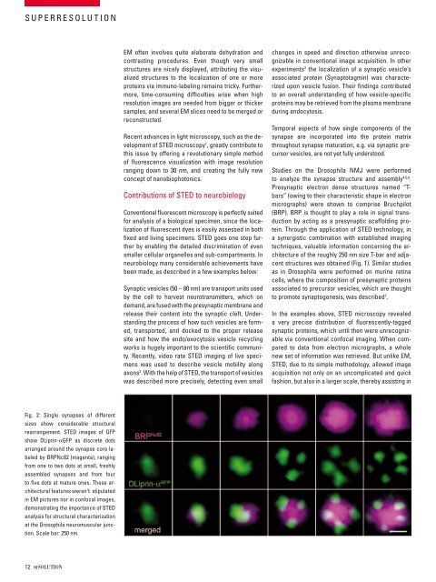
fig. 2: single synapses of different<br />
sizes show considerable structural<br />
rearrangement. sted images of gfP<br />
show dliprin-αgfP as discrete dots<br />
arranged around the synapse core labeled<br />
by brPnc82 (magenta), ranging<br />
from one to two dots at small, freshly<br />
assembled synapses and from four<br />
to five dots at mature ones. these architectural<br />
features weren‘t stipulated<br />
in em pictures nor in confocal images,<br />
demonstrating the importance of sted<br />
analysis for structural characterization<br />
at the drosophila neuromuscular junction.<br />
scale bar: 250 nm.<br />
12 resolutioN<br />
em often involves quite elaborate dehydration and<br />
contrasting procedures. even though very small<br />
structures are nicely displayed, attributing the visualized<br />
structures to the localization of one or more<br />
proteins via immuno-labeling remains tricky. furthermore,<br />
time-consuming difficulties arise when high<br />
resolution images are needed from bigger or thicker<br />
samples, and several em slices need to be merged or<br />
reconstructed.<br />
recent advances in light microscopy, such as the development<br />
of sted microscopy 1 , greatly contribute to<br />
this issue by offering a revolutionary simple method<br />
of fluorescence visualization with image resolution<br />
ranging down to 30 nm, and creating the fully new<br />
concept of nanobiophotonics.<br />
contributions of sted to neurobiology<br />
conventional fluorescent microscopy is perfectly suited<br />
for analysis of a biological specimen, since the localization<br />
of fluorescent dyes is easily assessed in both<br />
fixed and living specimens. sted goes one step further<br />
by enabling the detailed discrimination of even<br />
smaller cellular organelles and sub-compartments. in<br />
neurobiology many considerable achievements have<br />
been made, as described in a few examples below:<br />
synaptic vesicles (50 – 80 nm) are transport units used<br />
by the cell to harvest neurotransmitters, which on<br />
demand, are fused with the presynaptic membrane and<br />
release their content into the synaptic cleft. understanding<br />
the process of how such vesicles are formed,<br />
transported, and docked to the proper release<br />
site and how the endo/exocytosis vesicle recycling<br />
works is hugely important to the scientific community.<br />
recently, video rate sted imaging of live specimens<br />
was used to describe vesicle mobility along<br />
axons 2 . With the help of sted, the transport of vesicles<br />
was described more precisely, detecting even small<br />
changes in speed and direction otherwise unrecognizable<br />
in conventional image acquisition. in other<br />
experiments 3 the localization of a synaptic vesicle’s<br />
associated protein (synaptotagmin) was characterized<br />
upon vesicle fusion. their findings contributed<br />
to an overall understanding of how vesicle-specific<br />
proteins may be retrieved from the plasma membrane<br />
during endocytosis.<br />
temporal aspects of how single components of the<br />
synapse are incorporated into the protein matrix<br />
throughout synapse maturation, e.g. via synaptic precursor<br />
vesicles, are not yet fully understood.<br />
studies on the drosophila nmJ were performed<br />
to analyze the synapse structure and assembly 4,5,6 .<br />
Presynaptic electron dense structures named “tbars”<br />
(owing to their characteristic shape in electron<br />
micrographs) were shown to comprise bruchpilot<br />
(brP). brP is thought to play a role in signal transduction<br />
by acting as a presynaptic scaffolding protein.<br />
through the application of sted technology, in<br />
a synergistic combination with established imaging<br />
techniques, valuable information concerning the architecture<br />
of the roughly 250 nm size t-bar and adjacent<br />
structures was obtained (fig. 1). similar studies<br />
as in drosophila were performed on murine retina<br />
cells, where the composition of presynaptic proteins<br />
associated to precursor vesicles, which are thought<br />
to promote synaptogenesis, was described 7 .<br />
in the examples above, sted microscopy revealed<br />
a very precise distribution of fluorescently-tagged<br />
synaptic proteins, which until then were unrecognizable<br />
via conventional confocal imaging. When compared<br />
to data from electron micrographs, a whole<br />
new set of information was retrieved. but unlike em,<br />
sted, due to its simple methodology, allowed image<br />
acquisition not only on an uncomplicated and quick<br />
fashion, but also in a larger scale, thereby assisting in