

Vol 39 # 2 June 2007 - Kma.org.kw
Vol 39 # 2 June 2007 - Kma.org.kw
Vol 39 # 2 June 2007 - Kma.org.kw
- No tags were found...

You also want an ePaper? Increase the reach of your titles
YUMPU automatically turns print PDFs into web optimized ePapers that Google loves.
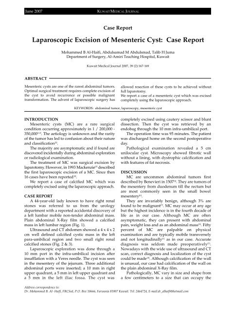
<strong>June</strong> <strong>2007</strong>KUWAIT MEDICAL JOURNALCase ReportLaparoscopic Excision of Mesenteric Cyst: Case ReportMohammed B Al-Haifi, Abdulsamad M Abdulsmad, Talib H JumaDepartment of Surgery, Al-Amiri Teaching Hospital, KuwaitKuwait Medical Journal <strong>2007</strong>, <strong>39</strong> (2):167-169ABSTRACTMesenteric cysts are one of the rarest abdominal tumors.Optimal surgical treatment requires complete excision ofthe cyst to avoid re c u r rence or possible malignanttransformation. The advent of laparoscopic surgery hasallowed resection of these cysts to be achieved withoutfull laparotomy.We report a case of a mesenteric cyst which was excisedcompletely using the laparoscopic approach.KEYWORDS: abdominal tumor, laparoscopy, mesenteric cystINTRODUCTIONMesenteric cysts (MC) are a rare surg i c a lcondition occurring approximately in 1 / 200,000 -350,000 [1,2] . The aetiology is unknown and the rarityof the tumor has led to confusion about their natureand classification [2] .The majority are asymptomatic and if found ared i s c o v e red incidentally during abdominal explorationor radiological examination.The treatment of MC was surgical excision byl a p a ro t o m y. However, in 1993 Mackenzie [ 3 ] d e s c r i b e dthe first laparoscopic excision of a MC. Since then16 cases have been reported [4] .We report a case of calcified MC which wascompletely excised using the laparoscopic appro a c h .CASE REPORTA 44-year-old lady known to have right renalstones was re f e r red to us from the uro l o g ydepartment with a reported accidental discovery ofa left lumbar mobile non-tender abdominal mass.Plain abdominal X-Ray film showed a calcifiedmass in left lumbar region (Fig. 1).Ultrasound and CT abdomen showed a 4 x 4 x 2cm well defined calcified cystic mass in the leftpara-umbilical region and two small right renalcalcified stones (Fig. 2 & 3).Laparoscopic exploration was done through a10 mm port in the infra-umbilical incision afterinsufflation with a Veres needle. The cyst was seenin the mesentery of the jejunum. Three additionalabdominal ports were inserted; a 10 mm in rightupper quadrant, a 5 mm in left upper quadrant anda 5 mm in the left iliac fossa. The cyst wascompletely excised using cautery scissor and bluntdissection. Then the cyst was retrieved by anendobag through the 10 mm infra-umbilical port.The operation time was 95 minutes. The patientwas discharged home on the second postoperativeday.Pathological examination revealed a 5 cmunilocular cyst. Microscopy showed fibrotic wallwithout a lining, with dystrophic calcification andwith features of fat necrosis.DISCUSSIONMC are uncommon abdominal tumors firstdescribed by Benevieri in 1507 [5] . They are tumors ofthe mesentery from duodenum till the rectum buta re most commonly seen in the small bowelmesentery [6] .They are invariably benign, although 3% arefound to be malignant [7] . MC may occur at any agebut the highest incidence is in the fourth decade oflife as in our case. Although MC are oftenasymptomatic, they can present with abdominalpain, weight loss and as an abdominal mass [8] . Fiftyp e rcent of MC are palpable on physicalexamination and are typically mobile transverselyand not longitudinally [1] as in our case. Accuratediagnosis was seldom made pre o p e r a t i v e l y [ 7 ] .Nowadays with the wide use of ultrasound and CTscan, correct diagnosis and localization of the cystcould be made [9] . Although calcification of the wallis unusual, our case had calcification of the wall onthe plain abdominal X-Ray film.Pathologically, MC vary in size and shape froma few centimeters to a size that can occupy theAddress correspondence to:Dr. Mohammed B. Al- Haifi, FRCSed, P.O. Box 18666, Farwania 81007 Kuwait. Tel: 2464724, E-mail:dr_alhaifi@hotmail.com


