IERG Abstracrt Book.indd - LV Prasad Eye Institute
IERG Abstracrt Book.indd - LV Prasad Eye Institute
IERG Abstracrt Book.indd - LV Prasad Eye Institute

Create successful ePaper yourself
Turn your PDF publications into a flip-book with our unique Google optimized e-Paper software.
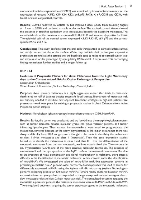
Basic Poster Sessions81mucosal epithelial transplantation (COMET) was examined by immunohistochemistry for theexpression of keratins (K3/12, K19, K14, K12), p63, p75, PAX6, Ki-67, CD31 and CD34, withlimbal, oral and conjunctival controls.Results: COMET followed by optical-PK has improved visual acuity from counting fingersat 15 cm to 20/40 and rendered a stable ocular surface. The excised corneal tissue showedthe presence of stratified epithelium with vasculatures beneath the basement membrane. Theendothelial cells of the vasculatures expressed CD31, CD34 and were rarely positive for Ki-67.The epithelial cells of the corneal button expressed K3, K19, Ki-67, p63, p75 and the corneaspecificPAX6 and K12.Conclusions: This study confirms that the oral cells transplanted to corneal surface surviveand stably reconstruct the ocular surface. While they maintain their native gene expressionprofiles and stemness at the ectopic site, the basal cells tend to respond to corneal cues in vivoand express an ocular phenotype by upregulating PAX6 and K12 expression. This encouragingfinding necessitates further studies and a longer follow up.IBP 034Evolution of Prognostic Markers for Uveal Melanoma from the Light Microscopydays to the Current microRNA: An Ocular Pathologist’s PerspectiveSubramanian KrishnakumarVision Research Foundation, Sankara Nethralaya, Chennai, India.Purpose: Uveal (ocular) melanoma is a highly aggressive cancer that leads to metastaticdeath in up to half of patients despite successful local therapy. Biomarkers of metastatic riskare critically needed to institute new adjuvant treatment strategies in high-risk patients. Wepresent our work over years for arriving at prognostic marker in Uveal Melanoma from IndianMelanoma tumor samplesMethods: Morphology, light microscopy, Immunohistochemistry, CISH, MicroRNAResults: Earlier, the tumor was enucleated and we looked into the morphological parameterssuch as tumor diameter, mitosis, nucleolar grade, cell types, vascular patterns and tumorinfiltrating lymphocytes. Then various immunomarkers were used to prognosticate themelanomas, however because of the heavy pigmentation in the Indian melanomas there wasalways a difficulty. Later HLA antigens were thought to be useful in classifying the melanomasto class I (Non metastatic) and class II (metastatic). Then the gene expression studieshelped us to classify the melanomas to class I and class II. For the differentiation of themetastatic melanoma from the non metastatic, we have standardized the Chromosomal insitu Hybridization (CISH), one of the more sensitive molecular techniques. The presence ofmonosomy–3 and the up regulation of the 8q22 confirm the metastatic melanoma. But dueto the presence of heavy pigmentation and clonal heterogeneity in melanoma, there arises adifficulty in the identification of metastatic melanoma. In this scenario enter the identificationof microRNA’s. We investigated the value of micro-RNA (miRNA) expression patterns inpredicting metastatic risk. A genome-wide, microarray-based approach was used to screen fordifferentially expressed miRNAs using the Agilent miRNA microarray (Agilent Technologies)platform containing probes for 470 human miRNAs. Tumors readily clustered based on miRNAexpression into two groups that corresponded to the gene expression-based subtypes: class 1(low metastatic risk) and class 2 (high metastatic risk).The upregulated oncomirs targeting themetastatic suppressor genes in the metastatic melanoma were miR-196a*, miR-549, miR-497*.The unregulated oncomirs targeting the tumor suppressor genes in the metastatic melanoma