Tour-de-Force
Tour-de-Force
Tour-de-Force
Create successful ePaper yourself
Turn your PDF publications into a flip-book with our unique Google optimized e-Paper software.
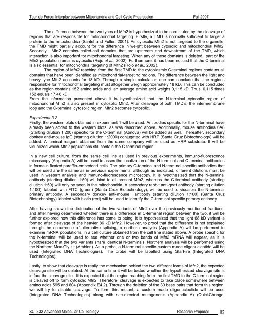
<strong>Tour</strong>-<strong>de</strong>-<strong>Force</strong>: Interplay between Mitochondria and Cell Cycle Progression Fall 2007The difference between the two types of Mfn2 is hypothesized to be constituted by the cleavage ofregions that are responsible for mitochondrial targeting. Firstly, a TMD is normally sufficient to target aprotein to the mitochondria (Santel and Fuller, 2001). As cytosolic Mfn2 is not targeted to the organelle,the TMD might partially account for the difference in weight between cytosolic and mitochondrial Mfn2.Secondly, Mfn2 contains coiled-coil domains that are upstream and downstream of the TMD, whichinteraction is also important for mitochondrial targeting. When any of these domains is <strong>de</strong>leted, part of theMfn2 population remains cytosolic (Rojo et al., 2002). Furthermore, it has been noticed that the C-terminalis also essential for mitochondrial targeting of Mfn2 (Rojo et al., 2002).The region of Mfn2 reaching from the first TMD to the cytoplasmic C-terminal regions contains alldomains that have been i<strong>de</strong>ntified as mitochondrial-targeting regions. The difference between the light andheavy type Mfn2 accounts for 18 kD. Through a simple calculation one can conclu<strong>de</strong> that the regionsresponsible for mitochondrial targeting must altogether weigh approximately 18 kD. This can be conclu<strong>de</strong>das the region contains 152 amino acids and an average amino acid weighs 0,115 kD. Thus, 0,115 times152 equals 17,48 kD.From the information presented above it is hypothesized that the N-terminal cytosolic region ofmitochondrial Mfn2 is also present in cytosolic Mfn2. After cleavage of both TMD’s, the intermembraneloop and the C-terminal cytosolic region, Mfn2 becomes cytosolic.Experiment 3.2Firstly, the western blots obtained in experiment 1 will be used. Antibodies specific for the N-terminal havealready been ad<strong>de</strong>d to the western blots, as was <strong>de</strong>scribed above. Additionally, mouse antibodies 6A8(Starting dilution 1:200) specific for the C-terminal (Abnova) will be ad<strong>de</strong>d as well. Thereafter, secondarydonkey anti-mouse IgG (starting dilution 1:2000) conjugated with HRP (Santa Cruz Biotechnology) will bead<strong>de</strong>d. A luminal reagent obtained from the same company will be used as HRP substrate. It will bevisualized which Mfn2 populations still contain the C-terminal region.In a new cell culture, from the same cell line as used in previous experiments, immuno-fluorescencemicroscopy (Appendix A) will be used to asses the localization of the N-terminal and C-terminal antibodiesin formalin fixated paraffin-embed<strong>de</strong>d cells. The primary C-terminal and N-terminal specific antibodies thatwill be used are the same as in previous experiments, although as indicated, different dilutions must beused in western analysis and immuno-fluorescence microscopy. It is hypothesized that the N-terminalantibody (starting dilution 1:50) will bind to all present Mfn2, whereas the C-terminal antibody (startingdilution 1:50) will only be seen in the mitochondria. A secondary rabbit anti-goat antibody (starting dilution1:100), labeled with FITC (green) (Santa Cruz Biotechnology), will be used to visualize the N-terminalprimary antibody. A secondary donkey anti-mouse antibody (starting dilution 1:100) (Santa CruzBiotechnology) labeled with biotin (red) will be used to i<strong>de</strong>ntify the C-terminal specific primary antibody.After having shown the distribution of the two variants of Mfn2 over the previously mentioned fractions,and after having <strong>de</strong>termined whether there is a difference in C-terminal region between the two, it will befurther explored how this difference has come to being. It is hypothesized that the light 68 kD variant isformed after cleavage of the heavier 86 kD Mfn2. However, to proof that the difference is not explainedthrough the occurrence of alternative splicing, a northern analysis (Appendix A) will be performed toexamine mRNA populations, in a cell culture obtained from the cell line stated above. A probe specific forthe N-terminal will be used to see whether one or two bands of Mfn2 mRNA will appear, as it ishypothesized that the two variants share i<strong>de</strong>ntical N-terminals. Northern analysis will be performed usingthe Northern Max-Gly kit (Ambion). As a probe, a N-terminal specific custom ma<strong>de</strong> oligonucleoti<strong>de</strong> will beused (Integrated DNA Technologies). The probe will be labelled using StarFire (Integrated DNATechnologies).Lastly, to show that cleavage is really the mechanism behind the two different forms of Mfn2, the expectedcleavage site will be <strong>de</strong>leted. At the same time it will be tested whether the hypothesized cleavage site isin fact the cleavage site. It is expected that the region reaching from the first TMD to the C-terminal regionis cleaved off to form cytosolic Mfn2. Therefore, cleavage is expected to take place somewhere betweenamino acids 595 and 604 (Appendix E4.2). Through the <strong>de</strong>letion of the 30 base pairs that form this region,we will try to disable cleavage. To form this mutant, a custom ma<strong>de</strong> oligonucleoti<strong>de</strong> will be used(Integrated DNA Technologies) along with site-directed mutagenesis (Appendix A) (QuickChange,SCI 332 Advanced Molecular Cell Biology Research Proposal 82


