Tour-de-Force
Tour-de-Force
Tour-de-Force
Create successful ePaper yourself
Turn your PDF publications into a flip-book with our unique Google optimized e-Paper software.
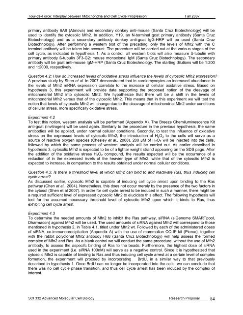
<strong>Tour</strong>-<strong>de</strong>-<strong>Force</strong>: Interplay between Mitochondria and Cell Cycle Progression Fall 2007primary antibody 6A8 (Abnova) and secondary donkey anti-mouse (Santa Cruz Biotechnology) will beused to i<strong>de</strong>ntify the cytosolic Mfn2. In addition, Y19, an N-terminal goat primary antibody (Santa CruzBiotechnology) and as a secondary antibody donkey anti-goat IgG-HRP will be used (Santa CruzBiotechnology). After performing a western blot of the preceding, only the levels of Mfn2 with the Cterminal antibody will be taken into account. The procedure will be carried out at the various stages of thecell cycle, as indicated in hypothesis 1. As a control, all western blots will also measure ß-tubulin withprimary antibody ß-tubulin 3F3-G2: mouse monoclonal IgM (Santa Cruz Biotechnology). The secondaryantibody will be goat anti-mouse IgM-HRP (Santa Cruz Biotechnology. The starting dilutions will be 1:200and 1:2000, respectively.Question 4.2: How do increased levels of oxidative stress influence the levels of cytosolic Mfn2 expression?A previous study by Shen et al. in 2007 <strong>de</strong>monstrated that in cardiomyocytes an increased abundance inthe levels of Mfn2 mRNA expression correlate to the increase of cellular oxidative stress. Based onhypothesis 3, this experiment will provi<strong>de</strong> data supporting the proposed notion of the cleavage ofmitochondrial Mfn2 into cytosolic Mfn2. We hypothesize that there will be a shift in the levels ofmitochondrial Mfn2 versus that of the cytosolic Mfn2. This means that in this experiment we will test thenotion that levels of cytosolic Mfn2 will change due to the cleavage of mitochondrial Mfn2 un<strong>de</strong>r conditionsof cellular stress, more specifically oxidative stress.Experiment 4.2To test this notion, western analysis will be performed (Appendix A). The Breeze Chemiluminescence Kitanti-goat (Invitrogen) will be used again. Similarly to the procedure in the previous hypothesis, the sameantibodies will be applied, un<strong>de</strong>r normal cellular conditions. Secondly, to test the influence of oxidativestress on the expressed levels of cytosolic Mfn2, the introduction of H 2 O 2 to the cells will serve as asource of reactive oxygen species (Chesley et al., 2000). 200 µM of H 2 O 2 will be injected into the cells,followed by which the same process of western analysis will be carried out. As earlier <strong>de</strong>scribed inhypothesis 3, cytosolic Mfn2 is expected to be of a lighter weight strand appearing on the SDS page. Afterthe addition of the oxidative stress H 2 O 2 compound, the results expected will be the occurrence of areduction of in the expressed levels of the heavier type of Mfn2, while that of the cytosolic Mfn2 isexpected to increase, in comparison to the results obtained un<strong>de</strong>r normal cellular conditions.Question 4.3: Is there a threshold level at which Mfn2 can bind to and inactivate Ras, thus inducing cellcycle arrest?As discussed earlier, cytosolic Mfn2 is capable of inducing cell cycle arrest upon binding to the Raspathway (Chen et al., 2004). Nonetheless, this does not occur merely by the presence of the two factors inthe cytosol (Shen et al 2007). In or<strong>de</strong>r for cell cycle arrest to be induced in such a manner, there might bea required sufficient level of expressed cytosolic Mfn2 to elucidate this effect. The following hypothesis willtest for the assumed necessary threshold level of cytosolic Mfn2 upon which it binds to Ras, thusexhibiting cell cycle arrest.Experiment 4.3To <strong>de</strong>termine the nee<strong>de</strong>d amounts of Mfn2 to inhibit the Ras pathway, siRNA (siGenome SMARTpool,Dharmacon) against Mfn2 will be used. The used amounts of siRNA against Mfn2 will correspond to thosementioned in hypothesis 2, in Table 4.1, titled un<strong>de</strong>r Mfn2 wt. Followed by each of the administered dosesof siRNA, co-immunoprecipitation (Appendix A) with the use of mammalian CO-IP kit (Pierce), togetherwith the rabbit polyclonal Mfn2 antibody H68 (Santa Cruz Biotechnology) will help assess the formedcomplex of Mfn2 and Ras. As a blank control we will conduct the same procedure, without the use of Mfn2antibody, to assess the aspecifc binding of Ras to the beads. Furthermore, the highest dose of siRNAused in the experiment (i.e. siRNA 100nM) will serve as a negative control. Since it is hypothesized thatcytosolic Mfn2 is capable of binding to Ras and thus inducing cell cycle arrest at a certain level of complexformation, the experiment will proceed by incorporating BrdU, in a similar way to that previously<strong>de</strong>scribed in hypothesis 1. Once BrdU can no longer be incorporated into the cells, we can conclu<strong>de</strong> thatthere was no cell cycle phase transition, and thus cell cycle arrest has been induced by the complex ofinterest.SCI 332 Advanced Molecular Cell Biology Research Proposal 84


