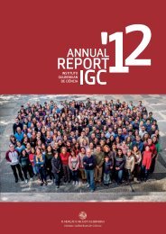
organisation - the Instituto Gulbenkian de Ciência
organisation - the Instituto Gulbenkian de Ciência
organisation - the Instituto Gulbenkian de Ciência
- No tags were found...

Create successful ePaper yourself
Turn your PDF publications into a flip-book with our unique Google optimized e-Paper software.
CELL IMAGING<br />
UNIT<br />
José Feijó Head<br />
PhD in Cell Biology, Universida<strong>de</strong> <strong>de</strong> Lisboa, 1995<br />
PI of Cell Biophysics and Development Group<br />
Head of Facility since 2003<br />
link to facility website<br />
The main goal of <strong>the</strong> Cell Imaging Unit is to provi<strong>de</strong> access to high-end technology<br />
and cutting-edge technical support for bioscience research. We provi<strong>de</strong><br />
a unique facility that allows ready access to a wi<strong>de</strong> range of technologies and<br />
expertise in an integrated manner that helps drive research forward efficiently.<br />
The unit currently stands as an international reference laboratory for confocal<br />
and multi-photon microscopy, as well as for high-throughput cell sorting.<br />
The unit is well equipped, with two cell sorters, three confocal microscopes,<br />
a DeltaVision <strong>de</strong>convolution microscope system and two multi-photon microscopes,<br />
besi<strong>de</strong>s a dozen more subsidiary and custom-ma<strong>de</strong> pieces of equipment.<br />
Researchers are trained through regular workshops on basic and advanced light<br />
microscopy techniques as well as in flow cytometry and image acquisition software.<br />
Technical assistance is available when necessary to ensure collection of<br />
high quality images and analysis of data. All users receive basic training in <strong>the</strong><br />
systems in use, in troubleshooting, and advice on experimental <strong>de</strong>sign. We have<br />
<strong>de</strong>veloped a strong and broad base of users and continue to train new users.<br />
Because microscopy is currently in high <strong>de</strong>mand and new systems and techniques<br />
are continuously being <strong>de</strong>veloped to meet increasing scientific needs, we try to<br />
expand <strong>the</strong> facilities, to keep accessibility problems low, and introduce <strong>the</strong> latest<br />
innovations (in microscopy and flow cytometry) to <strong>the</strong> research community.<br />
FACILITY STAFF<br />
Carlos Tadokoro (Multi-photon Microscopy Applications Manager /<br />
Research Fellow)<br />
Rui Gardner (Flow Cytometry Lab Manager)<br />
Nuno Moreno (General Coordinator Microscopy)<br />
Telma Lopes (Technician - Cell Sorting)<br />
Francisco Henrique (Technician - Microscopy)<br />
Pedro Almada (Technician - Microscopy)<br />
MAJOR PROJECTS AND ACCOMPLISHMENTS<br />
INTEGRATION OF A SCREENING MICROSCOPE<br />
This microscope is fully automated and works with open source software, being<br />
a good platform for on-<strong>the</strong>-fly analysis. The equipment uses fast filter wheels<br />
and shutters which, toge<strong>the</strong>r with a sensitive CCD camera, enable three colour<br />
images in a 96 well plate in few minutes. For now, it has been used for automatic<br />
stitching, producing images of full brain mice or fish slices.<br />
UPGRADE OF MULTIPHOTON MICROSCOPY SYSTEM<br />
The installation of a Prairie Ultima multiphoton microscope procee<strong>de</strong>d, with<br />
individual user training being provi<strong>de</strong>d. A gui<strong>de</strong>book for correct use of this<br />
microscope was <strong>de</strong>veloped, and technical support for microscope adjustment<br />
to individual scientific projects (different types of samples) was carried out.<br />
EQUIPMENT AND INFRASTRUCTURE<br />
INSPECTION WIDE-FIELD LIGHT MICROSCOPES<br />
• Olympus BH2;<br />
• Olympus IMT-2;<br />
• Leica DMLB2.<br />
RESEARCH WIDEFIELD LIGHT MICROSCOPES<br />
• Leica inverted DMIRE2;<br />
• Leica upright DMRA2;<br />
• Zeiss AxioImager M1.<br />
Funding Calouste <strong>Gulbenkian</strong> Foundation and Fundação para a Ciência e a Tecnologia,<br />
Portugal<br />
CONFOCAL MICROSCOPES<br />
• Leica SP5 (with resonant fast scanhead);<br />
• Zeiss LSM 510 (with Meta <strong>de</strong>tector);<br />
• Andor Spinning disk (with EM-CCD intensified camera).<br />
Funding Calouste <strong>Gulbenkian</strong> Foundation, Fundação para a Ciência e a Tecnologia<br />
(FCT), Portugal, European Molecular Biology Organisation (EMBO).<br />
TwoCellEmbryos - Mouse embryos at <strong>the</strong> two-cell stage.<br />
IGC ANNUAL REPORT ‘11<br />
FACILITIES AND SERVICES<br />
89