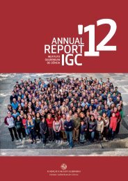
organisation - the Instituto Gulbenkian de Ciência
organisation - the Instituto Gulbenkian de Ciência
organisation - the Instituto Gulbenkian de Ciência
- No tags were found...

Create successful ePaper yourself
Turn your PDF publications into a flip-book with our unique Google optimized e-Paper software.
HISTOPATHOLOGY<br />
UNIT<br />
Miguel P. Soares Head<br />
PhD in Science, University of Louvain, Belgium, 1995<br />
PI of Inflammation Group<br />
Head of Facility since 2011<br />
The Histopathology Unit provi<strong>de</strong>s a wi<strong>de</strong> range of services related to tissue<br />
preparation. These inclu<strong>de</strong> collection, fixation, processing, embedding, sectioning<br />
and staining of animal tissue samples. The unit also provi<strong>de</strong>s microscopy<br />
assistance as well as training to new users in sample preparation and sectioning.<br />
FACILITY STAFF<br />
Ana Margarida Santos (Technician, left in October)<br />
Joana Rodrigues (Technician)<br />
EQUIPMENT AND INFRASTRUCTURE<br />
• Tissue processor: takes <strong>the</strong> fixed tissues through a series of gra<strong>de</strong>d alcohol<br />
baths, to <strong>de</strong>hydrate, <strong>the</strong>n into xylene and finally paraffin will penetrate <strong>the</strong><br />
tissues;<br />
• Paraffin embedding station and water bath: used to effect <strong>the</strong> paraffin<br />
inclusion of cytological and histological samples;<br />
• Microtome: sectioning instrument that allows for <strong>the</strong> cutting of extremely<br />
thin slices of material. These sections are <strong>the</strong>n observed un<strong>de</strong>r transmitted<br />
light. Steel bla<strong>de</strong>s are used to prepare sections of animal or plant tissues<br />
for light microscopy histology. Microtome sections have thickness between<br />
0.05 and 10 µm;<br />
• Cryostat: <strong>de</strong>vice used to maintain cold cryogenic temperatures of samples<br />
and to cut histological sli<strong>de</strong>s from samples fixed and frozen in OCT;<br />
• Vibratome: similar to a microtome but uses a vibrating razor bla<strong>de</strong> to cut<br />
through tissue. The vibration amplitu<strong>de</strong>, <strong>the</strong> speed, and <strong>the</strong> angle of <strong>the</strong><br />
bla<strong>de</strong> can all be controlled. Fixed or fresh tissue pieces are embed<strong>de</strong>d in<br />
low gelling temperature agarose. The resulting agarose block containing<br />
<strong>the</strong> tissue piece is <strong>the</strong>n glued to a metal block and sectioned while submerged<br />
in a water or buffer bath. Individual sections are <strong>the</strong>n collected<br />
with a fine brush and transferred to sli<strong>de</strong>s or multiwell plates for staining.<br />
Funding Calouste <strong>Gulbenkian</strong> Foundation, Portugal<br />
B<br />
HYALINE CARTILAGE.<br />
Hyaline cartilage, mouse. Luxol fast blue (400x).<br />
OVARIAN METASTASIS.<br />
Ovarian mestastasis of breast cancer, mouse. Hematoxylin and eosin (200x).<br />
IGC ANNUAL REPORT ‘11<br />
FACILITIES AND SERVICES<br />
94