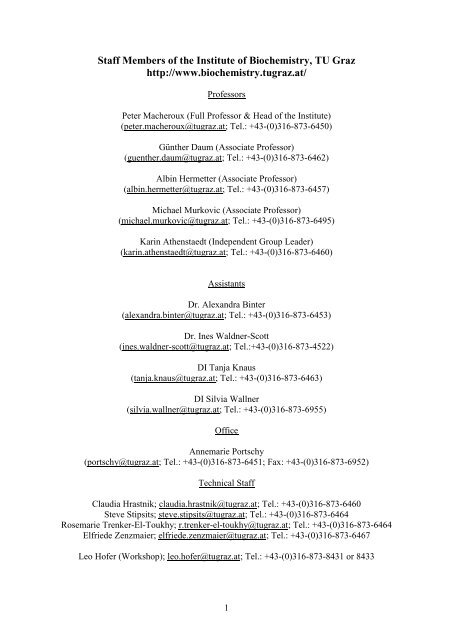
Staff Members of the Institute of Biochemistry, TU - Institut für ...
Staff Members of the Institute of Biochemistry, TU - Institut für ...
Staff Members of the Institute of Biochemistry, TU - Institut für ...

Create successful ePaper yourself
Turn your PDF publications into a flip-book with our unique Google optimized e-Paper software.
<strong>Staff</strong> <strong>Members</strong> <strong>of</strong> <strong>the</strong> <strong><strong>Institut</strong>e</strong> <strong>of</strong> <strong>Biochemistry</strong>, <strong>TU</strong> Graz<br />
http://www.biochemistry.tugraz.at/<br />
Pr<strong>of</strong>essors<br />
Peter Macheroux (Full Pr<strong>of</strong>essor & Head <strong>of</strong> <strong>the</strong> <strong><strong>Institut</strong>e</strong>)<br />
(peter.macheroux@tugraz.at; Tel.: +43-(0)316-873-6450)<br />
Gün<strong>the</strong>r Daum (Associate Pr<strong>of</strong>essor)<br />
(guen<strong>the</strong>r.daum@tugraz.at; Tel.: +43-(0)316-873-6462)<br />
Albin Hermetter (Associate Pr<strong>of</strong>essor)<br />
(albin.hermetter@tugraz.at; Tel.: +43-(0)316-873-6457)<br />
Michael Murkovic (Associate Pr<strong>of</strong>essor)<br />
(michael.murkovic@tugraz.at; Tel.: +43-(0)316-873-6495)<br />
Karin A<strong>the</strong>nstaedt (Independent Group Leader)<br />
(karin.a<strong>the</strong>nstaedt@tugraz.at; Tel.: +43-(0)316-873-6460)<br />
Assistants<br />
Dr. Alexandra Binter<br />
(alexandra.binter@tugraz.at; Tel.: +43-(0)316-873-6453)<br />
Dr. Ines Waldner-Scott<br />
(ines.waldner-scott@tugraz.at; Tel.:+43-(0)316-873-4522)<br />
DI Tanja Knaus<br />
(tanja.knaus@tugraz.at; Tel.: +43-(0)316-873-6463)<br />
DI Silvia Wallner<br />
(silvia.wallner@tugraz.at; Tel.: +43-(0)316-873-6955)<br />
Office<br />
Annemarie Portschy<br />
(portschy@tugraz.at; Tel.: +43-(0)316-873-6451; Fax: +43-(0)316-873-6952)<br />
Technical <strong>Staff</strong><br />
Claudia Hrastnik; claudia.hrastnik@tugraz.at; Tel.: +43-(0)316-873-6460<br />
Steve Stipsits; steve.stipsits@tugraz.at; Tel.: +43-(0)316-873-6464<br />
Rosemarie Trenker-El-Toukhy; r.trenker-el-toukhy@tugraz.at; Tel.: +43-(0)316-873-6464<br />
Elfriede Zenzmaier; elfriede.zenzmaier@tugraz.at; Tel.: +43-(0)316-873-6467<br />
Leo H<strong>of</strong>er (Workshop); leo.h<strong>of</strong>er@tugraz.at; Tel.: +43-(0)316-873-8431 or 8433<br />
1
Brief History <strong>of</strong> <strong>the</strong> <strong><strong>Institut</strong>e</strong> <strong>of</strong> <strong>Biochemistry</strong><br />
The <strong><strong>Institut</strong>e</strong> <strong>of</strong> <strong>Biochemistry</strong> and Food Chemistry was born out <strong>of</strong> <strong>the</strong> division <strong>of</strong> <strong>the</strong> <strong><strong>Institut</strong>e</strong><br />
<strong>of</strong> Biochemical Technology, Food Chemistry and Microchemistry <strong>of</strong> <strong>the</strong> former School <strong>of</strong><br />
Technology Graz. Toge<strong>the</strong>r with all <strong>the</strong> o<strong>the</strong>r chemistry institutes, it was located in <strong>the</strong> old<br />
Chemistry Building on Baron Mandell's ground, corner Technikerstrasse-Mandellstrasse.<br />
1929 The <strong><strong>Institut</strong>e</strong> <strong>of</strong> Technical <strong>Biochemistry</strong> and Microbiology moved to <strong>the</strong> building <strong>of</strong><br />
<strong>the</strong> Fürstlich-Dietrichstein-Stiftung, Schlögelgasse 9, in which all <strong>the</strong> biosciences were<br />
<strong>the</strong>n concentrated.<br />
1945 Georg GORBACH - initially in <strong>the</strong> rank <strong>of</strong> a docent and soon <strong>the</strong>reafter as a.o.<br />
Pr<strong>of</strong>essor - took over to lead <strong>the</strong> institute. The institute was renamed <strong><strong>Institut</strong>e</strong> <strong>of</strong><br />
Biochemical Technology and Food Chemistry.<br />
1948 G. GORBACH was nominated full pr<strong>of</strong>essor and head <strong>of</strong> <strong>the</strong> institute. In succession <strong>of</strong><br />
<strong>the</strong> famous Graz School <strong>of</strong> Microchemistry founded by PREGL and EMICH, Pr<strong>of</strong>.<br />
GORBACH was one <strong>of</strong> <strong>the</strong> most prominent and active leaders in <strong>the</strong> fields <strong>of</strong><br />
microchemistry, microbiology and nutritional sciences. After World War II, questions<br />
<strong>of</strong> water quality and waste water disposal became urgent; hence, <strong>the</strong> group <strong>of</strong> Pr<strong>of</strong>. K.<br />
S<strong>TU</strong>NDL, which at that time was part <strong>of</strong> <strong>the</strong> institute, was gaining importance. In<br />
addition, a division to fight dry-rot supervised by Dr. KUNZE and after his demise by<br />
H. SALOMON, was also affiliated with <strong>the</strong> institute.<br />
1955 In honour <strong>of</strong> <strong>the</strong> founder <strong>of</strong> microchemistry and former pr<strong>of</strong>essor at <strong>the</strong> Graz<br />
University <strong>of</strong> Technology, <strong>the</strong> extended laboratory was called EMICH-Laboratories.<br />
At <strong>the</strong> same time, <strong>the</strong> institute was renamed <strong><strong>Institut</strong>e</strong> <strong>of</strong> Biochemical Technology,<br />
Food Chemistry and Microchemistry.<br />
Lectures and laboratory courses were held in biochemistry, biochemical technology, food<br />
chemistry and food technology, technical microscopy and microchemistry. In addition, <strong>the</strong><br />
institute covered technical microbiology toge<strong>the</strong>r with biological and bacteriological analysis<br />
- with <strong>the</strong> exception <strong>of</strong> pathogenic microorganisms - and a lecture in organic raw materials<br />
sciences.<br />
1970 After <strong>the</strong> decease <strong>of</strong> Pr<strong>of</strong>. GORBACH, Pr<strong>of</strong>. GRUBITSCH was appointed head <strong>of</strong> <strong>the</strong><br />
institute. Towards <strong>the</strong> end <strong>of</strong> <strong>the</strong> sixties, <strong>the</strong> division for water and waste water<br />
disposal headed by Pr<strong>of</strong>. S<strong>TU</strong>NDL was drawn out <strong>of</strong> <strong>the</strong> institute and established as<br />
an independent institute. Pr<strong>of</strong>. SPITZY was nominated pr<strong>of</strong>essor <strong>of</strong> general chemistry,<br />
micro- and radiochemistry. This division was also drawn out <strong>of</strong> <strong>the</strong> mo<strong>the</strong>r institute<br />
and at <strong>the</strong> end <strong>of</strong> <strong>the</strong> sixties moved to a new building.<br />
1973 Division <strong>of</strong> <strong>the</strong> <strong><strong>Institut</strong>e</strong> for Biochemical Technology, Food Technology and<br />
Microchemistry took place. At first, biochemical technology toge<strong>the</strong>r with food<br />
technology formed a new institute now called <strong><strong>Institut</strong>e</strong> <strong>of</strong> Biotechnology and Food<br />
Chemistry headed by Pr<strong>of</strong>. LAFFERTY.<br />
1973 Dr. F. PALTAUF, docent at <strong>the</strong> Karl-Franzens-University Graz, was appointed<br />
pr<strong>of</strong>essor and head <strong>of</strong> <strong>the</strong> newly established <strong><strong>Institut</strong>e</strong> <strong>of</strong> <strong>Biochemistry</strong>. The interest <strong>of</strong><br />
Pr<strong>of</strong>. PALTAUF in studying biological membranes and lipids laid <strong>the</strong> foundation for<br />
2
<strong>the</strong> future direction <strong>of</strong> research. G. DAUM, S. D. KOHLWEIN, and A. HERMETTER<br />
joined <strong>the</strong> institute. All three young scientists were given <strong>the</strong> chance to work as post<br />
docs in renown laboratories in Switzerland and <strong>the</strong> USA: G. DAUM with <strong>the</strong> groups<br />
<strong>of</strong> G. Schatz (Basel, Switzerland) and R. Schekman (Berkeley, USA), A.<br />
HERMETTER with J. R. Lakowicz (Baltimore, USA) and S. D. KOHLWEIN with S.<br />
A. Henry (New York, USA). Consequently, independent research groups specialized<br />
in cell biology (G. D.), biophysics (A. H.) and molecular biology (S. D. K.) evolved at<br />
<strong>the</strong> institute in Graz, with <strong>the</strong> group <strong>of</strong> Pr<strong>of</strong>. F. PALTAUF still focusing on <strong>the</strong><br />
chemistry and biochemistry <strong>of</strong> lipids.<br />
Teaching was always a major task <strong>of</strong> <strong>the</strong> institute. Lectures, seminars and laboratory courses<br />
in basic biochemistry were complemented by special lectures, seminars, and courses held by<br />
<strong>the</strong> assistants who became docents in 1985 (G. D.), 1987 (A. H.), and 1992 (S. D. K.).<br />
Lectures in food chemistry and technology were held by C. WEBER and H. SALOMON.<br />
Hence <strong>the</strong> institute was renamed <strong><strong>Institut</strong>e</strong> <strong>of</strong> <strong>Biochemistry</strong> and Food Chemistry.<br />
1990 The institute moved to a new building at Petersgasse 12. The move was accompanied<br />
by <strong>the</strong> expansion <strong>of</strong> individual research groups and <strong>the</strong> acquisition <strong>of</strong> new equipment<br />
essential for <strong>the</strong> participation in novel collaborative efforts at <strong>the</strong> national and<br />
international level. Thus, <strong>the</strong> <strong><strong>Institut</strong>e</strong> <strong>of</strong> <strong>Biochemistry</strong>, toge<strong>the</strong>r with partner institutes<br />
from <strong>the</strong> Karl-Franzens-University was <strong>the</strong> driving force to establish Graz as a centre<br />
<strong>of</strong> competence in lipid research.<br />
1993 W. PFANNHAUSER was appointed as pr<strong>of</strong>essor <strong>of</strong> food chemistry. Through his own<br />
enthusiasm and engagement and that <strong>of</strong> his collaborators, this new section <strong>of</strong> <strong>the</strong><br />
institute rapidly developed and <strong>of</strong>fered students additional opportunities to receive a<br />
timely education.<br />
2000 The two sections, biochemistry and food chemistry, being independent <strong>of</strong> each o<strong>the</strong>r<br />
with respect to personnel, teaching, and research, were separated into <strong>the</strong> <strong><strong>Institut</strong>e</strong> <strong>of</strong><br />
<strong>Biochemistry</strong> (Head Pr<strong>of</strong>. PALTAUF) and <strong>the</strong> new <strong><strong>Institut</strong>e</strong> <strong>of</strong> Food Chemistry and<br />
Technology (Head Pr<strong>of</strong>. PFANNHAUSER).<br />
2001 After F. PALTAUF’s retirement, in September 2001, G. DAUM was elected head <strong>of</strong><br />
<strong>the</strong> institute. S. D. KOHLWEIN was appointed full pr<strong>of</strong>essor <strong>of</strong> biochemistry at <strong>the</strong><br />
Karl-Franzens University Graz.<br />
2003 P. MACHEROUX was appointed full pr<strong>of</strong>essor <strong>of</strong> biochemistry in September 2003<br />
and head <strong>of</strong> <strong>the</strong> <strong><strong>Institut</strong>e</strong> <strong>of</strong> <strong>Biochemistry</strong> in January 2004. His research interests<br />
revolve around topics in protein biochemistry and enzymology and shall streng<strong>the</strong>n<br />
<strong>the</strong> already existing activities in this area.<br />
2007 K. ATHENSTAEDT, a long-time associate <strong>of</strong> Pr<strong>of</strong>. DAUM, received <strong>the</strong> venia<br />
legendi for biochemistry. Karin is <strong>the</strong> first woman to complete <strong>the</strong> traditional<br />
habilitation at <strong>the</strong> <strong><strong>Institut</strong>e</strong> <strong>of</strong> <strong>Biochemistry</strong>!<br />
2009 After <strong>the</strong> retirement <strong>of</strong> Pr<strong>of</strong>. PFANNHAUSER in 2008, <strong>the</strong> <strong><strong>Institut</strong>e</strong> <strong>of</strong> Food<br />
Chemistry and Technology was disbanded and <strong>the</strong> research group <strong>of</strong> Pr<strong>of</strong>. M.<br />
MURKOVIC joined <strong>the</strong> <strong><strong>Institut</strong>e</strong> <strong>of</strong> <strong>Biochemistry</strong> increasing <strong>the</strong> number <strong>of</strong><br />
independent research groups to five.<br />
3
Highlights <strong>of</strong> 2011<br />
This was clearly <strong>the</strong> year for <strong>the</strong> flavin enthusiasts in <strong>the</strong> Macheroux group: <strong>the</strong><br />
17 th International Symposium on Flavins and Flavoproteins was hosted by <strong>the</strong> University <strong>of</strong><br />
California at Berkeley, USA, in July. Silvia Wallner, Thomas Bergner and Peter Macheroux<br />
presented posters revolving around “yellow enzymes”.<br />
4<br />
From left to right:<br />
Katharina Durchschein, Silvia<br />
Wallner, Thomas Bergner,<br />
Thomas Stoisser, Barbara Holl-<br />
Macheroux, Karl Gruber &<br />
Pauline Macheroux<br />
In September, Peter Macheroux was nominated as one <strong>of</strong> <strong>the</strong> top ten lecturers <strong>of</strong> Graz<br />
University <strong>of</strong> Technology. The <strong>of</strong>ficial ceremony took place on November 8 th in <strong>the</strong> aula <strong>of</strong><br />
<strong>the</strong> university. At <strong>the</strong> board meeting in October, <strong>the</strong> Austrian Science Fund (FWF) approved a<br />
new research project to investigate <strong>the</strong> mechanism <strong>of</strong> bacterial bioluminescence (Project<br />
P24189: “Bacterial bioluminescence”). Two former members <strong>of</strong> <strong>the</strong> group received<br />
fellowships to continue <strong>the</strong>ir scientific career: Dr. Andreas Winkler, currently a postdoctoral<br />
fellow at <strong>the</strong> Max-Planck <strong><strong>Institut</strong>e</strong> <strong>of</strong> Molecular Medicine in Heidelberg received an “Erwin-<br />
Schrödinger” stipend for 2012, and Dr. Sonja Sollner, currently at <strong>the</strong> <strong><strong>Institut</strong>e</strong> <strong>of</strong> Molecular<br />
Pathology in Vienna, was awarded a “Hertha-Firnberg” stipend. Both alumni were members<br />
<strong>of</strong> <strong>the</strong> PhD program “Molecular Enzymology”. Despite contracting budgets and stiff<br />
competition, this PhD program received excellent reviews and was extended for a second time<br />
until 2014. Three groups <strong>of</strong> <strong>the</strong> institute (Gün<strong>the</strong>r Daum, Albin Hermetter and Peter<br />
Macheroux) participate in this NAWI Graz interuniversity program.<br />
Norman Medal Award<br />
Ceremony at <strong>the</strong> 9 th Euro Fed<br />
Lipid Congress in Rotterdam,<br />
The Ne<strong>the</strong>rlands. Left K. Schurz,<br />
President <strong>of</strong> <strong>the</strong> DGF, right G.<br />
Daum.<br />
In 2011, three new PhD students<br />
joined <strong>the</strong> group <strong>of</strong> Gün<strong>the</strong>r<br />
Daum: Claudia Schmidt,<br />
Barbara Koch and Birgit Ploier<br />
started <strong>the</strong>ir studies on topics<br />
related to neutral lipid<br />
metabolism in <strong>the</strong> yeast.<br />
In 2011, Gün<strong>the</strong>r Daum started his third three year term as a Board Member <strong>of</strong> <strong>the</strong> Austrian<br />
Science Fund FWF. He also continued his work as President <strong>of</strong> <strong>the</strong> International Conference
on <strong>the</strong> Bioscience <strong>of</strong> Lipids (ICBL) and Chairman <strong>of</strong> <strong>the</strong> Yeast Lipid Conference (YLC).<br />
Among <strong>the</strong> various invitations to research institutions and conferences a visit at <strong>the</strong> Central<br />
<strong><strong>Institut</strong>e</strong> <strong>of</strong> Medicinal and Aromatic Plants in Lucknow and to <strong>the</strong> Jawaharla Nehru<br />
University, Delhi, India, was definitely one highlight. In September 2011, Gün<strong>the</strong>r Daum<br />
received <strong>the</strong> Normann Medal, <strong>the</strong> highest Award <strong>of</strong> <strong>the</strong> Deutsche Gesellschaft <strong>für</strong><br />
Fettwissenschaft (DGF), for his long term studies on biochemistry and cell biology <strong>of</strong> yeast<br />
lipids and biomembranes.<br />
Basic research in Albin Hermetter’s group focused on lipid<br />
(patho)physiology in human and animal cells. The respective<br />
projects were components <strong>of</strong> <strong>the</strong> research consortia GOLD/ GEN-<br />
AU (FFG), OXPHOS/ EuroMEMBRANE (ESF, FWF) and <strong>the</strong> PhD<br />
program “Molecular Enzymology” (FWF). A. Hermetter was<br />
invited to co-edit a special issue entitled “Oxidized phospholipids<strong>the</strong>ir<br />
properties and interactions with proteins” for Biochim.<br />
Biophys. Acta. Lingaraju Marlingapla Halasiddappa who is a PhD<br />
student in <strong>the</strong> PhD program “Molecular Enzymology” spent eight<br />
months in <strong>the</strong> Department <strong>of</strong> Biological Chemistry at Weizman<br />
<strong><strong>Institut</strong>e</strong> in Rehovot, Israel, to do research in <strong>the</strong> field <strong>of</strong> sphingolipid<br />
enzymology. He received an award for his oral presentation<br />
“Ceramide Synthases in Oxidized Phospholipid-induced RAW<br />
264.7 Macrophage Cell Death” at <strong>the</strong> International Charleston<br />
Ceramide Conference in Villars, Switzerland, March 2011.<br />
5<br />
Lingaraju Marlingapla<br />
Halasiddappa,<br />
PhD student<br />
In 2011 Karin A<strong>the</strong>nstaedt continued her work at our<br />
institute as an independent researcher. Her work is devoted to<br />
lipid metabolism in yeast. One <strong>of</strong> <strong>the</strong> highlights in Karin<br />
A<strong>the</strong>nstaedt’s scientific work in 2011 was attendance <strong>of</strong> <strong>the</strong><br />
Gordon Conference (Molecular & Cellular Biology <strong>of</strong> Lipids)<br />
in Waterville Valley, NH, USA, where she had <strong>the</strong><br />
opportunity to meet many experts in <strong>the</strong> field and to set <strong>the</strong><br />
stage for collaborative activities.<br />
In <strong>the</strong> group Chemistry <strong>of</strong> Functional Foods directed by<br />
Michael Murkovic in 2011 a PhD <strong>the</strong>sis was ongoing in which<br />
<strong>the</strong> polymerization <strong>of</strong> furfuryl alcohol is investigated. Furfuryl<br />
alcohol is formed during heating <strong>of</strong> foods, e.g. during roasting<br />
<strong>of</strong> c<strong>of</strong>fee. It can be activated metabolically which might result<br />
in <strong>the</strong> induction <strong>of</strong> tumors. The polymerization removes <strong>the</strong><br />
furfuryl alcohol from <strong>the</strong> foods and reduces <strong>the</strong> bioavailability.<br />
In 2011 several international students (Lithuania, Egypt,<br />
Hungary, Slovak Republic) were coming to <strong>the</strong> lab for carrying<br />
out specific experiments related to, e.g. new technologies <strong>of</strong><br />
heating, oxidation <strong>of</strong> oils in <strong>the</strong> bulk phase and in emulsions,<br />
and <strong>the</strong> application <strong>of</strong> asparaginase for reduction <strong>of</strong> <strong>the</strong><br />
acrylamide formation.
<strong>Biochemistry</strong> Group<br />
Group leader: Peter Macheroux<br />
Secretary: Annemarie Portschy<br />
Senior scientists: Alexandra Binter, Ines Waldner-Scott<br />
PhD students: Thomas Bergner, Bastian Daniel, Venugopal Gudipati, Tanja Knaus, Wolf-<br />
Dieter Lienhart, Silvia Wallner<br />
Master students: Corinna Dully, Karin Koch, Julia Koop, Katharina Lukas, Nicole Sudi,<br />
Marlene Tösch<br />
Technicians: Sabrina Moratti, Eva Maria Pointner, Steve Stipsits, Rosemarie Trenker-El-<br />
Toukhy<br />
General description<br />
The fundamental questions in <strong>the</strong> study <strong>of</strong> enzymes, <strong>the</strong> bio-catalysts <strong>of</strong> all living organisms,<br />
revolve around <strong>the</strong>ir ability to select a substrate (substrate specificity) and subject this<br />
substrate to a predetermined chemical reaction (reaction and regio-specificity). In general,<br />
only a few amino acid residues in <strong>the</strong> "active site" <strong>of</strong> an enzyme are involved in this process<br />
and hence provide <strong>the</strong> key to <strong>the</strong> processes taking place during enzyme catalysis. Therefore,<br />
<strong>the</strong> focus <strong>of</strong> our research is to achieve a deeper understanding <strong>of</strong> <strong>the</strong> functional role <strong>of</strong> amino<br />
acids in <strong>the</strong> active site <strong>of</strong> enzymes with regard to substrate-recognition and stereo- and<br />
regiospecificity <strong>of</strong> <strong>the</strong> chemical transformation. In addition, we are also interested in<br />
substrate-triggered conformational changes and how enzymes utilize c<strong>of</strong>actors (flavin,<br />
nicotinamide) to achieve catalysis. Towards <strong>the</strong>se aims we employ a multidisciplinary<br />
approach encompassing kinetic, <strong>the</strong>rmodynamic, spectroscopic and structural techniques. In<br />
addition, we use site-directed mutagenesis to generate mutant enzymes to probe <strong>the</strong>ir<br />
functional role in <strong>the</strong> mentioned processes. Fur<strong>the</strong>rmore, we collaborate with our partners in<br />
academia and industry to develop inhibitors for enzymes, which can yield important new<br />
insights into enzyme mechanisms and can be useful as potential lead compounds in <strong>the</strong> design<br />
<strong>of</strong> new drugs.<br />
The methods established in our laboratory comprise kinetic (stopped-flow and rapid quench<br />
analysis <strong>of</strong> enzymatic reactions), <strong>the</strong>rmodynamic (iso<strong>the</strong>rmal titration microcalorimetry) and<br />
spectroscopic (fluorescence, circular dichroism and UV/VIS absorbance) methods. We also<br />
frequently use MALDI-TOF and ESI mass spectrometry, protein purification techniques<br />
(chromatography and electrophoresis) and modern molecular biology methods to clone and<br />
express genes <strong>of</strong> interest. A brief description <strong>of</strong> our current research projects is given below.<br />
Berberine bridge enzyme & o<strong>the</strong>r flavin-dependent plant oxidases<br />
Berberine bridge enzyme (BBE) is a central enzyme in <strong>the</strong> biosyn<strong>the</strong>sis <strong>of</strong> berberine, a<br />
pharmaceutically important alkaloid. The enzyme possesses a covalently attached FAD<br />
moiety, which is essential for catalysis. The reaction involves <strong>the</strong> oxidation <strong>of</strong> <strong>the</strong> N-methyl<br />
group <strong>of</strong> <strong>the</strong> substrate (S)-reticuline by <strong>the</strong> enzyme-bound flavin and concomitant formation<br />
<strong>of</strong> a carbon-carbon bond (<strong>the</strong> “berberine bridge”). The ultimate acceptor <strong>of</strong> <strong>the</strong> substratederived<br />
electrons is dioxygen, which reoxidizes <strong>the</strong> flavin to its resting state:<br />
6
The BBE-catalysed oxidative carbon-carbon bond formation is a new example <strong>of</strong> <strong>the</strong><br />
versatility <strong>of</strong> <strong>the</strong> flavin c<strong>of</strong>actor in biochemical reactions. Our goal is to understand <strong>the</strong><br />
oxidative cyclization reaction by a biochemical and structural approach.<br />
We have developed a new expression system for BBE (using cDNA from Eschscholzia<br />
california, gold poppy) in Pichia pastoris, which produces large amounts <strong>of</strong> <strong>the</strong> protein (ca.<br />
500 mg from a 10-L culture). The availability <strong>of</strong> suitable quantities <strong>of</strong> BBE enabled us to<br />
crystallize <strong>the</strong> protein and to solve <strong>the</strong> structure in collaboration with Pr<strong>of</strong>. Karl Gruber at <strong>the</strong><br />
Karl-Franzens University Graz (see below).<br />
Based on <strong>the</strong> three-dimensional structure <strong>of</strong> BBE, we have performed a site-directed<br />
mutagenesis program to investigate <strong>the</strong> role <strong>of</strong> amino acids present in <strong>the</strong> active site <strong>of</strong> <strong>the</strong><br />
enzyme. In conjunction with o<strong>the</strong>r experiments, this has led to <strong>the</strong> formulation <strong>of</strong> a new<br />
reaction mechanism for <strong>the</strong> enzyme (<strong>the</strong>sis project <strong>of</strong> Andreas Winkler). Currently, Silvia<br />
Wallner (PhD student) studies <strong>the</strong> functional role <strong>of</strong> several amino acid residues interacting<br />
with <strong>the</strong> isoalloxazine ring <strong>of</strong> <strong>the</strong> flavin c<strong>of</strong>actor. In addition, Silvia and Corinna Dully<br />
7
(master student) investigate whe<strong>the</strong>r alternative covalent modifications in <strong>the</strong> 8�-position are<br />
feasible, i.e. whe<strong>the</strong>r aspartate or tyrosine can form a covalent bond to <strong>the</strong> 8�-methyl group.<br />
In collaboration with Pr<strong>of</strong>. Toni Kutchan at <strong>the</strong> Donald Danforth Plant Science Center in St.<br />
Louis, we have identified o<strong>the</strong>r plant genes that apparently encode flavin-dependent oxidases.<br />
Dipeptidylpeptidase III<br />
Dipeptidyl-peptidases III (DPPIII; EC 3.4.14.4) are Zn-dependent enzymes with molecular<br />
masses <strong>of</strong> ca. 80-85 kDa that specifically cleave <strong>the</strong> first two amino acids from <strong>the</strong> Nterminus<br />
<strong>of</strong> different length peptides. All known DPPIII sequences contain <strong>the</strong> unique motif<br />
HEXXGH, which enabled <strong>the</strong> recognition <strong>of</strong> <strong>the</strong> dipeptidyl-peptidase III family as a distinct<br />
evolutionary metallopeptidase family (M49). In mammals, DPPIII is associated with<br />
important physiological functions such as pain regulation, and hence <strong>the</strong> enzyme is a potential<br />
drug target. Previously, Sigrid Deller and Nina Jajcanin-Jozic have successfully expressed,<br />
purified and characterized <strong>the</strong> recombinant yeast enzyme, and Pravas Baral in Karl Gruber’s<br />
laboratory at <strong>the</strong> Karl-Franzens-University has elucidated <strong>the</strong> crystal structure <strong>of</strong> <strong>the</strong> yeast<br />
protein. This work revealed that yeast DPPIII features a novel protein topology.<br />
Structure <strong>of</strong> human DPPIII in its open form (right) and peptide liganded (closed) form<br />
In collaboration with a structural genomics group in Toronto led by Dr. Sirano Dhe-Paganon,<br />
Gustavo Arruda (PhD student in Karl Gruber’s group) solved <strong>the</strong> structure <strong>of</strong> <strong>the</strong> human<br />
enzyme both in its open (right, above) and closed conformation (left, above). The latter<br />
structure was obtained by co-crystallization <strong>of</strong> an inactive variant <strong>of</strong> human DPPIII with a<br />
tightly bound peptide. These two new structures constitute a major breakthrough in our effort<br />
to understand <strong>the</strong> physiological role <strong>of</strong> <strong>the</strong> enzyme and pave <strong>the</strong> way for <strong>the</strong> development <strong>of</strong><br />
8
potentially useful inhibitors <strong>of</strong> <strong>the</strong> enzyme (this project is supported by Alexandra Binter in<br />
our group).<br />
Luciferase and LuxF<br />
The emission <strong>of</strong> light by biological species (bioluminescence) is a fascinating process found<br />
in diverse organisms such as bacteria, fungi, insects, fish, limpets and nematodes. In all cases<br />
<strong>the</strong> bioluminescent process is based on a chemiluminescent reaction in which <strong>the</strong> chemical<br />
energy is (partially) transformed into light energy ("cold light"). All bioluminescent processes<br />
require a luciferase, i.e. an enzyme catalyzing <strong>the</strong> chemiluminescent reaction, and a luciferin,<br />
which can be considered a coenzyme. During <strong>the</strong> bioluminescent reaction <strong>the</strong> luciferin is<br />
generated in an excited state and serves as <strong>the</strong> emitter <strong>of</strong> light energy. In our laboratory, we<br />
are interested in <strong>the</strong> bioluminescence <strong>of</strong> marine photobacteria. In <strong>the</strong>se bacteria, luciferase is<br />
composed <strong>of</strong> an alpha/beta-heterodimeric protein, which binds reduced flavinmononucleotide<br />
(FMN) as <strong>the</strong> luciferin. The reduced FMN reacts with molecular dioxygen to a hydroperoxide<br />
intermediate with subsequent oxidation <strong>of</strong> a long-chain fatty aldehyde (e.g. tetradecanal) to<br />
<strong>the</strong> corresponding fatty acid (e.g. myristic acid). During this oxidation process, an excited<br />
flavin intermediate is generated which emits light. Some marine photobacteria possess an<br />
additional protein called LuxF which was found in complex with a myristylated flavin<br />
derivative where <strong>the</strong> C-3 atom <strong>of</strong> myristic acid is covalently attached to <strong>the</strong> 6-position <strong>of</strong> <strong>the</strong><br />
flavin ring system. It was postulated that this flavin adduct is generated in <strong>the</strong> luciferase<br />
catalyzed bioluminescent reaction. Fur<strong>the</strong>rmore, it was speculated that LuxF sequesters <strong>the</strong><br />
myristylated flavin adduct in order to prevent inhibition <strong>of</strong> <strong>the</strong> bioluminescent reaction.<br />
However, both hypo<strong>the</strong>ses have not been tested on a biochemical or physiological level yet.<br />
Hence, in this study we will design and perform experiments to examine <strong>the</strong> putative<br />
generation <strong>of</strong> myristylated FMN through <strong>the</strong> luciferase reaction (<strong>the</strong>sis project <strong>of</strong> Thomas<br />
Bergner)<br />
Nikkomycin biosyn<strong>the</strong>sis<br />
Nikkomycins are produced by several species <strong>of</strong> Streptomyces and exhibit fungicidal,<br />
insecticidal and acaricidal properties due to <strong>the</strong>ir strong inhibition <strong>of</strong> chitin synthase.<br />
Nikkomycins are promising compounds in <strong>the</strong> cure <strong>of</strong> <strong>the</strong> immunosuppressed, such as AIDS<br />
patients, organ transplant recipients and cancer patients undergoing chemo<strong>the</strong>rapy.<br />
Nikkomycin Z (R1= uracil & R2= OH, see below) is currently in clinical trial for its<br />
antifungal activity. Structurally, nikkomycins can be classified as peptidyl nucleosides<br />
containing two unusual amino acids, i.e. hydroxypyridylhomothreonine (HPHT) and<br />
aminohexuronic acid with an N-glycosidically linked base:<br />
9<br />
Structure <strong>of</strong> a LuxF dimer in <strong>the</strong><br />
absence (red) and presence (blue)<br />
<strong>of</strong> <strong>the</strong> myristylated flavin<br />
derivative (pdb code 1NFP)
Although <strong>the</strong> chemical structures <strong>of</strong> nikkomycins have been known since <strong>the</strong> 1970s, only a<br />
few biosyn<strong>the</strong>tic steps have been investigated in detail. The steps leading to <strong>the</strong> syn<strong>the</strong>sis <strong>of</strong><br />
aminohexuronic acid are unclear. The biosyn<strong>the</strong>sis commences with <strong>the</strong> attachment <strong>of</strong> an<br />
enolpyruvyl moiety to <strong>the</strong> 3´-position <strong>of</strong> UMP. Since none <strong>of</strong> <strong>the</strong> nikkomycins syn<strong>the</strong>sized<br />
possess an enolpyruvyl group in this position <strong>of</strong> <strong>the</strong> sugar moiety, it must be concluded that<br />
<strong>the</strong> resulting 3’-enolpyruvyl-UMP is subject to rearrangement reactions where <strong>the</strong><br />
enolpyruvyl is detached from its 3’-position and transferred to <strong>the</strong> 5’-position <strong>of</strong> <strong>the</strong> ensuing<br />
aminohexuronic acid moiety. Co-crystallization <strong>of</strong> NikO with fosfomycin yielded rod – like<br />
crystals diffracting up to 2.5 Å. A synchrotron dataset was measured at <strong>the</strong> Swiss Light<br />
Source and <strong>the</strong> structure was solved by molecular replacement using UDP-Nacetylglucosamine<br />
enolpyruvyl transferase (PDB code: 2rl1) as a model. Two molecules were<br />
found in <strong>the</strong> asymmetric unit exhibiting an inverse α,β-barrel fold with helices forming <strong>the</strong><br />
tightly packed core and sheets shielding <strong>the</strong> hydrophobic core from <strong>the</strong> solvent:<br />
Each chain is comprised <strong>of</strong> two inverse α,β-barrel subunits, which are connected by a hinge<br />
region. The final structure was refined to final R/Rfree values <strong>of</strong> 17% and 19%, respectively.<br />
The genes that are co-transcribed with nikO have been reported and are designated as<br />
nikI, nikJ, nikK, nikL, nikM and nikN. In order to investigate <strong>the</strong> role <strong>of</strong> <strong>the</strong> encoded proteins<br />
in <strong>the</strong> biosyn<strong>the</strong>sis <strong>of</strong> <strong>the</strong> aminohexuronic acid moiety, <strong>the</strong>se genes were cloned, expressed<br />
and <strong>the</strong> proteins purified for biochemical characterization and crystallization. Fur<strong>the</strong>rmore,<br />
nikkomycin intermediates produced by Streptomyces mutants featuring disruptions <strong>of</strong> <strong>the</strong><br />
respective genes were analyzed, but no nikkomycins could be detected in <strong>the</strong>ir fermentation<br />
media. Comparisons <strong>of</strong> Nik enzyme sequences to those <strong>of</strong> known enzymes in <strong>the</strong> databases<br />
allow some predictions <strong>of</strong> <strong>the</strong> reactions each nik enzyme might catalyze. None <strong>of</strong> <strong>the</strong> enzymes<br />
was observed to convert 3´-EPUMP, <strong>the</strong> product <strong>of</strong> <strong>the</strong> NikO catalyzed reaction, so far. NikK<br />
10
is a pyridoxal-5-phosphate dependent aminotransferase, which is expected to introduce <strong>the</strong><br />
amino group into <strong>the</strong> aminohexuronic acid moiety. Several amino acids were shown to serve<br />
as amino group donors, and L-glutamate was <strong>the</strong> most efficient one. NikJ possesses an ironsulfur<br />
cluster and probably belongs to <strong>the</strong> family <strong>of</strong> radical SAM enzymes. Its role in<br />
nikkomycin biosyn<strong>the</strong>sis is unclear, but it is expected to function as an oxidoreductase.<br />
Cleavage <strong>of</strong> S-adenosylmethionine could not be observed. Fur<strong>the</strong>rmore, NikS was expressed,<br />
an enzyme that is expected to play a role in <strong>the</strong> assembly <strong>of</strong> nikkomycins (<strong>the</strong>sis project <strong>of</strong><br />
Alexandra Binter and Gustav Oberdorfer in Karl Gruber’s laboratory, master <strong>the</strong>sis <strong>of</strong> Nicole<br />
Sudi).<br />
Lot6p – a redox regulated switch <strong>of</strong> <strong>the</strong> proteasome<br />
During our previous studies <strong>of</strong> bacterial quinone reductases, we have also investigated <strong>the</strong><br />
biochemical properties <strong>of</strong> <strong>the</strong> yeast homolog Lot6p. Despite <strong>the</strong> availability <strong>of</strong> a threedimensional<br />
structure for Lot6p (1T0I), <strong>the</strong> physiological role <strong>of</strong> <strong>the</strong> enzyme was unclear.<br />
Our recent studies have now demonstrated that <strong>the</strong> enzyme rapidly reduces quinones at <strong>the</strong><br />
expense <strong>of</strong> a reduced nicotinamide c<strong>of</strong>actor, ei<strong>the</strong>r NADH or NADPH. In order to fur<strong>the</strong>r<br />
characterize <strong>the</strong> cellular role <strong>of</strong> Lot6p, we have carried out pull-down assays and identified<br />
<strong>the</strong> 20S core particle <strong>of</strong> <strong>the</strong> yeast proteasome as interaction partner. Fur<strong>the</strong>r studies revealed<br />
that this complex recruits Yap4p, a member <strong>of</strong> <strong>the</strong> b-Zip transcription factor family, but only<br />
when <strong>the</strong> flavin-c<strong>of</strong>actor <strong>of</strong> Lot6p is in its reduced state. Oxidation <strong>of</strong> <strong>the</strong> flavin leads to<br />
dissociation <strong>of</strong> <strong>the</strong> transcription factor and relocalization to <strong>the</strong> nucleus (see scheme below).<br />
reduced<br />
Lot6p<br />
�<br />
�<br />
��<br />
��<br />
Yap4p<br />
Oxidation by<br />
e.g.<br />
A similar system is known from mammalian cells, where a homologous quinone reductase<br />
(NQO1) binds to <strong>the</strong> 20S proteasome and recruits important tumor suppressor proteins such as<br />
p53 and p73�. Hence, <strong>the</strong> discovery <strong>of</strong> a homologous protein interaction in yeast provides an<br />
interesting model system to investigate <strong>the</strong> molecular basis for protein complex formation and<br />
regulation <strong>of</strong> proteasomal degradation <strong>of</strong> transcription factors (<strong>the</strong>sis project <strong>of</strong> Wolf-Dieter<br />
Lienhart and Venugopal Gudipati, master <strong>the</strong>sis <strong>of</strong> Karin Koch).<br />
11<br />
oxidized<br />
Lot6p<br />
Very slow with oxygen O2 nucleus<br />
oxygen<br />
��<br />
�<br />
�<br />
��<br />
Yap4p<br />
Yap4p
Zn-dependent Alkylsulfatases<br />
Hydrolysis <strong>of</strong> alkylsulfates is an important pathway for soil and o<strong>the</strong>r bacteria to mobilize<br />
sulfur. Three classes <strong>of</strong> sulfatases - divided according to <strong>the</strong>ir reaction mechanism - have been<br />
discovered, and <strong>the</strong> recently elucidated structure <strong>of</strong> SdsA1 from Pseudomonas aeruginosa is<br />
ano<strong>the</strong>r example <strong>of</strong> <strong>the</strong> widely occurring family <strong>of</strong> metallo-ß-lactamases. This group <strong>of</strong><br />
alkylsulfatases is characterized by two Zn 2+ atoms in <strong>the</strong> active site which activate a water<br />
molecule for nucleophilic attack on <strong>the</strong> sulfate group. SdsA1 mainly cleaves long-chain<br />
primary alkylsulfates (preferred substrate is dodecylsulfate) by stereoinversion. In o<strong>the</strong>r<br />
words, <strong>the</strong> hydroxyl group attacks <strong>the</strong> carbon atom in <strong>the</strong> course <strong>of</strong> <strong>the</strong> reaction. Recently, a<br />
novel enzyme could be identified in Pseudomonas DSM 6611 (termed PISA1 = Pseudomonas<br />
inverting alkylsulfatase 1) which mainly cleaves secondary alkylsulfates, for example 2octylsulfate<br />
exhibiting stereopreference for <strong>the</strong> (R)-stereoisomer. In contrast to <strong>the</strong> majority <strong>of</strong><br />
hydrolases, which do not alter <strong>the</strong> stereochemistry <strong>of</strong> <strong>the</strong> substrate during catalysis, PISA1 is<br />
an attractive enzyme for <strong>the</strong> deracemisation <strong>of</strong> sec-alcohols.<br />
Analysis <strong>of</strong> <strong>the</strong> crystal structures <strong>of</strong> PISA1 and SdsA1 showed that <strong>the</strong> overall structure <strong>of</strong><br />
both proteins is virtually identical and both enzymes largely share <strong>the</strong> same active-site<br />
architecture, such as a sulfate binding site (composed <strong>of</strong> two Arg), a nucleophile site<br />
composed <strong>of</strong> a binuclear Zn 2+ -cluster typical for metallo-ß-lactamases and an Asn/Thrhydrogen<br />
binding network for substrate positioning. However, <strong>the</strong> active site <strong>of</strong> PISA1<br />
features several conspicuous amino acid exchanges (see figure below: in blue active side<br />
residues in SdsA1 and green those in PISA1).<br />
Phe/Gly Tyr/His<br />
Ala/Ile<br />
Tyr/Ser<br />
Met/Ser<br />
These amino acids are now subject <strong>of</strong> an extensive mutagenesis program to define <strong>the</strong>ir role<br />
in governing substrate preference <strong>of</strong> <strong>the</strong> reaction. This project is a close collaboration with<br />
Pr<strong>of</strong>s. Faber (biocatalysis) and Wagner (structure determination) from <strong>the</strong> University <strong>of</strong> Graz<br />
(<strong>the</strong>sis project <strong>of</strong> Tanja Knaus in our laboratory and Markus Schober in Pr<strong>of</strong>. Faber’s<br />
laboratory).<br />
12<br />
Met/Ser<br />
Tyr/Ser<br />
Leu/Pro<br />
Ala/Ile
Doctoral <strong>the</strong>sis completed<br />
Alexandra Binter: Enzymes <strong>of</strong> nikkomycin biosyn<strong>the</strong>sis<br />
Nikkomycins are peptide nucleosides which can be isolated from <strong>the</strong> fermentation broth <strong>of</strong><br />
Streptomyces tendae and S. ansochromogenes. Due to <strong>the</strong>ir inhibition <strong>of</strong> chitin synthase <strong>the</strong>y<br />
have fungicidal properties and a great potential as antibiotics. The peptide moiety is formed<br />
by hydroxypyridylhomothreonine, <strong>the</strong> nucleoside moiety consists <strong>of</strong> aminohexuronic acid<br />
with an N-glycosidically linked uracil or 4-formyl-4-imidazoline-2-one base. This work is<br />
intended to shed light onto <strong>the</strong> biosyn<strong>the</strong>tic pathway that leads to <strong>the</strong> aminohexuronic acid<br />
moiety <strong>of</strong> nikkomycins. Commencing with <strong>the</strong> transfer <strong>of</strong> an enolpyruvyl moiety to <strong>the</strong> 3´position<br />
<strong>of</strong> UMP, which is catalyzed by NikO, a series <strong>of</strong> reactions leads to a rearrangement<br />
<strong>of</strong> <strong>the</strong> carbon skeleton and finally to <strong>the</strong> formation <strong>of</strong> aminohexuronic acid. The enzymes<br />
involved in <strong>the</strong>se reactions are encoded on <strong>the</strong> nikIJKLMNO operon <strong>of</strong> S. tendae. The<br />
enzymes NikI, NikJ, NikK, NikL, NikM, and NikO were expressed heterologously in<br />
Escherichia coli, in order to characterize <strong>the</strong>m biochemically and determine <strong>the</strong>ir role in <strong>the</strong><br />
biosyn<strong>the</strong>sis <strong>of</strong> nikkomycins. Fur<strong>the</strong>rmore, nikkomycin intermediates produced by<br />
Streptomyces mutants featuring disruptions <strong>of</strong> <strong>the</strong> respective genes should be analyzed, but no<br />
nikkomycins could be detected in <strong>the</strong>ir fermentation media. Comparisons <strong>of</strong> Nik enzyme<br />
sequences to those <strong>of</strong> known enzymes in <strong>the</strong> databases allow some predictions on <strong>the</strong><br />
reactions each nik enzyme might catalyze. None <strong>of</strong> <strong>the</strong> enzymes was observed to convert 3´-<br />
EPUMP, <strong>the</strong> product <strong>of</strong> <strong>the</strong> NikO catalyzed reaction, so far. NikK is a pyridoxal-5-phosphate<br />
dependent aminotransferase, which is expected to introduce <strong>the</strong> amino group into <strong>the</strong><br />
aminohexuronic acid moiety. Several amino acids were shown to serve as amino group<br />
donors, and L-glutamate was <strong>the</strong> most efficient one. NikJ possesses an iron-sulfur cluster and<br />
probably belongs to <strong>the</strong> family <strong>of</strong> radical SAM enzymes. Its role in nikkomycin biosyn<strong>the</strong>sis<br />
is unclear, but it is expected to function as an oxidoreductase. Cleavage <strong>of</strong> Sadenosylmethionine<br />
could not be observed. Fur<strong>the</strong>rmore, NikS was expressed, an enzyme that<br />
is expected to play a role in <strong>the</strong> assembly <strong>of</strong> nikkomycins.<br />
International cooperations<br />
Maria Abramic, Ruder Boskovic <strong><strong>Institut</strong>e</strong> Zagreb, Croatia<br />
Steve Ealick, Cornell University, Ithaca, U.S.A.<br />
Toni Kutchan, Donald Danforth Plant Science Center, St. Louis, U.S.A.<br />
Shwu Liaw, National Yang-Ming University, Taipei, Taiwan<br />
Matthias Mack, Hochschule Mannheim, Germany<br />
Bruce Palfey, University <strong>of</strong> Michigan, Ann Arbor, U.S.A.<br />
Research projects<br />
FWF P24189: “Bacterial bioluminescence”<br />
FWF P22361: “Mechanism <strong>of</strong> redox controlled protein degradation”<br />
FWF P19858: “Enzymes <strong>of</strong> nikkomycin biosyn<strong>the</strong>sis”<br />
FWF-Doktoratskolleg “Molecular Enzymology”<br />
WTZ Austria-Croatia “Structure-function relationships in metallopeptidases <strong>of</strong> <strong>the</strong> M49<br />
family”<br />
13
Invited Lectures<br />
1) A genomic and structural view <strong>of</strong> flavin-dependent proteins. International conference on<br />
c<strong>of</strong>actors, 14. July 2011, Turku, Finland<br />
2) From structure-based flavoenzymology to functional flavogenomics. Colloquium at <strong>the</strong><br />
Max F. Perutz Laboratory - Vienna Biocentre, 27. October 2011, Vienna, Austria<br />
Publications<br />
1) Schrittwieser, J. H., Resch, V., Sattler, J., Lienhart, W.-D., Durchschein, K., Winkler,<br />
A., Gruber, K., Macheroux, P., and Kroutil, W.: Biocatalytic enantioselective oxidative<br />
C-C coupling by aerobic C-H activation <strong>of</strong> N-CH3 to yield novel berberine alkaloid<br />
derivatives, Angew. Chem. Int. Ed., 2011, 50:1068-1071.<br />
2) Schittmayer, M., Glieder, A., Uhl, M. K., Winkler, A., Zach, S., Schrittwieser, J.,<br />
Kroutil, W., Macheroux, P., Gruber, K., Kambourakis, S., Rozzell, J. D., Winkler, M.:<br />
Old Yellow Enzyme catalyzed dehydrogenation <strong>of</strong> saturated ketones, Adv. Synth.<br />
Catal., 2011, 353:268-274.<br />
3) Durchschein, K., Fabian, W. M. F., Macheroux, P., Zangger, K., Trimmel, G., Faber,<br />
K.: Reductive biotransformation <strong>of</strong> nitroalkenes via nitroso-intermediates to oxazetes<br />
catalyzed by xenobiotic reductase A (XenA), Org. Biomol. Chem., 2011, 9:3364-3369.<br />
4) Gesell, A., Chavez, M. L. D., Kramell, R., Piotrowski, M., Macheroux, P., and Kutchan,<br />
T. M.: Heterologous expression <strong>of</strong> two FAD-dependent oxidases with (S)tetrahydroprotoberberine<br />
oxidase activity from Argemonas mexicana and Berberis<br />
wilsoniae in insect cells, Planta, 2011, 233:1185-1197.<br />
5) Macheroux, P., Kappes, B., and Ealick, S. E.: Flavogenomics: A genomic and structural<br />
view on flavin-dependent proteins, FEBS J., 2011, 278:2625-2634.<br />
6) Schrittwieser, J. H., Resch, V., Wallner, S., Lienhart, W.-D., Sattler, J. H., Resch, J.,<br />
Macheroux, P., Kroutil, W.: Biocatalytic organic syn<strong>the</strong>sis <strong>of</strong> optically pure (S)scoulerine<br />
and novel berbine and benzylisoquinoline alkaloids, J. Org. Chem., 2011,<br />
76:6703-6714.<br />
7) Schober, M., Gadler, P., Knaus, T., Kayer, H., Birner-Grünberger, R., Gülly, C.,<br />
Macheroux, P., Wagner, U., Faber, K.: A stereoselective inverting sec-alkylsulfatase for<br />
<strong>the</strong> deracemisation <strong>of</strong> sec-alcohols, Org. Lett., 2011, 13:4296-4299.<br />
8) Resch, V., Schrittwieser, J. H., Wallner, S., Macheroux, P., Kroutil, W.: Biocatalytic<br />
oxidative carbon-carbon bond formation catalysed by berberine bridge enzyme: optimal<br />
reaction conditions, Adv. Synth. Catal., 2011, 353:2377-2383.<br />
9) Kappes, B., Tews, I., Binter, A., Macheroux, P.: PLP-dependent enzymes as potential<br />
drug targets for protozoan diseases, Biophys. Biochem. Acta, 2011, 1814:1567-1576.<br />
10) Binter, A., Oberdorfer, G., H<strong>of</strong>zumahaus, S., Ners<strong>the</strong>imer, S., Altenbacher, G., Gruber,<br />
K., Macheroux, P.: NikK is a pyridoxal 5’-phosphate dependent enzyme in nikkomycin<br />
biosyn<strong>the</strong>sis, FEBS J., 2011, 278:4122-4135.<br />
11) Jankowitsch, F., Kühm, C., Kellner, R., Kalinowski, J., Pelzer, S., Macheroux, P., and<br />
Mack, M.: RosA is a novel N,N-8-amino-8-demethyl-D-rib<strong>of</strong>lavin dimethyltransferase<br />
catalyzing <strong>the</strong> two terminal steps <strong>of</strong> rose<strong>of</strong>lavin biosyn<strong>the</strong>sis in Streptomyces<br />
davawensis, J. Biol. Chem., 2011, 286:38275-38285.<br />
14
Cell Biology Group<br />
Group leader: Gün<strong>the</strong>r Daum<br />
Postdoctoral Fellow: Karlheinz Grillitsch (ACIB)<br />
PhD students: Miroslava Spanova (till March 2011), Susanne Horvath, Martina Gsell, Vid V.<br />
Flis, Vasyl’ Ivashov, Lisa Klug, Claudia Schmidt, Barbara Koch, Birgit Ploier<br />
Master students: Brigitte Wagner, Gerald Mascher, Stefanie Horvath<br />
Technicians: Claudia Hrastnik (half time), Alma Ljubijankic (half time)<br />
Guest students: Giulia De Giglio (University <strong>of</strong> Torino, Italy); Suresh Nair (University <strong>of</strong><br />
New South Wales, Sydney, Australia)<br />
General description<br />
Organelles divide <strong>the</strong> cell in different compartments which harbor distinct enzymatic steps<br />
and metabolic pathways. Therefore, organelles are <strong>the</strong> basis for regulated processes within a<br />
cell. All organelles are surrounded by membranes which do not only protect <strong>the</strong> interior from<br />
access but also govern communication within <strong>the</strong> cell. Our laboratory has a long standing<br />
tradition to study biogenesis and maintenance <strong>of</strong> biological membranes and assembly <strong>of</strong> lipids<br />
into organelle membranes using <strong>the</strong> yeast as an experimental model system. We combine<br />
biochemical, molecular and cell biological methods addressing problems <strong>of</strong> lipid metabolism,<br />
lipid depot formation and membrane biogenesis.<br />
Specific aspects studied recently in our laboratory are (i) biosyn<strong>the</strong>sis <strong>of</strong><br />
phosphatidylethanolamine (PE) in yeast organelles with emphasis on <strong>the</strong> role <strong>of</strong> <strong>the</strong> major PE<br />
syn<strong>the</strong>sizing enzyme, <strong>the</strong> mitochondrial phosphatidylserine decarboxylase 1; (ii) non-polar<br />
lipid metabolism in <strong>the</strong> yeast with emphasis on lipid particle/droplet formation and<br />
mobilization <strong>of</strong> lipid depots by lipases and esterases; and (iii) isolation and characterization <strong>of</strong><br />
organelle membranes from <strong>the</strong> yeast Pichia pastoris.<br />
Phosphatidylethanolamine, a key component <strong>of</strong> yeast organelle membranes<br />
Phosphatidylethanolamine (PE) is one <strong>of</strong> <strong>the</strong> major phospholipids <strong>of</strong> yeast membranes. It is<br />
highly important for membrane stability and integrity and thus also for cell function and<br />
proliferation. PE syn<strong>the</strong>sis in <strong>the</strong> yeast is accomplished by four different pathways, namely<br />
(i) syn<strong>the</strong>sis <strong>of</strong> phosphatidylserine (PS) in <strong>the</strong> endoplasmic reticulum and decarboxylation by<br />
mitochondrial phosphatidylserine decarboxylase 1 (Psd1p); (ii) syn<strong>the</strong>sis <strong>of</strong> PS and<br />
conversion to PE by <strong>the</strong> Golgi localized Psd2p, (iii) <strong>the</strong> CDP-ethanolamine pathway<br />
(Kennedy pathway) in <strong>the</strong> endoplasmic reticulum, and (iv) <strong>the</strong> lysophospholipid acylation<br />
route catalyzed by Ale1p and Tgl3p. To obtain more insight into biosyn<strong>the</strong>sis, assembly and<br />
homeostasis <strong>of</strong> PE, single and multiple yeast mutants bearing defects in <strong>the</strong> respective<br />
pathways are required.<br />
15
Pathways <strong>of</strong> phosphatidylethanolamine formation in <strong>the</strong> yeast<br />
Previous investigations in our laboratory were aimed at <strong>the</strong> molecular biological<br />
identification <strong>of</strong> novel components involved in PE homeostasis <strong>of</strong> <strong>the</strong> yeast Saccharomyces<br />
cerevisiae. For this purpose, genetic screenings and DNA microarray analysis were<br />
performed which led to <strong>the</strong> discovery <strong>of</strong> a number <strong>of</strong> candidate genes. Grouping <strong>the</strong><br />
respective gene products into functional categories revealed that PE formation by Psd1p<br />
influenced <strong>the</strong> expression <strong>of</strong> genes involved in diverse cellular pathways including transport,<br />
carbohydrate metabolism and stress response. The most promising candidates <strong>of</strong> this<br />
screening were investigated in some detail. Deletion mutants as well as overexpressing<br />
strains were used to analyze <strong>the</strong> influence <strong>of</strong> <strong>the</strong> novel gene products on lipid metabolism,<br />
membrane proliferation and cell growth. This study which is still in progress will contribute<br />
to our understanding <strong>of</strong> <strong>the</strong> complex network <strong>of</strong> phospholipid syn<strong>the</strong>sis in <strong>the</strong> yeast.<br />
PE homeostasis in <strong>the</strong> yeast cell is linked to traffic <strong>of</strong> this phospholipid between various<br />
compartments. Currently, <strong>the</strong> link between PE metabolism and peroxisome proliferation is<br />
subject <strong>of</strong> investigation with emphasis on <strong>the</strong> role <strong>of</strong> enzymes and lipid transport routes<br />
involved. Previous studies had suggested that PE formed through all four pathways (see<br />
above) in different subcellular membranes can be supplied to peroxisomes with comparable<br />
efficiency. However, mechanisms involved in <strong>the</strong>se translocation processes are still unclear.<br />
By means <strong>of</strong> various mutants and employing cell biological methods such as <strong>the</strong> use <strong>of</strong><br />
permeabilized yeast cells PE transport to peroxisomes can be studied. The contribution <strong>of</strong> <strong>the</strong><br />
different PE biosyn<strong>the</strong>tic pathways to <strong>the</strong> supply <strong>of</strong> PE to peroxisomes and mechanisms <strong>of</strong><br />
PE translocation between organelles are central aspects <strong>of</strong> this study. More recently, <strong>the</strong>se<br />
investigations were extended to <strong>the</strong> supply <strong>of</strong> phosphatidylcholine (PC) to peroxisomes. Also<br />
in this case <strong>the</strong> emphasis is on <strong>the</strong> contribution <strong>of</strong> different PC forming pathways (PE<br />
methylation and CDP-choline pathway) and modes <strong>of</strong> PC translocation to this organelle.<br />
During <strong>the</strong> last year a major aspect <strong>of</strong> this project was characterization <strong>of</strong> <strong>the</strong> mitochondrial<br />
Psd1p with respect to its molecular properties. Like most mitochondrial proteins, Psd1p is<br />
syn<strong>the</strong>sized on free cytosolic ribosomes and imported into mitochondria where processing<br />
occurs. The Psd1-proenzyme contains a mitochondrial targeting sequence, an internal sorting<br />
sequence, and an alpha- and a beta-subunit which are linked through an LGST cleavage site.<br />
Cleavage at this site leads to <strong>the</strong> mature and active form <strong>of</strong> <strong>the</strong> enzyme generating a pyruvoyl<br />
group at <strong>the</strong> N-terminus <strong>of</strong> <strong>the</strong> alpha subunit. In recent studies performed in a most fruitful<br />
collaboration with <strong>the</strong> laboratory <strong>of</strong> Pr<strong>of</strong>. N. Pfanner and colleagues, Freiburg, Germany, we<br />
investigated (i) <strong>the</strong> precise import route <strong>of</strong> Psd1p through mitochondrial membranes, (ii) <strong>the</strong><br />
16
specific role <strong>of</strong> <strong>the</strong> LGST cleavage site on <strong>the</strong> import, assembly and maturation <strong>of</strong> <strong>the</strong><br />
enzyme, (iii) <strong>the</strong> topology <strong>of</strong> Psd1p in <strong>the</strong> inner mitochondrial membrane, (iv) <strong>the</strong> effect <strong>of</strong><br />
mitochondrial processing peptidases on protein maturation, and (v) possible complex<br />
formation <strong>of</strong> mature Psd1p.<br />
Assembly <strong>of</strong> Psd1p into mitochondrial membranes<br />
We also studied <strong>the</strong> link between PE metabolism and non-polar lipid storage. In <strong>the</strong> yeast<br />
Saccharomyces cerevisiae triacylglycerols (TG) are syn<strong>the</strong>sized by <strong>the</strong> acyl-CoA dependent<br />
acyltransferases Dga1p, Are1p, Are2p and <strong>the</strong> acyl-CoA independent phospholipid:<br />
diacylglycerol acyltransferase (PDAT) Lro1p which uses PE as a preferred acyl donor. In this<br />
study we investigated a link between TG and PE metabolism by analyzing <strong>the</strong> contribution <strong>of</strong><br />
<strong>the</strong> four different PE biosyn<strong>the</strong>tic pathways (see above) to TG formation. In<br />
cki1∆dpl1∆eki1∆ mutants bearing defects in <strong>the</strong> CDP-ethanolamine pathway <strong>of</strong> PE syn<strong>the</strong>sis<br />
both <strong>the</strong> cellular and <strong>the</strong> microsomal levels <strong>of</strong> PE were markedly decreased, whereas in o<strong>the</strong>r<br />
mutants <strong>of</strong> PE biosyn<strong>the</strong>tic routes depletion <strong>of</strong> this aminoglycerophospholipid was less<br />
pronounced in microsomes. This observation is important because Lro1p similar to <strong>the</strong><br />
enzymes <strong>of</strong> <strong>the</strong> CDP-ethanolamine pathway is a component <strong>of</strong> <strong>the</strong> ER. We concluded from<br />
<strong>the</strong>se results that in cki1∆dpl1∆eki1∆ insufficient supply <strong>of</strong> PE to <strong>the</strong> PDAT Lro1p was a<br />
major reason for <strong>the</strong> strongly reduced TG level. Moreover, we found that Lro1p activity was<br />
lower in cki1∆dpl1∆eki1∆, although transcription <strong>of</strong> LRO1 was not affected. Our findings<br />
imply that TG and PE syn<strong>the</strong>sis in <strong>the</strong> yeast are tightly linked, and TG formation by <strong>the</strong><br />
PDAT Lro1p strongly depends on PE syn<strong>the</strong>sis through <strong>the</strong> CDP-ethanolamine pathway.<br />
17
Storage <strong>of</strong> non-polar lipids in lipid droplets and mobilization<br />
Yeast cells like most o<strong>the</strong>r cell types have <strong>the</strong> capacity to store non-polar lipids. In <strong>the</strong> case <strong>of</strong><br />
Saccharomyces cerevisiae triacylglycerols (TG) and steryl esters (SE) are <strong>the</strong> predominant<br />
lipid storage molecules which accumulate in subcellular structures named lipid<br />
particles/droplets. Upon requirement, TG and SE can be mobilized and serve as building<br />
blocks for membrane biosyn<strong>the</strong>sis. In a long-standing project <strong>of</strong> our laboratory, we investigate<br />
<strong>the</strong> characterization <strong>of</strong> enzymatic steps which lead to formation and mobilization <strong>of</strong> TG and<br />
SE depots.<br />
Life cycle <strong>of</strong> yeast neutral lipids: Genes involved in non-polar lipid formation and<br />
degradation.<br />
Previous studies in our laboratory had identified Tgl3p, Tgl4p and Tgl5p as major yeast TG<br />
lipases. In addition, however, a number <strong>of</strong> candidate gene products with potential<br />
lipase/esterase activity were also identified. One <strong>of</strong> <strong>the</strong>se polypeptides is Ldh1p, a lipid<br />
droplet associated hydrolytic enzyme. In collaboration with <strong>the</strong> laboratory <strong>of</strong> R. Erdmann,<br />
Bochum, Germany, we demonstrated that recombinant Ldh1p exerts esterase and TG lipase<br />
activities. The enzyme activity was abolished upon mutation <strong>of</strong> <strong>the</strong> conserved GXSXG-type<br />
lipase motif <strong>of</strong> <strong>the</strong> protein. Saccharomyces cerevisiae deleted <strong>of</strong> LDH1 was characterized by<br />
<strong>the</strong> appearance <strong>of</strong> giant lipid droplets and accumulation <strong>of</strong> non-polar lipids and phospholipids<br />
in LDs, indicative <strong>of</strong> a role <strong>of</strong> Ldh1p in maintaining lipid homeostasis.<br />
A major focus <strong>of</strong> our non-polar lipid project was <strong>the</strong> biochemical characterization <strong>of</strong> <strong>the</strong> three<br />
yeast TG lipases, Tgl3p, Tgl4p and Tgl5p. Previous work from our laboratory had<br />
demonstrated that deletion <strong>of</strong> TGL3 encoding <strong>the</strong> major yeast TG lipase resulted in decreased<br />
mobilization <strong>of</strong> TG, a sporulation defect and a changed pattern <strong>of</strong> fatty acids, especially<br />
increased amounts <strong>of</strong> C22:0 and C26:0 very long chain fatty acids in <strong>the</strong> TG fraction.<br />
Surprising evidence was obtained when <strong>the</strong> enzymology <strong>of</strong> <strong>the</strong> three TG lipases Tgl3p, Tgl4p<br />
18
and Tgl5p was studied. Motif search analysis indicated that Tgl3p, Tgl4p and Tgl5p do not<br />
only contain <strong>the</strong> TG lipase but also an acyltransferase motif. Indeed, all three enzymes exhibit<br />
lipase activity but also catalyze acylation <strong>of</strong> lysophosphatidylethanolamine and<br />
lysophosphatidic acid, respectively. Experiments using variants <strong>of</strong> lipases demonstrated that<br />
<strong>the</strong> two enzymatic activities act independently <strong>of</strong> each o<strong>the</strong>r. These results demonstrated that<br />
yeast lipases may play a dual role in lipid metabolism contributing to both anabolic and<br />
catabolic processes. This finding led us to investigate more deeply <strong>the</strong> biochemistry <strong>of</strong> yeast<br />
lipases. Currently, molecular properties <strong>of</strong> <strong>the</strong>se enzymes including <strong>the</strong>ir topology on lipid<br />
droplets and <strong>the</strong> functional link between non-polar lipid formation and degradation are<br />
studied.<br />
Several years ago we identified through a mass spectrometric approach for <strong>the</strong> first time <strong>the</strong><br />
major lipid droplet proteins from Saccharomyces cerevisiae. This approach was a milestone in<br />
<strong>the</strong> field because it identified a number <strong>of</strong> new gene products and <strong>the</strong>ir function and provided<br />
valuable hints for processes associated with lipid particles/droplets. Recently, a more precise<br />
yeast lipid droplet proteome analysis was initiated in collaboration with M. Karas, Frankfurt,<br />
Germany. This proteome study was combined with a lipidome investigation performed in<br />
collaboration with H. Köfeler, Medical University <strong>of</strong> Graz, Austria. In this study, we<br />
compared lipid droplet components from cells grown on glucose or oleate. This approach<br />
identified a number <strong>of</strong> lipid droplet proteins that were already known but also some novel<br />
polypeptide candidates. We also realized through this approach that <strong>the</strong>re were some<br />
differences in <strong>the</strong> lipid droplet proteome from cells grown on glucose or oleate. Finally, mass<br />
spectrometric analyses revealed marked differences in <strong>the</strong> lipidome <strong>of</strong> lipid particles from<br />
cells grown on <strong>the</strong> two different carbon sources. This study sets <strong>the</strong> stage for fur<strong>the</strong>r<br />
investigations <strong>of</strong> protein-lipid interaction on <strong>the</strong> surface <strong>of</strong> lipid droplets and provides<br />
fundamental evidence for <strong>the</strong> coordinated biosyn<strong>the</strong>sis <strong>of</strong> lipid and protein components during<br />
biogenesis <strong>of</strong> this compartment.<br />
Identification <strong>of</strong> novel lipid droplet proteins from cell grown on glucose (YPD) or oleate<br />
(YPO).<br />
Ano<strong>the</strong>r non-polar storage lipid is squalene. This component belongs to <strong>the</strong> group <strong>of</strong><br />
isoprenoids and is precursor for <strong>the</strong> syn<strong>the</strong>sis <strong>of</strong> sterols, steroids and ubiquinons. In a previous<br />
study we had demonstrated that squalene accumulates in yeast strains bearing a deletion <strong>of</strong> <strong>the</strong><br />
HEM1 gene. In such strains, <strong>the</strong> vast majority <strong>of</strong> squalene is stored in lipid particles/droplets<br />
toge<strong>the</strong>r with TG and SE. In mutants lacking <strong>the</strong> ability to form lipid droplets, however,<br />
substantial amounts <strong>of</strong> squalene accumulate in organelle membranes. In a recent study, we<br />
19
investigated <strong>the</strong> effect <strong>of</strong> squalene on biophysical properties <strong>of</strong> lipid particles and membranes<br />
and compared <strong>the</strong>se results to artificial membranes. Our experiments showed that squalene<br />
lowered <strong>the</strong> order <strong>of</strong> SE which form concentric shells in lipid droplets from wild type. In<br />
biological and artificial membranes fluorescence spectroscopy studies revealed that changes<br />
in membrane fluidity/rigidity are not result <strong>of</strong> absolute squalene levels, but are ra<strong>the</strong>r affected<br />
by <strong>the</strong> squalene to ergosterol ratio. In a fluid membrane environment squalene induces rigidity<br />
<strong>of</strong> <strong>the</strong> membrane, whereas in rigid membrane <strong>the</strong>re is almost no additive effect <strong>of</strong> squalene.<br />
Although squalene is not a typical membrane lipid it may be regarded as a mild modulator <strong>of</strong><br />
biophysical membrane properties.<br />
Pichia pastoris organelles and lipids<br />
Doctoral Thesis completed<br />
Miroslava Spanova: Neutral Lipid Storage in Yeast<br />
The yeast Pichia pastoris is an important experimental<br />
system for heterologous expression <strong>of</strong> proteins.<br />
Never<strong>the</strong>less, surprisingly little is known about organelles <strong>of</strong><br />
this microorganism. For this reason, we started a systematic<br />
biochemical and cell biological study to establish<br />
standardized methods <strong>of</strong> Pichia pastoris organelle isolation<br />
and characterization. Recent work focused on <strong>the</strong><br />
biochemical characterization <strong>of</strong> <strong>the</strong> plasma membrane and<br />
secretory organelles from Pichia pastoris. Moreover, lipid<br />
droplets from Pichia pastoris were isolated and analyzed<br />
with respect to <strong>the</strong>ir lipids and proteins. Mutants bearing<br />
defects in non-polar lipid formation <strong>of</strong> Pichia pastoris were<br />
constructed. Detailed investigations with <strong>the</strong>se strains are<br />
currently in progress. Methods <strong>of</strong> Pichia pastoris organelle<br />
characterization include standardized techniques <strong>of</strong> lipidome<br />
and proteome analyses.<br />
Transmission electron micrographs <strong>of</strong> Pichia pastoris wild<br />
type cells grown on glucose (A), glycerol (B), and methanol<br />
(C). Pictures shown be courtesy <strong>of</strong> Dr. G. Zellnig, University<br />
<strong>of</strong> Graz, Austria<br />
Squalene, a natural triterpene, is a key intermediate in ergosterol syn<strong>the</strong>sis <strong>of</strong> <strong>the</strong> yeast and<br />
present in <strong>the</strong> cell only at minor amounts under standard conditions. For <strong>the</strong> present study we<br />
constructed strains <strong>of</strong> Saccharomyces cerevisiae accumulating squalene to perform<br />
fundamental research but also as a possible source for biotechnological production <strong>of</strong> this<br />
lipid. We first addressed localization <strong>of</strong> squalene within <strong>the</strong> cell and its possible lipotoxic<br />
effect in <strong>the</strong> yeast. We showed that <strong>the</strong> highly hydrophobic squalene preferentially localizes to<br />
lipid particles/droplets. In this compartment it decreases <strong>the</strong> order which is created by steryl<br />
esters forming concentric layers around a core <strong>of</strong> triacylglycerols. We also observed that<br />
squalene did not exhibit a lipotoxic effect even in mutant strains which are unable to form<br />
lipid particles. In strains devoid <strong>of</strong> lipid particles large amounts <strong>of</strong> squalene were stored in<br />
20
membranes. This finding was surprising because squalene is not a typical membrane lipid and<br />
cannot actively contribute to membrane formation per se due to its hydrophobic nature. This<br />
observation led us to investigate <strong>the</strong> influence <strong>of</strong> squalene on membrane stability. We showed<br />
that endoplasmic reticulum membranes became more rigid when enriched with squalene,<br />
whereas plasma membrane samples became s<strong>of</strong>ter. The latter finding was in line with<br />
increased sensitivity <strong>of</strong> cells to high and low pH, high salt or detergent concentration.<br />
However, <strong>the</strong> combination <strong>of</strong> squalene with low or high amounts <strong>of</strong> sterols in a membrane<br />
seems to be important for <strong>the</strong> squalene effect. In summary, our results demonstrated that<br />
squalene (i) can be well accommodated in yeast lipid particles and organelle membranes<br />
without causing deleterious effects; and (ii) although not being a typical membrane lipid may<br />
be regarded as a mild modulator <strong>of</strong> biophysical membrane properties.<br />
Master Theses completed<br />
Brigitte Wagner: Cloning and characterization <strong>of</strong> novel hydrolases in Saccharomyces<br />
cerevisiae<br />
This Master Thesis was focused on <strong>the</strong> search <strong>of</strong> novel hydrolases in <strong>the</strong> yeast Saccharomyces<br />
cerevisiae. Previous studies provided evidence for several candidate enzymes <strong>of</strong> this type.<br />
Thus, <strong>the</strong> aim <strong>of</strong> this work was to identify and to characterize some <strong>of</strong> <strong>the</strong>m. It was<br />
hypo<strong>the</strong>sized that <strong>the</strong> enzymes investigated might also be involved in neutral lipid metabolism<br />
<strong>of</strong> S. cerevisiae. During this investigation efforts were focused on <strong>the</strong> gene YBR056w which<br />
was successfully cloned into <strong>the</strong> E. coli expression vector pET21b. Hydrophobicity programs<br />
predicted no transmembrane regions for YBR056wp and proposed ra<strong>the</strong>r a globular protein<br />
without any signal peptides. The recombinant protein was overexpressed and purified via a 6x<br />
Histidin-tag. Finally, enzymatic analysis was performed. Enzyme activity measurements<br />
showed that YBR056wp indeed possesses hydrolytic activity with a vmax <strong>of</strong> 0.18<br />
µmol/min/mg and a Km <strong>of</strong> 5.07 mM with p-nitrophenol-butyrate as substrate. The addition <strong>of</strong><br />
different ions did not improve <strong>the</strong> enzyme activity. Fur<strong>the</strong>rmore, <strong>the</strong> pH optimum was<br />
determined to be pH 6.0, which lies in <strong>the</strong> normal range <strong>of</strong> most enzymes. Moreover, <strong>the</strong><br />
disturbing effect <strong>of</strong> detergents on enzyme activity was shown. An influence <strong>of</strong> YBR056wp on<br />
neutral lipid metabolism <strong>of</strong> <strong>the</strong> yeast could not be demonstrated. Recently, <strong>the</strong> protein Ldh1p<br />
was identified as a novel yeast hydrolase by Debelyy et al. (Eukaryot. Cell, 10, 776–781,<br />
2011). Thus, Ldh1p served as a positive control for hydrolase activity measurements. For this<br />
purpose, also Ldh1p was overexpressed and purified. The maximal reaction rate <strong>of</strong> 0.36<br />
µmol/min/mg as well as <strong>the</strong> Km value <strong>of</strong> 0.89 mM correlated reasonably with <strong>the</strong> published<br />
data. Fur<strong>the</strong>r characterization <strong>of</strong> this enzyme showed that Ldh1p has a pH optimum <strong>of</strong> pH 7.2<br />
and detergents had a harmful effect on enzyme activity. Finally, it was shown that ions had no<br />
positive effect on enzyme activity. In summary, YBR056wp and Ldh1p are two novel<br />
hydrolases <strong>of</strong> <strong>the</strong> yeast S. cerevisiae with similar properties.<br />
Gerald Mascher: Gph1p, Rsb1p and Gpm2p: investigation <strong>of</strong> <strong>the</strong>ir connection to<br />
phospholipid metabolism in <strong>the</strong> yeast Saccharomyces cerevisiae<br />
Phosphatidylethanolamine (PE) is one <strong>of</strong> <strong>the</strong> major phospholipids in membranes <strong>of</strong> <strong>the</strong> yeast<br />
Saccharomyces cerevisiae. Its syn<strong>the</strong>sis is accomplished by a network <strong>of</strong> reactions which<br />
comprises four different pathways. Psd1p (phosphatidylserine decarboxylase 1) which<br />
contributes most to PE formation in yeast is localized to <strong>the</strong> inner mitochondrial membrane.<br />
21
To study <strong>the</strong> general role <strong>of</strong> PE in yeast and effects <strong>of</strong> an unbalanced PE level, genome wide<br />
effects <strong>of</strong> a ∆psd1 deletion were analyzed in a DNA microarray study. In <strong>the</strong> deletion mutant<br />
54 genes were up-regulated compared to wild type. Three <strong>of</strong> <strong>the</strong>se genes, GPH1, RSB1 and<br />
GPM2 were analyzed in more detail during <strong>the</strong> present study. To study possible physiological<br />
functions <strong>of</strong> <strong>the</strong> gene products, yeast strains lacking and overexpressing <strong>the</strong> respective genes<br />
were investigated for growth phenotype on different carbon sources and phospholipid<br />
patterns. Additionally, <strong>the</strong> localization <strong>of</strong> <strong>the</strong> GFP-tagged gene products was studied by<br />
fluorescence microscopy, and <strong>the</strong> role <strong>of</strong> Gph1p on cell structure was analyzed by electron<br />
microscopy. Fur<strong>the</strong>rmore, <strong>the</strong> influence <strong>of</strong> <strong>the</strong> genetic wild type background on <strong>the</strong><br />
phospholipid composition was part <strong>of</strong> this study. GPH1 which encodes a glycogen<br />
phosphorylase seems to be essential for osmotic stability, resistance to SDS, formation <strong>of</strong><br />
lipid particles and a balanced phospholipid metabolism. The predicted role in osmotic stability<br />
and resistance to SDS is linked to <strong>the</strong> genetic background. The two wild type strains BY4741<br />
and BY4742 resulted in a different phospholipid composition <strong>of</strong> <strong>the</strong> plasma membrane. The<br />
higher PE level <strong>of</strong> BY4741 combined with a GPH1 deletion caused osmotic instability and<br />
strong sensitivity to SDS. Lipid analyses showed that Gph1p may be involved in syn<strong>the</strong>sis<br />
and transport <strong>of</strong> phosphatidylcholine (PC) and play also a role in neutral lipid storage. All<br />
<strong>the</strong>se findings confirmed that Gph1p is a global player in lipid metabolism, but its specific<br />
function remained unclear. The second gene <strong>of</strong> interest, RSB1, does not play a special role in<br />
phospholipid metabolism. Fluorescence microscopic analysis showed that Rsb1p is enriched<br />
in PAM (plasma membrane associated membranes <strong>of</strong> <strong>the</strong> ER) where it may be involved in<br />
LCB (sphingoid long chain base) transport and topology in bilayer membranes. These<br />
functions seem to be important for maintaining membrane integrity, for example during<br />
depletion <strong>of</strong> PE. A connection <strong>of</strong> GPM2 to lipid metabolism was not observed. Gpm2p is<br />
predicted to be a non-functional homologue <strong>of</strong> Gpm1p which acts as phosphoglycerate<br />
mutase during glycolysis and gluconeogenesis.<br />
International cooperations<br />
N. Pfanner, <strong><strong>Institut</strong>e</strong> <strong>of</strong> <strong>Biochemistry</strong> and Molecular Biology, ZBMZ, University <strong>of</strong> Freiburg,<br />
Germany<br />
R. Erdmann, <strong><strong>Institut</strong>e</strong> <strong>of</strong> Physiological Chemistry, Ruhr-University Bochum, Germany<br />
M. Karas, <strong><strong>Institut</strong>e</strong> <strong>of</strong> Pharmaceutical Chemistry, Johann Wolfgang Goe<strong>the</strong> University,<br />
Frankfurt, Germany<br />
C. F. Clarke, Department <strong>of</strong> Chemistry and <strong>Biochemistry</strong>, University <strong>of</strong> California, Los<br />
Angeles, CA, USA<br />
S. Moye-Rowley, Molecular Physiology and Biophysics, University <strong>of</strong> Iowa, Iowa City, IA,<br />
USA<br />
Research projects<br />
FWF P21429: Phosphatidylserine decarboxylase<br />
FWF P23029 Lipases <strong>of</strong> <strong>the</strong> yeast Saccharomyces cerevisiae<br />
FWF TRP 009 Pichia Lipidomics (Translational Research)<br />
FWF PhD Program: Molecular Enzymology<br />
Austrian Center <strong>of</strong> Industrial Biotechnology (ACIB): Pichia pastoris Cell Factory and Protein<br />
Production<br />
Invited Lectures<br />
22
1. G. Daum (Guest lecturer)<br />
University Rovira i Virgili Campus Sescelades Marcel, Tarragona, Spain<br />
Lecture Series: Yeast Lipids (Master Program). 14-18 February 2011<br />
2. G. Daum<br />
Central <strong><strong>Institut</strong>e</strong> <strong>of</strong> Medicinal and Aromatic Plants, Lucknow, India<br />
Yeast as a model system for genetics, biochemistry and cell biology from <strong>the</strong> viewpoint<br />
<strong>of</strong> a lipidologist. 14 March 2011<br />
3. G. Daum<br />
Central <strong><strong>Institut</strong>e</strong> <strong>of</strong> Medicinal and Aromatic Plants, Lucknow, India<br />
Cell biology <strong>of</strong> lipid storage and mobilization in <strong>the</strong> yeast. 15 March 2011<br />
4. G. Daum<br />
Council <strong>of</strong> Scientific and Industrial Research, Jawaharla Nehru University, Delhi, India<br />
Cell biology <strong>of</strong> lipid storage and mobilization in <strong>the</strong> yeast. 25 March 2011<br />
5. M. Spanova, D. Zweytick, K. Lohner, E. Leitner, A. Hermetter and G. Daum<br />
10 th Yeast Lipid Conference, Oulu, Finland, 26-29 May 2011<br />
Squalene - ano<strong>the</strong>r storage lipid <strong>of</strong> <strong>the</strong> yeast.<br />
6. G. Daum (Normann Medal Award Lecture)<br />
9 th Euro Fed Lipid Congress, Oils, Fats and Lipids for a Healthy and Sustainable World,<br />
Rotterdam, The Ne<strong>the</strong>rlands, 18-21 September 2011<br />
Lipid Storage: Yeast we can!<br />
7. G. Daum<br />
Amyris, Lipidfest Symposium, Emeryville, CA, USA<br />
Lipid Storage: Yeast we can! 11 October 2011<br />
8. G. Daum<br />
Department <strong>of</strong> Molecular and Cell Biology, U. C. Berkeley, CA, USA<br />
Lipids <strong>of</strong> <strong>the</strong> yeast. 13 October 2011<br />
Publications<br />
1. Grillitsch, K. and Daum, G.<br />
Triacylglycerol lipases <strong>of</strong> <strong>the</strong> yeast<br />
Front. Biol. 6 (2011) 219-230<br />
2. Debelyy, M. O., Thoms, S., Connerth, M., Daum, G. and Erdmann, R.<br />
Involvement <strong>of</strong> <strong>the</strong> yeast hydrolase Ldh1p in lipid homeostasis.<br />
Eukaryot. Cell 10 (2011) 776-781<br />
3. Thoms, S., Debelyy, M. O., Connerth, M., Daum, G. and Erdmann, R.<br />
The putative yeast hydrolase Ldh1p is localized to lipid droplets.<br />
Eukaryot. Cell 10 (2011) 770-775<br />
4. Grillitsch, K., Connerth, M., Köfeler, H., Arrey, T. N., Rietschel, B., Wagner, B., Karas,<br />
M. and Daum, G.:<br />
Lipid particles/droplets <strong>of</strong> <strong>the</strong> yeast Saccharomyces cerevisiae revisited: Lipidome<br />
meets Proteome.<br />
Biochim. Biophys. Acta 1811 (2011) 1165-1176<br />
5. Horvath, S. E., Wagner, A., Steyrer, E. and Daum, G.<br />
Metabolic link between phosphatidylethanolamine and triacylglycerol metabolism in <strong>the</strong><br />
yeast Saccharomyces cerevisiae.<br />
Biochim. Biophys. Acta 1811 (2011) 1030-1037<br />
23
6. Spanova, M. and Daum, G.<br />
Squalene – <strong>Biochemistry</strong>, Molecular Biology, Process Biotechnology and Applications<br />
Eur. J. Lipid Sci. Technol. 113 (2011) 1299-1320<br />
7. A<strong>the</strong>nstaedt, K. and Daum, G.<br />
Lipid storage: Yeast we can!<br />
Eur. J. Lipid Sci. Technol. 113 (2011) 1188-1197<br />
Awards<br />
Karlheinz Grillitsch<br />
Award for best poster<br />
Characterization <strong>of</strong> Pichia pastoris plasma membrane<br />
Non-conventional yeasts in <strong>the</strong> Postgenomic Era (NCY-2011), Lviv, Ukraine, 11-14<br />
September 2011<br />
Gün<strong>the</strong>r Daum<br />
Normann Medal <strong>of</strong> <strong>the</strong> German Society <strong>of</strong> Lipid Research (DGF)<br />
Award Lecture: Lipid Storage: Yeast we can!<br />
9 th Euro Fed Lipid Congress, Oils, Fats and Lipids for a Healthy and Sustainable<br />
World, Rotterdam, The Ne<strong>the</strong>rlands, 18-21 September 2011<br />
Susanne Horvath spent a research semester at <strong>the</strong> <strong><strong>Institut</strong>e</strong> <strong>of</strong> <strong>Biochemistry</strong> and Molecular<br />
Biology, ZBMZ, University <strong>of</strong> Freiburg, Germany, in <strong>the</strong> laboratory <strong>of</strong> N. Pfanner. This<br />
stay abroad was supported by <strong>the</strong> FWF PhD Program Molecular Enzymology.<br />
24
Molecular Biology Group<br />
Group leader: Karin A<strong>the</strong>nstaedt<br />
Two triacylglycerol synthases <strong>of</strong> <strong>the</strong> oleaginous yeast Yarrowia lipolytica<br />
The oleaginous yeast Yarrowia lipolytica has an outstanding capacity to accumulate huge<br />
amounts <strong>of</strong> triacylglycerols (TAG). Along with o<strong>the</strong>r neutral lipids, TAG are stored in lipid<br />
particles, cell compartments with a ra<strong>the</strong>r simple structure. A neutral lipid core mainly formed<br />
<strong>of</strong> TAG and steryl esters is surrounded by a phospholipid monolayer with few proteins<br />
embedded. By growing Yarrowia lipolytica cells in media containing, e.g., industrial fats or<br />
glycerol as a carbon source, <strong>the</strong> amount <strong>of</strong> TAG can be increased up to 40% <strong>of</strong> cell dry<br />
weight. This ability leads to its application in biotechnological processes such as single cell<br />
oil production or production <strong>of</strong> nutrients enriched in essential fatty acids which can serve as<br />
nutritional complements. Despite its industrial application information about TAG (lipid)<br />
metabolism in Yarrowia lipolytica is ra<strong>the</strong>r limited. Homology searches with TAG synthases<br />
<strong>of</strong> o<strong>the</strong>r yeast species as queries highlighted two candidate gene-products <strong>of</strong> <strong>the</strong> oleaginous<br />
yeast potentially catalyzing <strong>the</strong> formation <strong>of</strong> TAG. In <strong>the</strong> respective single deletion mutants<br />
<strong>the</strong> amount <strong>of</strong> TAG is significantly decreased. To investigate whe<strong>the</strong>r <strong>the</strong>se candidate genes<br />
encode true TAG synthases <strong>the</strong>se genes were heterologously expressed in cells <strong>of</strong> a<br />
Saccharomyces cerevisiae mutant defective in neutral lipid syn<strong>the</strong>sis and as a consequence<br />
lacking lipid particles. Heterologous expression <strong>of</strong> both polypeptides restored <strong>the</strong> formation <strong>of</strong><br />
lipid particles in host cells. Lipid analyses revealed that <strong>the</strong> neutral lipid core <strong>of</strong> lipid particles<br />
in <strong>the</strong> respective host cells was formed <strong>of</strong> TAG (Fig. 1).<br />
A<br />
B<br />
S<br />
STE<br />
E<br />
S + STE<br />
TAG<br />
E<br />
1 2 3 4 5<br />
Biosyn<strong>the</strong>sis <strong>of</strong> phosphatidic acid in yeast<br />
Figure 1: Heterologous expression <strong>of</strong> Yarrowia<br />
lipolytica TAG synthases restores <strong>the</strong> formation <strong>of</strong><br />
TAG (lanes 4 and 5) in mutant cells <strong>of</strong> <strong>the</strong> budding<br />
yeast Saccharomyces cerevisiae o<strong>the</strong>rwise lacking<br />
neutral lipids (lane 3). Lane 1: standard containing<br />
ergosterol (E), triacylglycerols (TAG) and sterol esters<br />
(STE); lane 2: neutral lipid pattern <strong>of</strong> <strong>the</strong><br />
corresponding Saccharomyces cerevisiae wild-type<br />
strain. S: squalene.<br />
Phosphatidic acid is <strong>of</strong> utmost importance, since it is <strong>the</strong> key intermediate for <strong>the</strong> formation <strong>of</strong><br />
all glycerolipids, namely glycerophospholipids (membrane lipids) and triacylglycerols<br />
(storage lipids). Fur<strong>the</strong>rmore, this lipid functions in cell signaling. In all eukaryotic organisms<br />
enzymes catalyzing phosphatidic acid biosyn<strong>the</strong>sis occur in redundancy. One reason for <strong>the</strong><br />
existence <strong>of</strong> isoenzymes may be <strong>the</strong> formation <strong>of</strong> different phosphatidic acid pools supplying<br />
different pathways. In this project this hypo<strong>the</strong>sis is tested in <strong>the</strong> model organism yeast<br />
Saccharomyces cerevisiae, which contains two glycerol-3-phosphate acyltransferases, Gat1p<br />
25
and Gat2p, and two major 1-acyl glycerol-3-phosphate acyltransferases, Slc1p and Ale1p.<br />
Glycerol-3-phosphate acyltransferases catalyze <strong>the</strong> first and rate limiting step in de novo<br />
syn<strong>the</strong>sis <strong>of</strong> phosphatidic acid via <strong>the</strong> glycerol-3-phosphate pathway, namely <strong>the</strong> acylation <strong>of</strong><br />
glycerol-3-phosphate yielding 1-acyl glycerol-3-phosphate (lyso-phosphatidic acid). A<br />
subsequent acylation converts lyso-phosphatidic acid to phosphatidic acid and is catalyzed by<br />
a 1-acyl glycerol-3-phosphate acyltransferase.<br />
Localization studies revealed that Gat1p is associated with lipid particles and <strong>the</strong> endoplasmic<br />
reticulum, whereas Gat2p is exclusively localized to <strong>the</strong> latter compartment in wild-type<br />
background (Fig. 2).<br />
A<br />
B<br />
LP<br />
Most interestingly, <strong>the</strong> distribution pattern <strong>of</strong> both glycerol-3-phosphate acyltransferases<br />
changes in <strong>the</strong> absence <strong>of</strong> <strong>the</strong> respective counterpart. Gene expression analyses revealed that<br />
several genes conversely respond to <strong>the</strong> presence or absence <strong>of</strong> GAT1 and GAT2 indicating<br />
that phosphatidic acid formed by Gat1p or Gat2p has different functions. These indicated<br />
differences in metabolism <strong>of</strong> phosphatidic acid species as well as potential interactions <strong>of</strong><br />
GAT-proteins with candidate proteins are currently under investigation.<br />
International Cooperations<br />
V. Zaremberg, Department <strong>of</strong> Biological Sciences, University <strong>of</strong> Calgary, Canada<br />
J. Goodman, Department <strong>of</strong> Pharmacology, University <strong>of</strong> Texas Southwestern Medical<br />
School, USA<br />
Research project<br />
FWF P21251: Biosyn<strong>the</strong>sis <strong>of</strong> phosphatidic acid in yeast<br />
Publications<br />
LP<br />
Wild-type + GFP-Gat1p<br />
Wild-type + GFP-Gat2p<br />
Figure 2: Fluorescence microscopy <strong>of</strong> Gat1p<br />
and Gat2p, respectively, tagged with green<br />
fluorescent protein (GFP) in wild-type<br />
background.<br />
A: GFP-Gat1p localizes to <strong>the</strong> endoplasmic<br />
reticulum and lipid particles (indicated by<br />
arrow heads). In contrast, GFP-Gat2p is only<br />
present in <strong>the</strong> endoplasmic reticulum (B).<br />
1) A<strong>the</strong>nstaedt, K.<br />
YALI0E32769g (DGA1) and YALI0E16797g (LRO1) encode major triacylglycerol<br />
synthases <strong>of</strong> <strong>the</strong> oleaginous yeast Yarrowia lipolytica.<br />
Biochim. Biophys. Acta 1811 (2011) 587-596<br />
2) A<strong>the</strong>nstaedt, K. and Daum, G.<br />
Lipid storage: Yeast we can!<br />
Eur. J. Lipid Sci. Technol. 113 (2011) 1188-1197<br />
26
Biophysical Chemistry Group<br />
Group leader: Albin Hermetter<br />
PhD students: Ute Stemmer, Daniel Koller, Claudia Ramprecht, Lingaraju Marlingapla<br />
Halasiddappa, Bojana Stojcic, Franziska Vogl<br />
Master students: Gabriel Pürstinger, Hannah Jaritz, Luise Britz<br />
Technician: Elfriede Zenzmaier<br />
General description<br />
Our research deals with <strong>the</strong> role <strong>of</strong> glycero(phospho)lipids and lipid modifying enzymes as<br />
components <strong>of</strong> membranes and lipoproteins, <strong>the</strong>ir function as mediators in cellular<br />
(patho)biochemistry, and <strong>the</strong>ir application as analytical tools in enzyme technology.<br />
Fluorescence spectroscopy is used as a main technique to investigate <strong>the</strong> behaviour <strong>of</strong> <strong>the</strong>se<br />
biomolecules in supramolecular systems.<br />
Section 1 <strong>of</strong> <strong>the</strong> following report summarizes our studies on <strong>the</strong> toxic effects <strong>of</strong> oxidized<br />
phospholipids on <strong>the</strong> cells <strong>of</strong> <strong>the</strong> vascular wall and on cancer cells with emphasis on ceramide<br />
as a lipid mediator <strong>of</strong> lipid toxicity.<br />
Section 2 describes <strong>the</strong> development <strong>of</strong> fluorescence methods for functional proteomic<br />
analysis <strong>of</strong> lipolytic enzymes in animal and human cells.<br />
1. Oxidized phospholipids and disease – Lipid toxicity<br />
Polyunsaturated phospholipids in cell membranes and plasma lipoproteins are modified under<br />
<strong>the</strong> conditions <strong>of</strong> oxidative stress. Oxidized phospholipids (OxPL) are formed, showing a<br />
great variety <strong>of</strong> biological activities. Many <strong>of</strong> <strong>the</strong>se compounds are toxic in cells. On <strong>the</strong> one<br />
hand, lipid toxicity may lead to pathophysiological consequences. In <strong>the</strong> cells <strong>of</strong> <strong>the</strong> arterial<br />
wall, OxPL are involved in <strong>the</strong> initiation and progress <strong>of</strong> a<strong>the</strong>rosclerosis. On <strong>the</strong> o<strong>the</strong>r hand,<br />
<strong>the</strong>se compounds might elicit beneficial effects. We recently found that some OxPL<br />
preferably kill tumor cells and <strong>the</strong>refore could possess <strong>the</strong>rapeutic potential in cancer<br />
treatment (patent application filed).<br />
1.1. Toxicity <strong>of</strong> oxidized phospholipids in vascular cells<br />
Interactions <strong>of</strong> oxidized lipoproteins with <strong>the</strong> cells <strong>of</strong> <strong>the</strong> arterial wall induce and influence <strong>the</strong><br />
progress <strong>of</strong> a<strong>the</strong>rosclerosis. Accumulation <strong>of</strong> foam cells originating from macrophages and<br />
excessive intimal growth <strong>of</strong> vascular smooth muscle cells (SMC) alternating with focal<br />
massive cell death are characteristics typical <strong>of</strong> <strong>the</strong> a<strong>the</strong>rosclerotic lesion. These phenomena<br />
are largely mediated by <strong>the</strong> oxidized phospholipid components <strong>of</strong> <strong>the</strong> modified particles that<br />
are generated under <strong>the</strong> conditions <strong>of</strong> oxidative stress. In <strong>the</strong> framework <strong>of</strong> <strong>the</strong> ESF project<br />
OXPHOS (EuroMEMBRANE), we investigate <strong>the</strong> “Toxicity <strong>of</strong> oxidized phospholipids in<br />
macrophages” and “The protein targets and apoptotic signaling <strong>of</strong> oxidized phospholipids”.<br />
These studies aim at identifying <strong>the</strong> molecular and cellular effects <strong>of</strong> an important subfamily<br />
<strong>of</strong> <strong>the</strong> oxidized phospholipids containing long hydrocarbon chains in position sn-1 and short<br />
27
polar acyl residues in position sn-2 <strong>of</strong> <strong>the</strong> glycerol backbone. The respective compounds<br />
trigger an intracellular signaling network including sphingomyelinases, (MAP) kinases and<br />
transcription factors <strong>the</strong>reby inducing proliferation or apoptosis <strong>of</strong> vascular cells. Both<br />
phenomena largely depend on <strong>the</strong> specific action <strong>of</strong> sphingolipid mediators that are acutely<br />
formed upon cell stimulation by <strong>the</strong> oxidized lipids. Fluorescence microscopic studies on<br />
labeled lipid analogs revealed that <strong>the</strong> short-chain phosphatidylcholines are easily transferred<br />
from <strong>the</strong> aqueous phase into <strong>the</strong> cell plasma membrane and eventually spread throughout <strong>the</strong><br />
cells. Under <strong>the</strong>se circumstances, <strong>the</strong> biologically active compounds are very likely to<br />
interfere directly with various signaling components inside <strong>the</strong> cells, which finally decide<br />
about cell growth or death. Investigations are being performed on <strong>the</strong> levels <strong>of</strong> <strong>the</strong><br />
sphingolipidome, <strong>the</strong> enzyme activities responsible for ceramide homeostasis, <strong>the</strong> proteome<br />
and <strong>the</strong> transcriptome to find <strong>the</strong> primary molecular targets and <strong>the</strong>ir downstream elements<br />
that are <strong>the</strong> functional components <strong>of</strong> lipid-induced cell death.<br />
Sphingomyelin<br />
acid<br />
Sphingomyelinase<br />
Ceramide<br />
JNK<br />
p38 MAPK<br />
Caspase 3<br />
Oxidized -<br />
O<br />
Phospholipids<br />
H<br />
O<br />
O<br />
O<br />
O<br />
O<br />
O<br />
O P<br />
O<br />
O<br />
OH<br />
+<br />
N<br />
O<br />
O<br />
O<br />
O<br />
O P<br />
O<br />
OH<br />
N +<br />
O<br />
ERK<br />
AKT/PKB<br />
NFkB<br />
PROLIFERATION<br />
SURVIVAL<br />
OXPHOS-EuroMEMBRANE<br />
1.2. Toxicity <strong>of</strong> oxidized phospholipids in cancer cells<br />
Oxidized phospholipids generate apoptotic<br />
blebs in melanoma cells<br />
Cancer is a complex disease characterized by genetic<br />
mutations that lead to uncontrolled cell growth and<br />
spread <strong>of</strong> abnormal cells. One in every three cancers<br />
diagnosed is skin cancer, which is skin growth <strong>of</strong><br />
melanocytic and non-melanocytic cells with<br />
differing causes and varying degrees <strong>of</strong> malignancy.<br />
Melanomas which derive from pigment-producing<br />
melanocytes in <strong>the</strong> basal layer <strong>of</strong> <strong>the</strong> epidermis<br />
represent <strong>the</strong> most dangerous form <strong>of</strong> skin cancer<br />
and although not being <strong>the</strong> most common skin<br />
cancer type, <strong>the</strong>y are responsible for most skin<br />
cancer deaths. These tumors have a high chance <strong>of</strong><br />
metastasizing and becoming lethal. Surgical removal<br />
<strong>of</strong> <strong>the</strong> tumor is only possible at a very early stage,<br />
and chemo<strong>the</strong>rapy as an overall strategy is not very<br />
effective in treating melanomas. Only 15 % to 20 % <strong>of</strong> patients respond to chemo<strong>the</strong>rapy<br />
which typically works for less than a year and has little or no effect on survival time. In<br />
28
collaboration with Dr. Schaider from <strong>the</strong> Department <strong>of</strong> Dermatology at Medical University<br />
Graz, we found that oxidized phospholipids induce apoptosis in various types <strong>of</strong> skin cancer<br />
cells, including melanomas and non-melanoma skin cancer. To understand <strong>the</strong> basis <strong>of</strong> lipidinduced<br />
cell death, we studied <strong>the</strong> uptake and stability <strong>of</strong> <strong>the</strong>se lipids in different skin cancer<br />
cell lines. In addition, we analyzed apoptotic signaling pathways associated with sphingolipid<br />
metabolism and screened for potential protein and membrane lipid targets. In summary, <strong>the</strong><br />
toxic lipid effects were much more pronounced in cancer cells, especially on metastatic<br />
melanoma cells, than in healthy cells. Therefore, <strong>the</strong> results <strong>of</strong> this study can be considered a<br />
useful basis <strong>of</strong> a new generalized approach for <strong>the</strong> <strong>the</strong>rapy <strong>of</strong> skin cancer, which allows<br />
bypassing <strong>the</strong> genetic heterogeneity <strong>of</strong> tumor cells.<br />
European patent application filed by <strong>TU</strong> Graz.<br />
2. Functional proteomic analysis <strong>of</strong> lipolytic enzymes<br />
The functional properties <strong>of</strong> lipases and phospholipases are <strong>the</strong> subject <strong>of</strong> our studies in <strong>the</strong><br />
framework <strong>of</strong> <strong>the</strong> joint research program GOLD-Genomics <strong>of</strong> Lipid-associated Disorders at<br />
KFU Graz. Lipolytic enzymes catalyze intra- and extracellular lipid degradation.<br />
Dysfunctions in lipid metabolism may lead to various diseases including obesity, diabetes or<br />
a<strong>the</strong>rosclerosis. In chemistry, lipases and esterases are important biocatalysts for<br />
(stereo)selective reactions on syn<strong>the</strong>tic and natural substrates leading to defined products for<br />
pharmaceutical or agrochemical use.<br />
2.1. Fluorescent suicide inhibitors for in-gel analysis <strong>of</strong> lipases and phospholipases<br />
Fluorescent suicide inhibitors have been developed as activity recognition probes (ARPs) for<br />
qualitative and quantitative analysis <strong>of</strong> active lipolytic enzymes in complex biological<br />
samples (industrial enzyme preparations, serum, cells, tissues). Since inhibitor binding to <strong>the</strong><br />
active sites <strong>of</strong> lipases and esterases is specific and stoichiometric, accurate information can be<br />
obtained about <strong>the</strong> type <strong>of</strong> enzyme and <strong>the</strong> moles <strong>of</strong> active protein (active sites) in<br />
electrophoretically pure or heterogeneous enzyme preparations.<br />
Labelling <strong>of</strong> enzymes with an ARP specific for serine hydrolases<br />
Inactive<br />
Active<br />
Inactive<br />
Inactive<br />
Inactive<br />
Inhibited<br />
BG<br />
BG<br />
RG<br />
RG<br />
NBD<br />
NBD<br />
RG:<br />
R e a c t i v e p h o s p h o n a t e g r o u p ,<br />
irreversibly inhibits <strong>the</strong> catalytic serine<br />
BG:<br />
Binding group: is specifically recognized<br />
by <strong>the</strong> active enzyme<br />
NBD : fluorescent tag l : 488 nm; l : 540 nm<br />
29<br />
ex em<br />
Fluorescent inhibitors are currently applied to proteomic analysis <strong>of</strong> <strong>the</strong> lipolytic enzymes in<br />
human and animal cells. These studies are performed in <strong>the</strong> framework <strong>of</strong> <strong>the</strong> joint project<br />
GOLD (Genomics <strong>of</strong> Lipid-associated Disorders/ coordinated by KFU Graz) which aims at
discovering novel genes, processes and pathways that regulate lipid homeostasis in humans,<br />
mice and yeast. This is one <strong>of</strong> <strong>the</strong> projects in <strong>the</strong> field <strong>of</strong> functional genomics (GEN-<br />
AU,GENome research in AUstria) funded by <strong>the</strong> Austrian Federal Ministry for Education,<br />
Science and Culture (bm:bwk).<br />
Green: wt Red: ko Red: wt Green: ko<br />
30<br />
DABGE Analysis <strong>of</strong><br />
lipases and esterases<br />
in mouse adipose tissue<br />
<strong>of</strong> wt and lipase ko mice<br />
Differential activity-based gel electrophoresis (DABGE) was developed for comparative<br />
analysis <strong>of</strong> two lipolytic proteomes in one polyacrylamide gel. For this purpose, <strong>the</strong> active<br />
lipases/esterases <strong>of</strong> two different samples are labelled with fluorescent inhibitors that possess<br />
identical substrate analogous structures but carry different cyanine dyes as reporter<br />
fluorophores. After sample mixing and protein separation by 1-D or 2-D PAGE, <strong>the</strong> enzymes<br />
carrying <strong>the</strong> sample-specific colors are detected and quantified. This technique can be used<br />
for <strong>the</strong> determination <strong>of</strong> differences in enzyme patterns, e.g. due to effects <strong>of</strong> genetic<br />
background, environment, metabolic state and disease.<br />
Doctoral Thesis completed:<br />
Ute Stemmer: Toxicity, uptake and targeting <strong>of</strong> oxidized phospholipids in cultured<br />
macrophages.<br />
The uptake <strong>of</strong> oxidized low-density lipoprotein (oxLDL) by <strong>the</strong> macrophages <strong>of</strong> <strong>the</strong> arterial<br />
wall and <strong>the</strong> accumulation <strong>of</strong> apoptotic cells in a<strong>the</strong>rosclerotic lesions are hallmarks <strong>of</strong><br />
a<strong>the</strong>rosclerosis. The harmful effects <strong>of</strong> oxLDL are largely mediated by <strong>the</strong> intrinsic toxicity <strong>of</strong><br />
a great variety <strong>of</strong> lipid oxidation products that are generated in LDL under <strong>the</strong> conditions <strong>of</strong><br />
oxidative stress. These compounds comprise oxidized sterols, oxidized fatty acid derivatives<br />
and oxidized glycerophospholipids. Oxidized phospholipids may contain a modified longchain<br />
carboxylic acid or a truncated acyl residue with a polar functional group at its ω-end.<br />
Typical derivatives <strong>of</strong> truncated phospholipids are <strong>the</strong> carboxy-phospholipid 1-palmitoyl-2glutaroyl-sn-glycero-3-phosphocholine<br />
(PGPC) and <strong>the</strong> chemically reactive aldehydophosphospholipid<br />
1-palmitoyl-2-(5-oxovaleroyl)-sn-glycero-3-phosphocholine (POVPC),<br />
which are oxidation products <strong>of</strong> arachidonoyl-phosphatidylcholine. The latter lipid can<br />
undergo Schiff base formation with amino groups <strong>of</strong> proteins, <strong>the</strong>reby modulating <strong>the</strong>ir<br />
properties and functions. It was <strong>the</strong> aim <strong>of</strong> <strong>the</strong> first part <strong>of</strong> this doctoral <strong>the</strong>sis to find out
whe<strong>the</strong>r and to what extent <strong>the</strong> biological effects <strong>of</strong> oxLDL are mediated by truncated<br />
diacylphospholipids in cultured macrophages. For this purpose, we used chemically defined<br />
PGPC and POVPC as well as <strong>the</strong>ir natural 1-O-hexadecyl analogs and determined <strong>the</strong>ir<br />
toxicity in <strong>the</strong> macrophage-like cell line RAW 264.7 and bone marrow-derived macrophages<br />
(BMM). PGPC and POVPC induced apoptosis in both cell types, <strong>the</strong> bone marrow-derived<br />
cells being slightly more susceptible to phospholipid toxicity than <strong>the</strong> RAW 264.7 cells.<br />
POVPC rapidly activated acid sphingomyelinase (aSMase) in cultured RAW 264.7<br />
macrophages. If <strong>the</strong> expression <strong>of</strong> aSMase was abolished by a specific inhibitor, <strong>the</strong> cells<br />
became more resistant to POVPC-induced apoptosis, showing that <strong>the</strong> activity <strong>of</strong> this enzyme<br />
is causally linked to POVPC toxicity in macrophages. The second part <strong>of</strong> this doctoral <strong>the</strong>sis<br />
was devoted to <strong>the</strong> cellular uptake and <strong>the</strong> primary molecular targets <strong>of</strong> <strong>the</strong> oxidized<br />
phosphatidylcholines. For this purpose, we used <strong>the</strong> fluorescently-labeled analogs <strong>of</strong> PGPC<br />
and POVPC for visualization <strong>of</strong> lipids in live cells and lipid-protein complexes in acrylamide<br />
or agarose gels. The fluorescent analogs <strong>of</strong> PGPC and POVPC were easily taken up by <strong>the</strong><br />
RAW 264.7 macrophages irrespective <strong>of</strong> <strong>the</strong> lipid presentation. Whereas POVPC was initially<br />
scavenged by covalent reaction with <strong>the</strong> components <strong>of</strong> <strong>the</strong> plasma membrane, PGPC was<br />
quickly internalized. Despite <strong>the</strong> covalent binding to its lipid and protein targets, POVPC is<br />
freely exchangeable between membranes and (lipo-) protein surfaces. As a consequence, this<br />
lipid represents a toxic compound which is active not only at <strong>the</strong> site <strong>of</strong> its formation but also<br />
in cells far distant from areas <strong>of</strong> oxidative stress. We isolated and identified <strong>the</strong> protein targets<br />
in cultured RAW 264.7 macrophages, which formed covalent Schiff base adducts with BY-<br />
POVPE. The respective polypeptides are involved in membrane transport, stress response,<br />
apoptosis and lipid metabolism. The identification <strong>of</strong> a considerable number <strong>of</strong> potential<br />
POVPC protein targets supports <strong>the</strong> assumption that POVPC interacts with multiple sites and<br />
not only with <strong>the</strong> traditional specific receptors (U. Stemmer et al., 2 manuscripts in revision).<br />
International cooperations:<br />
T. Futerman, Department <strong>of</strong> Biological Chemistry, Weizman <strong><strong>Institut</strong>e</strong>, Rehovot, Israel<br />
T. Hornemann, <strong><strong>Institut</strong>e</strong> for Clinical Chemistry, University Hospital Zürich; Zürich,<br />
Switzerland<br />
M. H<strong>of</strong>, Department <strong>of</strong> Biophysical Chemistry, Academy <strong>of</strong> Sciences <strong>of</strong> <strong>the</strong> Czech Republic,<br />
Prague<br />
I. Parmryd, Cell Biology, The Wenner-Gren <strong><strong>Institut</strong>e</strong>, Stockholm University, Stockholm,<br />
Sweden<br />
T. Hugel, IME<strong>TU</strong>M, <strong>TU</strong> München, Garching, Germany<br />
P. Kinnunen, Biophysics, Aalto University, Helsinki, Finland<br />
D. Russell, Department <strong>of</strong> Microbiology & Immunology, College <strong>of</strong> Veterinary Medicine;<br />
Cornell University, Ithaca, USA<br />
R.P. Kühnlein, Abteilung Molekulare Entwicklungsbiologie, Max-Planck-<strong>Institut</strong> <strong>für</strong><br />
Biophysikalische Chemie, Göttingen, Germany.<br />
Research projects:<br />
BM.W_F Genomforschung in Österreich (GEN-AU): Exploring <strong>the</strong>lipolytic proteome.<br />
Phospholipases (GOLD 3 project-Genomics <strong>of</strong> Lipid-associated Disorders, KFU Graz).<br />
FWF Special Research Program (SFB) LIPOTOX (KFU Graz): Toxicity <strong>of</strong> oxidized<br />
phospholipids in macrophages (until March 2011).<br />
31
FWF Doctoral Program Molecular Enzymology: Enzymes <strong>of</strong> ceramide signaling in response<br />
to oxidized phospholipids.<br />
FWF, ESF-EuroMEMBRANE-OXPHOS: Protein targets and apoptotic signaling <strong>of</strong> oxidized<br />
phospholipids<br />
Invited lecture:<br />
Toxicity <strong>of</strong> oxidized phospholipids in macrophages. Effects on ceramide syn<strong>the</strong>sis.<br />
COST Workshop CM0603 Free Radicals in Chemical Biology, June 14th – 17th, 2011<br />
Ruđer Boškovic <strong><strong>Institut</strong>e</strong> , Zagreb, Croatia<br />
Publications:<br />
1. Watschinger K, Fuchs JE, Yarov-Yarovoy V, Keller MA, Golderer G, Hermetter A,<br />
Werner-Felmayer G, Hulo N, Werner ER. Catalytic residues and a predicted structure <strong>of</strong><br />
tetrahydrobiopterin-dependent alkylglycerol monooxygenase. Biochem J, Epub ahead <strong>of</strong><br />
print, 2011.<br />
2. Birner-Gruenberger R, Lange J, Bickmeyer I, Hermetter A, Kollroser M, Rechberger<br />
GN, Kühnlein RP. Functional fat body proteomics and gene targeting reveal in vivo<br />
functions <strong>of</strong> drosophila melanogaster α-Esterase-7. Insect Biochem & Mol Biol, Epub<br />
ahead <strong>of</strong> print, 2011.<br />
3. Stemmer U, Ramprecht C, Zenzmaier E, Stojcic B, Rechberger G, Kollroser M,<br />
Hermetter A.<br />
Uptake and protein targeting <strong>of</strong> fluorescent oxidized phospholipids in cultured RAW<br />
264.7 macrophages.<br />
Biochim. Biophys .Acta, in press (2011)<br />
European patent application :<br />
Phospholipid compounds for use in cancer treatment.<br />
Inventors: A. Hermetter, C. Ramprecht, H. Schaider<br />
32
Group Chemistry <strong>of</strong> Functional Foods<br />
Group leader: Michael Murkovic<br />
PhD students: Yuliana Reni Swasti<br />
Diploma students: Vanessa Verient, Wolfgang Sindler, Evelina Dimitrova<br />
International students: Monika Ugintaite, Ausra Degutyte, György Gyana, Tarek Gamal<br />
Technician: Alma Ljubijankic<br />
Honorary Pr<strong>of</strong>essor: Klaus Gün<strong>the</strong>r, Forschungszentrum Jülich, Germany<br />
Visiting Pr<strong>of</strong>essors: Evangelos Katzojannos, TEJ <strong>of</strong> A<strong>the</strong>ns, Greece; Zhong Hayan, Central<br />
South University <strong>of</strong> Forestry and Technology, China; Zuzana Ciesarova, Food Research<br />
<strong><strong>Institut</strong>e</strong> Bratislava, Slovakia<br />
General description<br />
Antioxidants have different functions depending on <strong>the</strong> location <strong>of</strong> action. Is it <strong>the</strong> protection<br />
<strong>of</strong> biological systems maintaining <strong>the</strong> integrity <strong>of</strong> <strong>the</strong> system or <strong>the</strong> protection <strong>of</strong> foods against<br />
oxidation leading to health threatening substances? The exposure to oxidation products is<br />
ei<strong>the</strong>r described as oxidative stress or <strong>the</strong> oxidized substances have an acute or chronic<br />
toxicity or are carcinogenic. The production <strong>of</strong> healthier and safer foods is <strong>of</strong> primary interest<br />
<strong>of</strong> this research group.<br />
The antioxidants <strong>of</strong> interest are polyphenols including anthocyanins and carotenoids. The<br />
evaluation <strong>of</strong> <strong>the</strong>ir occurrence in food and <strong>the</strong>ir behavior during processing and cooking is<br />
important especially when <strong>the</strong>se substances are used as food additives. The safety evaluation<br />
<strong>of</strong> <strong>the</strong>se compounds includes <strong>the</strong> evaluation <strong>of</strong> possible degradation products.<br />
Heating <strong>of</strong> food is a process that is normally done to improve <strong>the</strong> safety and digestibility and<br />
improve <strong>the</strong> sensory attributes like texture, color, and aroma. During <strong>the</strong> heating reactions<br />
occur that lead to <strong>the</strong> degradation <strong>of</strong> nutritive constituents like carbohydrates, proteins, amino<br />
acids and lipids. Some <strong>of</strong> <strong>the</strong> reaction products are contributing to <strong>the</strong> nice aroma, color, and<br />
texture <strong>of</strong> <strong>the</strong> prepared food and many <strong>of</strong> <strong>the</strong>m are highly toxic and/or carcinogenic. However,<br />
<strong>the</strong>se hazardous compounds occur in ra<strong>the</strong>r low concentrations being normally not acute<br />
toxic. The substances have a very diverse chemical background like heterocyclic amines,<br />
polycondensated aromatic compounds, acrylamide or furan derivatives. The aim <strong>of</strong> <strong>the</strong><br />
research is to investigate <strong>the</strong> reaction mechanisms that lead to <strong>the</strong> formation <strong>of</strong> <strong>the</strong>se<br />
hazardous compounds and establish strategies to mitigate <strong>the</strong> formation and <strong>the</strong>reby reducing<br />
<strong>the</strong> alimentary exposure.<br />
Polymerization <strong>of</strong> furfuryl alcohol during roasting <strong>of</strong> c<strong>of</strong>fee<br />
Analysis <strong>of</strong> furfuryl alcohol oligomers from a model system and from roasted c<strong>of</strong>fee using<br />
Ion Trap MS: The dimeric and trimeric furfuryl alcohol were also analyzed with Ion Trap MS.<br />
From MS1 results showed that <strong>the</strong> abundant ion is m/z 161 and m/z 241 which refer to<br />
dimeric and trimeric furfuryl, respectively. Those 2 abundant ions were <strong>the</strong>n fragmented and<br />
showed <strong>the</strong> same way <strong>of</strong> fragmentation as in LC/MS analysis as can be seen in its MS2 and<br />
MS3. The dimeric furfuryl alcohol also found roasted c<strong>of</strong>fee that was roasted at 210 °C for 4<br />
minutes using a household roasting machine.<br />
33
Figure 1. MS2 <strong>of</strong> dimeric <strong>of</strong> furfuryl alcohol in ion tap analysis.<br />
Green c<strong>of</strong>fee does not contain any furfuryl alcohol. However, furfuryl alcohol is formed<br />
during roasting. After 3 min <strong>of</strong> roasting at 210 °C <strong>the</strong> formation <strong>of</strong> furfuryl alcohol starts with<br />
a significant increase during continued roasting and reduced at 5 min roasting. This means<br />
that a degradation <strong>of</strong> <strong>the</strong> 1,2-enediol (key intermediate in <strong>the</strong> isomerization reaction <strong>of</strong><br />
fructose and glucose) and <strong>the</strong> degradation <strong>of</strong> quinic acid could occur. Furfuryl alcohol<br />
oligomers are also found after 3 min <strong>of</strong> roasting and are also reduced after 5 min <strong>of</strong> roasting.<br />
The oligomers that were identified with LC-MS is <strong>the</strong> dimer and acid condition also occur<br />
during c<strong>of</strong>fee roasting. The dimer is present because during degradation a 1,2-enediol also<br />
form acid condition by producing formic acid. Therefore, furfuryl alcohol is able to<br />
polymerize under that conditions and contribute in <strong>the</strong> formation <strong>of</strong> brown color.<br />
Master Thesis completed<br />
Tajana Golukova: Formation <strong>of</strong> potentially toxic furan derivatives in foods<br />
During processing and especially roasting <strong>of</strong> food, many substances are formed by <strong>the</strong><br />
Maillard reaction and caramelization. Many <strong>of</strong> <strong>the</strong> formed substances contribute to <strong>the</strong> flavor,<br />
but some <strong>of</strong> <strong>the</strong>m can be harmful like HMF (5-hydroxymethylfurfural), HMFA (5hydroxymethyl-2-furoic<br />
acid), furfural and FA (furfuryl alcohol). In <strong>the</strong> present work HPLC<br />
methods for <strong>the</strong> determination <strong>of</strong> <strong>the</strong>se substances were established, optimized, and afterwards<br />
adapted to <strong>the</strong> analyses <strong>of</strong> food. C<strong>of</strong>fee is well known for its high content <strong>of</strong> Maillard<br />
Reaction Products (MRPs) and also for <strong>the</strong> high content <strong>of</strong> furan derivatives. Therefore a<br />
focus was laid on c<strong>of</strong>fee and <strong>the</strong> development <strong>of</strong> HMF, furfural and FA during <strong>the</strong> roasting<br />
process at 210 °C. Ano<strong>the</strong>r aim was to find <strong>the</strong> precursors <strong>of</strong> HMFA in c<strong>of</strong>fee with <strong>the</strong><br />
purpose to explain <strong>the</strong> high concentration in roasted c<strong>of</strong>fee. Furfural was derivatized with<br />
DNPH (2,4-dinitrophenylhydrazine) to get a better selectivity. Furfural was only found in<br />
small quantities in filter c<strong>of</strong>fee (6,36 µg/g) and balsamic vinegar (below <strong>the</strong> LQ). High<br />
contents <strong>of</strong> furfuryl alcohol were observed in filter c<strong>of</strong>fee (1430 µg/g) and instant c<strong>of</strong>fee (267<br />
µg/g). Fur<strong>the</strong>rmore a low concentration was found in pineapple juice (3,28 µg/g). HMF was<br />
detected in plum juice (5,98 µg/g), c<strong>of</strong>fee substitute (38,4 µg/g), balsamic vinegar (59,1<br />
µg/mL), caramel-syrup (33,9 µg/mL), filter c<strong>of</strong>fee (187 µg/g) and instant c<strong>of</strong>fee (869 µg/g).<br />
The roasting pr<strong>of</strong>ile shows that <strong>the</strong> maximum concentrations <strong>of</strong> HMF (152 µg/g) and furfural<br />
(3,51 µg/g) occur after 2 minutes. In contrast <strong>the</strong> highest concentration <strong>of</strong> furfuryl alcohol<br />
34
(1340 µg/g) was found after 5 minutes <strong>of</strong> roasting. To find an explanation <strong>of</strong> <strong>the</strong> high HMFAcontent<br />
in c<strong>of</strong>fee, several model systems were developed to simulate <strong>the</strong> processes <strong>of</strong> <strong>the</strong><br />
Maillard reaction during <strong>the</strong> roasting <strong>of</strong> c<strong>of</strong>fee. Following model systems were investigated:<br />
1. Glyceraldehyde + Na-pyruvate 2. Alanine + glyceraldehyde 3. Alanine + sucrose 4. Pectin<br />
5. Galacturonic acid 6. Glucuronic acid 7. Glucono-delta-lactone Only glucono-�-lactone and<br />
glyceraldehyde with Na-pyruvate generate HMFA. However, <strong>the</strong> HMFA concentrations were<br />
too low to explain <strong>the</strong> excessive formation in c<strong>of</strong>fee.<br />
International cooperations<br />
R. Venskutonis, <strong><strong>Institut</strong>e</strong> <strong>of</strong> Food Technology, Kaunas University <strong>of</strong> Technology, Lithuania<br />
T. Husoy, National <strong><strong>Institut</strong>e</strong> <strong>of</strong> Public Health, Olso, Norway<br />
H.R. Glatt, Deutsches <strong>Institut</strong> <strong>für</strong> Ernährungsforschung, Potsdam Rehbrücke, Germany<br />
E. Lazos, TEJ <strong>of</strong> A<strong>the</strong>ns, Greece<br />
R. Grujic, University <strong>of</strong> East Sarajevo, Bosnia and Herzegovina<br />
E. Habul, University <strong>of</strong> Sarajevo, Bosnia and Herzegovina<br />
E. Winkelhausen, University Ss Cyril and Methodius, Skopie, FRYM<br />
H. Pinheiro, <strong>Institut</strong>o Superior Tecnico, Lisboa, Portugal<br />
V. Piironen, Department <strong>of</strong> Applied Chemistry and Microbiology, Helsinki, Finland<br />
M.J. Cantalejo, Department <strong>of</strong> Food Technol., Public University <strong>of</strong> Navarre, Pamplona, Spain<br />
Z. Cieserova, Food Research <strong><strong>Institut</strong>e</strong>, Bratislava, Slovakia<br />
C. Thongraung, Prince <strong>of</strong> Songkla University, Hatyai, Thailand<br />
D.W. Marseno, Gadjah Mada University<br />
Research project<br />
European Network <strong>of</strong> Excellence: EuroFIR European Food Information Resource<br />
Global Network <strong>of</strong> University Chairs on Innovation (UNCHAIN)<br />
Mitigation <strong>of</strong> acrylamide in bread by application <strong>of</strong> asparaginase<br />
Invited Lectures<br />
1) Murkovic, M.:<br />
Pumpkin Seed Oil. - in: <strong><strong>Institut</strong>e</strong>s Seminar. Wuhan<br />
2) Murkovic, M.:<br />
Fat oxidation and antioxidant. - in: <strong><strong>Institut</strong>e</strong>s Seminar. University <strong>of</strong> Gadjah Mada,<br />
Yogyakarta<br />
3) Murkovic, M.:<br />
Antioxidants - possible implications on human health. - in: <strong><strong>Institut</strong>e</strong>s Seminar.<br />
Guangzhou<br />
4) Murkovic, M.:<br />
Fat oxidation and antioxidants. - in: <strong><strong>Institut</strong>e</strong>s Seminar. University <strong>of</strong> Palembang<br />
5) Murkovic, M.:<br />
Formation <strong>of</strong> carcinogenic substances during heating <strong>of</strong> foods. - in: <strong><strong>Institut</strong>e</strong>s Seminar.<br />
University <strong>of</strong> Palembang<br />
6) Murkovic, M.:<br />
Fat oxidation and antioxidants. - in: <strong><strong>Institut</strong>e</strong>s Seminar. University <strong>of</strong> Mataram<br />
35
7) Murkovic, M.:<br />
Formation <strong>of</strong> Carcinogens during Cooking. - in: Pharma and Food Lecture Series. Wien<br />
8) Murkovic, M.:<br />
Heat induced carcinogenic compounds in foods. - in: Recent Advances in Food<br />
Research. Bratislava<br />
9) Murkovic, M.:<br />
How antioxidants could contribute to a healthier food. - in: With Food to Health. Tuzla<br />
10) Murkovic, M.:<br />
Formation <strong>of</strong> carcinogenic substances during heating <strong>of</strong> foods. - in: 2nd International<br />
Congress: Engineering, Ecology and Materials in <strong>the</strong> Processing Industry. Jahorina<br />
11) Murkovic, M.; Zeb, A.:<br />
Mass spectrometric characterization <strong>of</strong> triglyceride oxidation. - in: 2nd MS Food Day.<br />
Trieste<br />
Publications<br />
1. Dimitrovska, M.; Bocevska, M.; Dimitrovski, D.; Murkovic, M.<br />
Anthocyanin composition <strong>of</strong> Vranec, Cabernet Sauvignon, Merlot and Pinot Noir<br />
grapes as indicator <strong>of</strong> <strong>the</strong>ir varietal differentiation. European Food Research and<br />
Technology 232 (2011) 591 - 600<br />
2. Zeb, A.; Murkovic, M.<br />
Carotenoids and Triacylglycerols Interactions during Thermal Oxidation <strong>of</strong> Refined<br />
Olive oil. Food Chemistry 127 (2011) 1584 – 1593<br />
3. Glatt, H.; Schneider, H.; Murkovic, M.; Monien, B. H.; Meinl, W.<br />
Hydroxymethyl-substituted furans: mutagenicity in Salmonella typhimurium strains<br />
engineered for expression <strong>of</strong> various human and rodent sulfotransferases. Mutagenesis<br />
(2011) in press<br />
36
Lectures and Laboratory Courses<br />
Aktuelle Trends und Ergebnisse in der<br />
Analytischen Chemie und Lebensmittelchemie<br />
2 VO Gün<strong>the</strong>r K<br />
Anleitung zu wissenschaftlichen Arbeiten aus 2 SE Daum G, Hermetter A,<br />
Biochemie und Molekularer Biomedizin<br />
Macheroux P<br />
Anleitung zu wissenschaftlichen Arbeiten aus 2 SE Daum G, Hermetter A,<br />
Biochemie und Molekularer Biomedizin<br />
Macheroux P<br />
Bioanalytik 2.25 VO Hermetter A, Klimant I<br />
Biochemie I 3.75 VO Macheroux P<br />
Biochemie II 1.5 VO Macheroux P<br />
Biophysikalische Chemie 1 2 PV Laggner P<br />
Biophysikalische Chemie 2 2 PV Laggner P<br />
Biophysikalische Chemie der Lipide 1 2 PV Hermetter A<br />
Biophysikalische Chemie der Lipide 2 2 PV Hermetter A<br />
Chemie und Technologie der Lebensmittel II 2 VO Murkovic M<br />
Chemische Veränderungen in Lebensmitteln I 2 PV Murkovic M<br />
Chemische Veränderungen in Lebensmittel II 2 PV Murkovic M<br />
Chemische Veränderungen in Lebensmitteln<br />
bei der Verarbeitung<br />
2 SE Murkovic M<br />
DissertantInnenseminar 1 1 SE Faculty<br />
DissertantInnenseminar 2 1 SE Faculty<br />
Enzymatische und mikrobielle Verfahren in der<br />
Lebensmittelherstellung<br />
2 VO Murkovic M<br />
Fisch- und Fischprodukte 1 VO Murkovic M<br />
Fluoreszenztechnologie 2 VO Hermetter A<br />
Fluoreszenztechnologie 1.5 LU Hermetter A<br />
GL Biochemie (BMT) 2 VO Macheroux P, Waldner-Scott I<br />
GL Chemie (BMT) 2 VO Hermetter A<br />
GL der Pharmakologie 2 VO Dittrich P<br />
Immunologische Methoden 2 VO Daum G<br />
Immunologische Methoden 2 LU Binter A, Daum G, Knaus T,<br />
Wallner S<br />
Immunologische Methoden 1 VO Daum G<br />
Lebensmittelbiotechnologie 1.3 VO Murkovic M<br />
Lebensmittelchemie/Technologie 4 VU Leitner E, Murkovic M<br />
LU aus Biochemie I 5.33 LU Binter A, Daum G, Hermetter<br />
A, Knaus T, Macheroux P,<br />
Waldner-Scott I, Wallner S<br />
LU aus Biochemie II 4 LU Binter A, Daum G, Macheroux<br />
P, Waldner-Scott I, Wallner S<br />
LU aus Molekularbiologie 3 LU Knaus T, Waldner-Scott I<br />
Membran Biophysik 1 2 PV Lohner K<br />
Membran Biophysik 2 2 PV Lohner K<br />
Membran-Mimetik 1 VO Lohner K<br />
Molekulare Enzymologie 2 VO Gruber K, Macheroux P,<br />
Nidetzky B<br />
Molekulare Enzymologie I 2 PV Macheroux P<br />
Molekulare Enzymologie II 2 PV Macheroux P<br />
37
Molekulare Gastronomie und<br />
Lebensmittelverarbeitung II<br />
1 EV Murkovic M<br />
Molekulare Physiologie 2 VO Macheroux P<br />
Physikalische Methoden in der Biochemie 2 LU Laggner P<br />
Physikalische Methoden in der Biochemie 1 VO Laggner P<br />
Projektlabor Bioanalytik 1 6 PR Daum G, Hermetter A,<br />
Macheroux P<br />
Projektlabor Bioanalytik 2 6 PR Daum G, Hermetter A,<br />
Macheroux P<br />
Projektlabor Biochemie und Molekulare 9 PR Macheroux P, Daum G,<br />
Biomedizin 1<br />
Hermetter A, Murkovic M,<br />
Waldner-Scott I<br />
Projektlabor Biochemie und Molekulare 9 PR Macheroux P, Daum G,<br />
Biomedizin 2<br />
Hermetter A, Murkovic M,<br />
Waldner-Scott I<br />
Projektlabor Lebensmittelbiotechnologie 1 6 PR Leitner E, Murkovic M<br />
Projektlabor Lebensmittelbiotechnologie 2 6 PR Leitner E, Murkovic M<br />
Seminar zu den LU aus Molekularbiologie 1 SE A<strong>the</strong>nstaedt K, Waldner-Scott I<br />
Spezielle Kapitel der Biochemie 1 VO Daum G, Hermetter A<br />
Spezielle Kapitel der Lebensmittelchem.- u.<br />
technologie I<br />
2 SE Murkovic M<br />
Spezielle Kapitel der Lebensmittelchemie- und<br />
techn.II<br />
2 SE Murkovic M<br />
Strukturanalyse in Biophysik u.<br />
Materialforschung<br />
2 VO Laggner P<br />
Unterrichtspraxis 2 SE Macheroux P<br />
Unterrichtspraxis 2 SE Macheroux P<br />
Wissenschaftliches Kolloquium <strong>für</strong><br />
DissertantInnen 1<br />
1 SE Faculty<br />
Wissenschaftliches Kolloquium <strong>für</strong><br />
DissertantInnen 2<br />
1 SE Faculty<br />
Zellbiologie 1.5 VO Daum G<br />
Zellbiologie 1 1 SE Daum G<br />
Zellbiologie 2 1 SE Daum G<br />
Zellbiologie der Lipide 1 2 PV Daum G<br />
Zellbiologie der Lipide 2 2 PV Daum G<br />
38