EACVI Echo Handbook - sample
Discover the EACVI Echo Handbook
Discover the EACVI Echo Handbook

You also want an ePaper? Increase the reach of your titles
YUMPU automatically turns print PDFs into web optimized ePapers that Google loves.
Chapter 1 Examination<br />
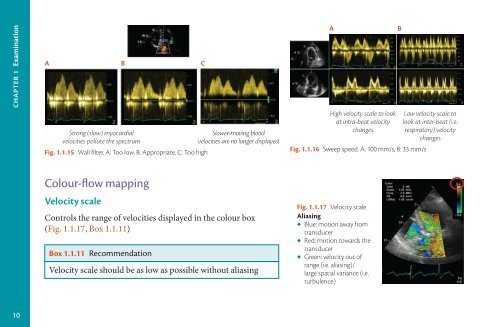
A B C<br />
Strong (slow) myocardial<br />
velocities pollute the spectrum<br />
Fig. 1.1.15 Wall filter. A: Too low, B: Appropriate, C: Too high<br />
Slower-moving blood<br />
velocities are no longer displayed<br />
A<br />
High velocity scale to look<br />
at intra-beat velocity<br />
changes<br />
Fig. 1.1.16 Sweep speed. A: 100 mm/s, B: 33 mm/s<br />
B<br />
Low velocity scale to<br />
look at inter-beat (i.e.<br />
respiratory) velocity<br />
changes<br />
Colour-flow mapping<br />
Velocity scale<br />
Controls the range of velocities displayed in the colour box<br />
(Fig. 1.1.17, Box 1.1.11)<br />
Box 1.1.11 Recommendation<br />
Velocity scale should be as low as possible without aliasing<br />
Fig. 1.1.17 Velocity scale<br />
Aliasing<br />
◆ Blue: motion away from<br />
transducer<br />
◆ Red: motion towards the<br />
transducer<br />
◆ Green: velocity out of<br />
range (i.e. aliasing)/<br />
large spatial variance (i.e.<br />
turbulence)<br />
10