FLEISCHWIRTSCHAFT international_04_2018
- No tags were found...

Create successful ePaper yourself
Turn your PDF publications into a flip-book with our unique Google optimized e-Paper software.
22<br />
Fleischwirtschaft <strong>international</strong> 4_<strong>2018</strong><br />
Hygiene<br />
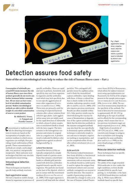
Fig. 1: Rapid<br />
agglutination assays<br />
show a negative<br />
result, when the<br />
original color<br />
retains; a positive<br />
result is indicated<br />
by distinct color<br />
agglutination.<br />
Detection assures food safety<br />
State-of-the-art microbiological tests help to reduce the risk of human illness cases – Part 2<br />
Consumption of minimally processed<br />
RTE-meats increases the risk<br />
of human illness cases since these<br />
products generally do not receive any<br />
further treatment before consumption.<br />
All raw meat can have some<br />
level of microbial contamination<br />
present. Modern microbiological<br />
methods are able to deliver detailed<br />
insight of a microbial contamination<br />
of meat or a meat product.<br />
By Akhilesh K. Verma,<br />
A. Prajapati and<br />
Pramila Umaraw<br />
Conventional or traditional methods<br />
for detecting microorganisms<br />
in meats are based on the<br />
incorporation of samples into a<br />
nutrient medium in which microorganisms<br />
can multiply, thus providing<br />
visual confirmation of their<br />
growth. These conventional test<br />
methods are simple, easily adaptable,<br />
very practical, and generally<br />
inexpensive. More and more new<br />
technolgies find their way into labs<br />
specialized in meat testing.<br />
Immunological detection<br />
methods<br />
These techniques are based on<br />
specific body-antibody reactions.<br />
Rapid agglutination assays<br />
The test is based on visible clumps<br />
shown by organisms in presence of<br />
specific antibodies. These are rapid<br />
and easy to perform. Sensitivity and<br />
specificity may vary from organism<br />
to organism and the antibodies<br />
used which may lack specificity due<br />
to non-specific agglutination of<br />
some other organisms (CHEES-<br />
BROUGH and DONNELLY, 1996).<br />
These tests are primarily used for<br />
screening of suspected bacterial<br />
colonies after culture isolation from<br />
selective agar plates. Latex agglutination<br />
assay tests are widely used<br />
for the rapid detection of Salmonella<br />
in which a drop of colony suspension<br />
or enrichment broth is mixed<br />
with the latex reagent. The latex<br />
remains in the homogenous suspension<br />
and retains its original<br />
color in a negative test. A positive<br />
result is indicated by distinct color<br />
agglutination against an altered<br />
background (Fig. 1).<br />
Lateral flow devices<br />
Lateral flow devices (LFD) are<br />
typically comprised of a simple<br />
dipstick made of a porous membrane<br />
that contains colored latex<br />
beads or colloidal gold particles<br />
coated with detection antibodies<br />
targeted toward a specific microorganism.<br />
The particles are found<br />
on the base of the dipstick, which is<br />
put in contact with the enrichment<br />
medium (POSTHUMA-TRUMPIE et<br />
al., 2009). If the target organism is<br />
present it will bind with the colored<br />
particles. This conjugated cell/<br />
particle moves by capillary action<br />
until it finds the immobilized<br />
capture antibodies. Upon binding<br />
with these, it forms a colored line<br />
that is clearly visible in the device<br />
window, indicating a positive result<br />
(BETTS and BLACKBURN, 2009). LFD<br />
also requires previous enrichment.<br />
Results are often available within<br />
24 h. False positive results may be<br />
observed during the reaction because<br />
of denaturation or degradation<br />
of the capture antibody and it is<br />
likely that the detection antibody or<br />
the enzyme-conjugated antibody<br />
may also bind the non-specifically<br />
to denatured capture antibody. The<br />
technique is extremely simple to<br />
use and easy to interpret, requires<br />
no washing or manipulation, and<br />
can be completed within 10 min<br />
after culture enrichment (ALDUS et<br />
al., 2003).<br />
ELISA and ELFA<br />
The Enzyme-Linked Immunosorbent<br />
Assay (ELISA) is a biochemical<br />
technique that combines an immunoassay<br />
with an enzymatic assay.<br />
An antibody bound to a solid matrix<br />
is used to capture the antigen from<br />
enrichment cultures and a second<br />
antibody conjugated to an enzyme is<br />
used for detection. The enzyme is<br />
capable of generating a product<br />
detectable by a change in color, or in<br />
the case of Enzyme-Linked Fluorescence<br />
Assay (ELFA) in fluorescence,<br />
which allows for indirect measurement<br />
using spectrophotometry (or<br />
fluorometry for ELFA) of the antigen<br />
present in the sample (microorganism<br />
or toxin) (COHEN and KERDAHI,<br />
1996; JASSON et al., 2010). The success<br />
of an immunoassay depends on<br />
the specificity of the antibody. The<br />
limit of detection for immunoassays<br />
is approximately 10 4 –10 5 CFU/g<br />
depending on the type of antibody<br />
and its affinity for the corresponding<br />
epitope, which means that one or<br />
two previous enrichment stages are<br />
always required (JASSON et al., 2010).<br />
High limits of sensitivity of<br />
>10 5 CFU/mL (COX, 1988), cross<br />
reactivity and changes to antigens<br />
due to acetylation and changing<br />
recognition by assay antibodies<br />
(KIM and SLAUCH, 1999) are the<br />
some disadvantages of ELISA<br />
methods.<br />
Molecular detection methods<br />
These techniques base on specific<br />
molecular reactions.<br />
DNA based methods<br />
The ability of two single stranded<br />
DNA-molecules in vitro, under the<br />
right conditions, to form double<br />
stranded DNA by specific base<br />
pairing, i.e. to hybridize, is the basis<br />
of all DNA-based detection methods<br />
(Fig. 2). While many different<br />
methods based on specific base