Dental Asia March/April 2022
For more than two decades, Dental Asia is the premium journal in linking dental innovators and manufacturers to its rightful audience. We devote ourselves in showcasing the latest dental technology and share evidence-based clinical philosophies to serve as an educational platform to dental professionals. Our combined portfolio of print and digital media also allows us to reach a wider market and secure our position as the leading dental media in the Asia Pacific region while facilitating global interactions among our readers.
For more than two decades, Dental Asia is the premium journal in linking dental innovators and manufacturers to its rightful audience. We devote ourselves in showcasing the latest dental technology and share evidence-based clinical philosophies to serve as an educational platform to dental professionals. Our combined portfolio of print and digital media also allows us to reach a wider market and secure our position as the leading dental media in the Asia Pacific region while facilitating global interactions among our readers.
- No tags were found...

You also want an ePaper? Increase the reach of your titles
YUMPU automatically turns print PDFs into web optimized ePapers that Google loves.
USER REPORT<br />
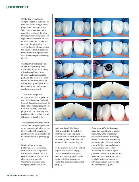
On the day of treatment,<br />
complete anterior isolation was<br />
then performed using a heavy<br />
gauge latex rubber dam, with<br />
W2A clamps attached to the<br />
premolars to secure the dam.<br />
Floss ligatures were placed and<br />
tightened around both central<br />
incisors to further retract the<br />
dam into the gingival sulcus<br />
with the benefit of suppressing<br />
the papilla, which is extremely<br />
useful when closing diastemas<br />
with direct composite bonding<br />
(Fig. 4).<br />
Fig. 4<br />
Fig. 6<br />
Fig. 5<br />
Fig. 7<br />
The teeth were cleaned with<br />
an Enhance polishing cone,<br />
followed by air abrasion using<br />
a Rønvig sandblaster with<br />
30-micron aluminium oxide<br />
particles. The teeth were then<br />
etched, followed by thorough<br />
rinsing, drying and application<br />
of a bonding agent that was<br />
carefully air dispersed.<br />
Fig. 8<br />
Fig. 9<br />
A pre-rolled composite<br />
increment was first applied to<br />
the UR1 and adapted freehand<br />
from all directions to achieve the<br />
ideal initial starting proportions.<br />
Care was taken to adapt the<br />
gingival portion to avoid gross<br />
overhangs. Good isolation made<br />
this much easier (Fig. 5).<br />
This increment was then cured<br />
and refined using interproximal<br />
strips to remove gingival excess<br />
(Fig. 6) and a Sof-Lex disc to<br />
gently contour the contact point<br />
to a smooth convex profile (Fig.<br />
7).<br />
Polytetrafluoroethylene<br />
(PTFE) tape was then placed<br />
over the UR1 and the process<br />
was repeated on the UL1. For<br />
optimal adaption of composite<br />
placement and smooth<br />
transitional junctions from<br />
composite to the tooth, a GC<br />
Fig. 10<br />
Fig. 12<br />
sculpting brush (Fig. 8) and<br />
Optrasculpt (Fig. 9) modelling<br />
instruments were employed to<br />
eliminate instrument indentations<br />
from being introduced into the<br />
composite increments (Fig. 10).<br />
Following final curing, the palatal<br />
aspect was re-checked (Fig.<br />
11) and a small overhang was<br />
removed by using an ultra-thin<br />
metal polishing strip passed<br />
under the interproximal contact<br />
(Fig. 12).<br />
Fig. 11<br />
Fig. 13<br />
Once again, effective isolation<br />
made this possible and is almost<br />
mandatory when attempting<br />
such cases freehand. Following<br />
rubber dam removal, the primary<br />
form was lightly adjusted using a<br />
coarse Sof-Lex disc. An Enhance<br />
polishing cone was used to<br />
reductively polish the marginal<br />
junctions. A medium grit flame bur<br />
was used in an electric handpiece<br />
in a light feathering motion to<br />
introduce tertiary anatomy into<br />
the restorations (Fig. 13).<br />
50 DENTAL ASIA MARCH / APRIL <strong>2022</strong>