Dental Asia March/April 2022
For more than two decades, Dental Asia is the premium journal in linking dental innovators and manufacturers to its rightful audience. We devote ourselves in showcasing the latest dental technology and share evidence-based clinical philosophies to serve as an educational platform to dental professionals. Our combined portfolio of print and digital media also allows us to reach a wider market and secure our position as the leading dental media in the Asia Pacific region while facilitating global interactions among our readers.
For more than two decades, Dental Asia is the premium journal in linking dental innovators and manufacturers to its rightful audience. We devote ourselves in showcasing the latest dental technology and share evidence-based clinical philosophies to serve as an educational platform to dental professionals. Our combined portfolio of print and digital media also allows us to reach a wider market and secure our position as the leading dental media in the Asia Pacific region while facilitating global interactions among our readers.
- No tags were found...

Create successful ePaper yourself
Turn your PDF publications into a flip-book with our unique Google optimized e-Paper software.
BEHIND THE SCENES<br />
Combining digital and conventional<br />
denture workflows: An immediate<br />
denture case report<br />
By Wendy Auclair Clark, DDS, MS; Ibrahim Duqum DDS, MS; with Chris Love, laboratory technician, CDT; at Absolute <strong>Dental</strong> Lab, Durham NC<br />
Emerging technologies and<br />
developing workflows have completely<br />
changed the way many clinicians<br />
and dental laboratory technicians<br />
approach complete dentures in<br />
the last decade. It is important<br />
to remember, however, that<br />
fundamental prosthodontic concepts<br />
have not changed. One of the most<br />
exciting things about digital dentures<br />
is the flexibility of the workflows; once<br />
you grasp both the conventional and<br />
digital complete denture concepts,<br />
your toolbox becomes vast. This<br />
immediate denture case exemplifies<br />
the integration of conventional and<br />
digital complete denture concepts.<br />
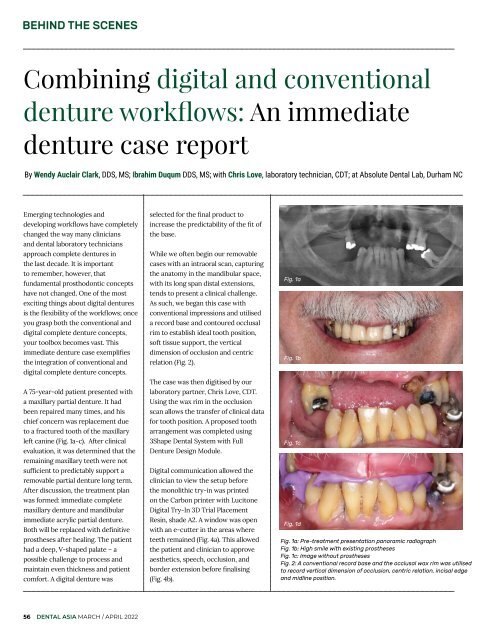
A 75-year-old patient presented with<br />
a maxillary partial denture. It had<br />
been repaired many times, and his<br />
chief concern was replacement due<br />
to a fractured tooth of the maxillary<br />
left canine (Fig. 1a-c). After clinical<br />
evaluation, it was determined that the<br />
remaining maxillary teeth were not<br />
sufficient to predictably support a<br />
removable partial denture long term.<br />
After discussion, the treatment plan<br />
was formed: immediate complete<br />
maxillary denture and mandibular<br />
immediate acrylic partial denture.<br />
Both will be replaced with definitive<br />
prostheses after healing. The patient<br />
had a deep, V-shaped palate – a<br />
possible challenge to process and<br />
maintain even thickness and patient<br />
comfort. A digital denture was<br />
selected for the final product to<br />
increase the predictability of the fit of<br />
the base.<br />
While we often begin our removable<br />
cases with an intraoral scan, capturing<br />
the anatomy in the mandibular space,<br />
with its long span distal extensions,<br />
tends to present a clinical challenge.<br />
As such, we began this case with<br />
conventional impressions and utilised<br />
a record base and contoured occlusal<br />
rim to establish ideal tooth position,<br />
soft tissue support, the vertical<br />
dimension of occlusion and centric<br />
relation (Fig. 2).<br />
The case was then digitised by our<br />
laboratory partner, Chris Love, CDT.<br />
Using the wax rim in the occlusion<br />
scan allows the transfer of clinical data<br />
for tooth position. A proposed tooth<br />
arrangement was completed using<br />
3Shape <strong>Dental</strong> System with Full<br />
Denture Design Module.<br />
Digital communication allowed the<br />
clinician to view the setup before<br />
the monolithic try-in was printed<br />
on the Carbon printer with Lucitone<br />
Digital Try-In 3D Trial Placement<br />
Resin, shade A2. A window was open<br />
with an e-cutter in the areas where<br />
teeth remained (Fig. 4a). This allowed<br />
the patient and clinician to approve<br />
aesthetics, speech, occlusion, and<br />
border extension before finalising<br />
(Fig. 4b).<br />
Fig. 1a<br />
Fig. 1b<br />
Fig. 1c<br />
Fig. 1d<br />
Fig. 1a: Pre-treatment presentation panoramic radiograph<br />
Fig. 1b: High smile with existing prostheses<br />
Fig. 1c: Image without prostheses<br />
Fig. 2: A conventional record base and the occlusal wax rim was utilised<br />
to record vertical dimension of occlusion, centric relation, incisal edge<br />
and midline position.<br />
56 DENTAL ASIA MARCH / APRIL <strong>2022</strong>