Dental Asia March/April 2022
For more than two decades, Dental Asia is the premium journal in linking dental innovators and manufacturers to its rightful audience. We devote ourselves in showcasing the latest dental technology and share evidence-based clinical philosophies to serve as an educational platform to dental professionals. Our combined portfolio of print and digital media also allows us to reach a wider market and secure our position as the leading dental media in the Asia Pacific region while facilitating global interactions among our readers.
For more than two decades, Dental Asia is the premium journal in linking dental innovators and manufacturers to its rightful audience. We devote ourselves in showcasing the latest dental technology and share evidence-based clinical philosophies to serve as an educational platform to dental professionals. Our combined portfolio of print and digital media also allows us to reach a wider market and secure our position as the leading dental media in the Asia Pacific region while facilitating global interactions among our readers.
- No tags were found...

You also want an ePaper? Increase the reach of your titles
YUMPU automatically turns print PDFs into web optimized ePapers that Google loves.
USER REPORT<br />
Simultaneous GBR and GTR in<br />
the posterior mandible area<br />
By Dr Cheng-Hsiang Hsu<br />
EXAMINATION<br />
A 58-year-old male patient had lost his<br />
lower-right first molar one month ago.<br />
It was extracted due to severe mobile<br />
and discomfort. He was told that a<br />
second premolar was to be taken to<br />
restore the posterior dentition with<br />
dental implants. He came to my clinic for<br />
a second opinion.<br />
The probing depths from mesial to distal<br />
were 3,2,3mm on the buccal surface and<br />
3,3,5mm on the lingual surface of the<br />
second premolar. A probing depth of<br />
7mm at the middle site of the posterior<br />
surface was also found. No mobility of<br />
teeth was found at this area although<br />
the attrition of the buccal cusp of the<br />
second premolar was found. No other<br />
symptoms and signs of inflammation<br />
were noted.<br />
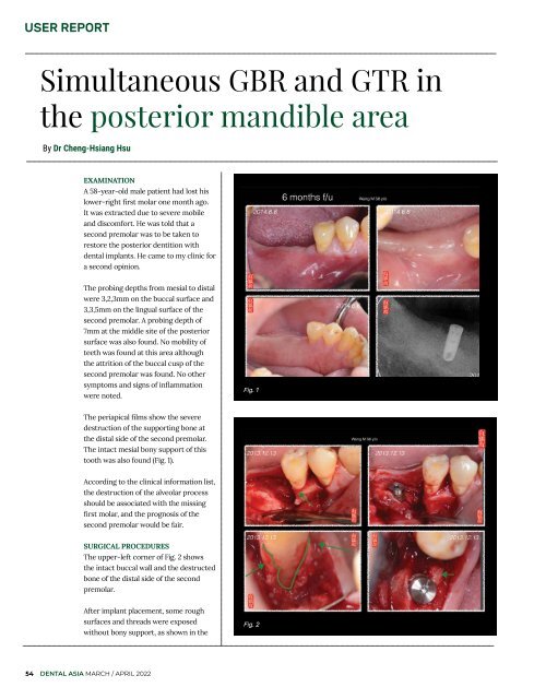
Fig. 1<br />
The periapical films show the severe<br />
destruction of the supporting bone at<br />
the distal side of the second premolar.<br />
The intact mesial bony support of this<br />
tooth was also found (Fig. 1).<br />
According to the clinical information list,<br />
the destruction of the alveolar process<br />
should be associated with the missing<br />
first molar, and the prognosis of the<br />
second premolar would be fair.<br />
SURGICAL PROCEDURES<br />
The upper-left corner of Fig. 2 shows<br />
the intact buccal wall and the destructed<br />
bone of the distal side of the second<br />
premolar.<br />
After implant placement, some rough<br />
surfaces and threads were exposed<br />
without bony support, as shown in the<br />
Fig. 2<br />
54 DENTAL ASIA MARCH / APRIL <strong>2022</strong>