institut für kernchemie universität mainz jahresbericht 2009
institut für kernchemie universität mainz jahresbericht 2009
institut für kernchemie universität mainz jahresbericht 2009

You also want an ePaper? Increase the reach of your titles
YUMPU automatically turns print PDFs into web optimized ePapers that Google loves.
Radioactive labeling of HPMA-based polymeric systems with fluorine-18 for<br />
in vivo imaging and evaluation by positron emission tomography (PET)<br />
Dorothea Moderegger 1 , Mareli Allmeroth 2 , Rudolf Zentel 2 , Frank Roesch 1<br />
1 Institute of Nuclear Chemistry, Johannes Gutenberg University of Mainz, D-55128 Mainz, Germany;<br />
2 Institute of Organic Chemistry, Johannes Gutenberg University of Mainz, D-55128 Mainz, Germany<br />
Introduction: During the last decades polymer<br />
therapeutics became more and more an emerging field of<br />
interest. 1 An example which has already been<br />
intensively studied in clinical trials is the biocompatible<br />
polymeric backbone N-(2-hydroxypropyl)-methacrylamide<br />
(HPMA). 2 Nevertheless, detailed knowledge<br />
about the biodistribution of polymeric drug-delivery<br />
systems in living organisms is still lacking. Especially<br />
information about the tumor accumulation in vivo due to<br />
the enhanced permeability and retention effect are of<br />
major interest. Here, positron emission tomography<br />
(PET) as non-invasive, molecular whole body imaging<br />
technique offers a great opportunity to visualize the in<br />
vivo behavior of radioactively labeled polymeric<br />
structures.<br />
Experimental: Well defined HPMA-based random<br />
copolymers of different molecular weights (Mw=12 kDa<br />
and 77 kDa) synthesized via the RAFT polymerization<br />
technique 3 were labeled with the positron emitting<br />
isotope fluorine-18 using the secondary labeling synthon<br />
2-[ 18 F]fluoroethyl-1-tosylate ([ 18 F]FETos). For labeling<br />
purposes, the polymeric precursors were functionalized<br />
with ~ 4% tyramine moieties thus offering a reactive site<br />
for the prosthetic labeling procedure using [ 18 F]FETos.<br />
The radioactive coupling step was performed using a<br />
solution of 3 mg polymer, 1 µL 5N NaOH and<br />
[ 18 F]FETos in 1 mL of DMSO (figure 1). The clear<br />
solution was kept at 120 °C for 15 min. The reaction<br />
mixture was purified using size exclusion chromatography<br />
(HiTrap Desalting Column, Sephadex G-25<br />
Superfine, column volume 5 mL; flow: 1 mL/min<br />
physiological saline) leading to a pure solution of the<br />
18<br />
F-labeled polymer. For kinetic PET studies, the<br />
animals (tumor bearing Copenhagen rats, R3327-AT1<br />
dunning prostate carcinoma) were anaesthetized with<br />
pentobarbital and a catheter was inserted into the left<br />
jugular vein for radiotracer application. Listmode<br />
acquisition was started with the injection of 25-35 MBq<br />
of the purified polymer solution. The tracer distribution<br />
was followed for 2h p.i. Thereafter, a whole body scan<br />
of the rat was performed. After the experiment, the rats<br />
were sacrificed and an urine sample was taken, and the<br />
radioactivity was measured with an �-counter.<br />
Figure 1. Radioactive labeling of polymers using [ 18 F]FETos.<br />
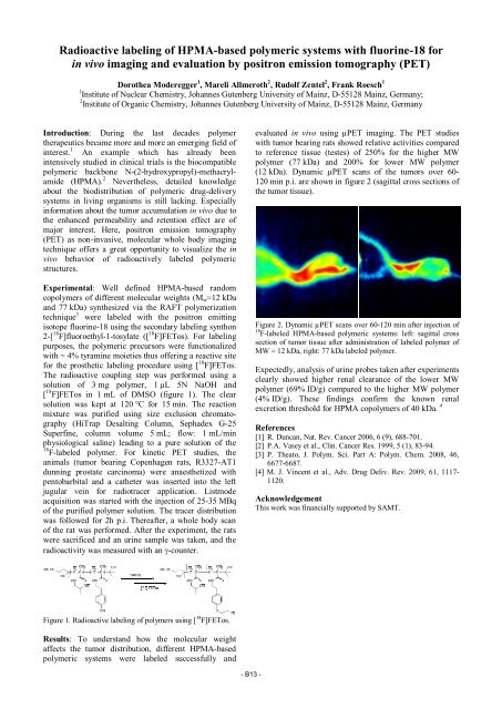
Results: To understand how the molecular weight<br />
affects the tumor distribution, different HPMA-based<br />
polymeric systems were labeled successfully and<br />
- B13 -<br />
evaluated in vivo using µPET imaging. The PET studies<br />
with tumor bearing rats showed relative activities compared<br />
to reference tissue (testes) of 250% for the higher MW<br />
polymer (77 kDa) and 200% for lower MW polymer<br />
(12 kDa). Dynamic µPET scans of the tumors over 60-<br />
120 min p.i. are shown in figure 2 (sagittal cross sections of<br />
the tumor tissue).<br />
Figure 2. Dynamic µPET scans over 60-120 min after injection of<br />
18 F-labeled HPMA-based polymeric systems: left: sagittal cross<br />
section of tumor tissue after administration of labeled polymer of<br />
MW = 12 kDa, right: 77 kDa labeled polymer.<br />
Expectedly, analysis of urine probes taken after experiments<br />
clearly showed higher renal clearance of the lower MW<br />
polymer (69% ID/g) compared to the higher MW polymer<br />
(4% ID/g). These findings confirm the known renal<br />
excretion threshold for HPMA copolymers of 40 kDa. 4<br />
References<br />
[1] R. Duncan, Nat. Rev. Cancer 2006, 6 (9), 688-701.<br />
[2] P.A. Vasey et al., Clin. Cancer Res. 1999, 5 (1), 83-94.<br />
[3] P. Theato, J. Polym. Sci. Part A: Polym. Chem. 2008, 46,<br />
6677-6687.<br />
[4] M. J. Vincent et al., Adv. Drug Deliv. Rev. <strong>2009</strong>, 61, 1117-<br />
1120.<br />
Acknowledgement<br />
This work was financially supported by SAMT.