institut für kernchemie universität mainz jahresbericht 2009
institut für kernchemie universität mainz jahresbericht 2009
institut für kernchemie universität mainz jahresbericht 2009

Create successful ePaper yourself
Turn your PDF publications into a flip-book with our unique Google optimized e-Paper software.
68 Ga-bisphosponates for imaging bone diseases<br />
M. Fellner 1 , R. P. Baum 2 , V. Kubicek 3 , P. Hermann 3 , V. Prasad 2 , F. Rösch 1<br />
1 Institute of Nuclear Chemistry, University of Mainz, Germany<br />
2 Department of Nuclear Medicine, Center for PET/CT, Zentralklinik Bad Berka, Germany<br />
3 Department of Inorganic Chemistry, University of Prague, Czech Republic<br />
99m<br />
Introduction: As Tc-bisphosphonates are well<br />
established tracers for the diagnosis of osteoblastic bone<br />
metastases using SPECT. Analogue attempts for PET<br />
68<br />
using Ga tracers would be potentially useful.<br />
Molecules combining a bisphosphonate bone-targeting<br />
structure and a macrocyclic complexing moiety for<br />
trivalent gallium could be considered as interesting<br />
vectors. A novel 68 Ga-DOTA-based bisphosphonate<br />
(BPAMD) was successfully synthesised and evaluated in<br />
vivo in humans.<br />
Experimentalteil: Using the cation exchanger method<br />
for purification and concentration of the 68 Ge/ 68 Ga<br />
generator eluate the ligand BPAMD was 68 Ga -labelled<br />
in 85% yield within 10 min. The product was purified<br />
using a Strata-X-C cartridge for removing small amounts<br />
of free 68 Ga. After purification the acidic pH was<br />
adjusted and the product was passed trough a sterile<br />
filter. Quality control was performed by different radio-<br />
TLC systems and HPLC.<br />
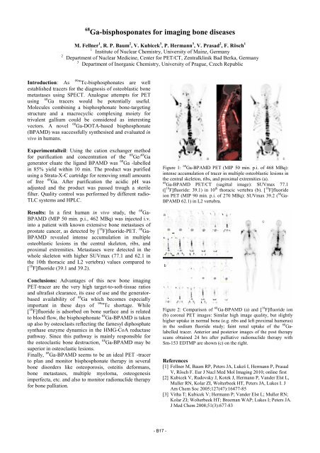
Results: In a first human in vivo study, the 68 Ga-<br />
BPAMD (MIP 50 min. p.i., 462 MBq) was injected i.v.<br />
into a patient with known extensive bone metastases of<br />
prostate cancer, as detected by [ 18 F]fluoride-PET. 68 Ga-<br />
BPAMD revealed intense accumulation in multiple<br />
osteoblastic lesions in the central skeleton, ribs, and<br />
proximal extremities. Metastases were detected in the<br />
whole skeleton with higher SUVmax (77.1 and 62.1 in<br />
the 10th thoracic and L2 vertebra) values compared to<br />
[ 18 F]fluoride (39.1 and 39.2).<br />
Conclusions: Advantages of this new bone imaging<br />
PET-tracer are the very high target-to-soft-tissue ratios<br />
and ultrafast clearance, its ease of use and the generatorbased<br />
availability of 68 Ga which becomes especially<br />
important in these days of 99m Tc shortage. While<br />
[ 18 F]fluoride is adsorbed on bone surface and is related<br />
to blood flow, the bisphosphonate 68 Ga-BPAMD is taken<br />
up also by osteoclasts reflecting the farnesyl diphosphate<br />
synthase enzyme dynamics in the HMG-CoA reductase<br />
pathway. Since this pathway is mainly responsible for<br />
the osteoclastic bone destruction, 68 Ga-BPAMD may be<br />
superior in osteoclastic lesions.<br />
Finally, 68 Ga-BPAMD seems to be an ideal PET -tracer<br />
to plan and monitor bisphosphonate therapy in several<br />
bone disorders like osteoporosis, osteitis deformans,<br />
bone metastases, multiple myeloma, osteogenesis<br />
imperfecta, etc. and also to monitor radionuclide therapy<br />
for bone palliation.<br />
- B17 -<br />
Figure 1: 68 Ga-BPAMD PET (MIP 50 min. p.i. of 468 MBq):<br />
intense accumulation of tracer in multiple osteoblastic lesions in<br />
the central skeleton, ribs, and proximal extremities (a).<br />
68 Ga-BPAMD PET/CT (sagittal image): SUVmax 77.1<br />
([ 18 F]fluoride: 39.1) in 10 th thoracic vertebra (b). [ 18 F]fluoride<br />
ion PET (MIP 90 min. p.i. of 270 MBq): SUVmax 39.2 ( 68 Ga-<br />
BPAMD 62.1) in L2 vertebra.<br />
Figure 2: Comparison of 68 Ga-BPAMD (a) and [ 18 F]fluoride ion<br />
(b) coronal PET images: Similar high image quality, but slightly<br />
higher uptake in normal bone (e.g. ribs and left proximal humerus)<br />
in the sodium fluoride study; faint renal uptake of the 68 Galabelled<br />
tracer. Anterior and posterior images of the post therapy<br />
scans obtained 24 hrs after palliative radionuclide therapy with<br />
Sm-153 EDTMP are shown (c) on the right.<br />
References<br />
[1] Fellner M, Baum RP, Peters JA, Lukeš I, Hermann P, Prasad<br />
V, Rösch F. Eur J Nucl Med Mol Imaging 2010; online first<br />
[2] Kubicek V, Rudovsky J, Kotek J, Hermann P, Vander Elst L,<br />
Muller RN, Kolar ZI, Wolterbeek HT, Peters JA, Lukes I. J<br />
Am Chem Soc 2005;127(47):16477-85<br />
[3] Vitha T; Kubicek V; Hermann P; Vander Elst L; Muller RN;<br />
Kolar ZI; Wolterbeek HT; Breeman WAP; Lukes I; Peters JA.<br />
J Med Chem 2008;51(3):677-83