institut für kernchemie universität mainz jahresbericht 2009
institut für kernchemie universität mainz jahresbericht 2009
institut für kernchemie universität mainz jahresbericht 2009

You also want an ePaper? Increase the reach of your titles
YUMPU automatically turns print PDFs into web optimized ePapers that Google loves.
Boron analysis of blood, tissue and cell samples by neutron autoradiography and<br />
prompt-gamma ray spectroscopy in combination with histological methods<br />
C. Schütz 1 , G.Hampel 1 , S.Werner 1 , T. Schmitz 1 , J.V. Kratz 1 , K. Appelmann 2 , R. Moss 2 ,<br />
C. Brochhausen 3 , J. Kirkpatrick 3 , H. Schmidberger 4 , B. Kuczewski 5 , G. Otto 6<br />
1 Institute for Nuclear Chemistry, University of Mainz, Fritz-Strassmann-Weg 2,D-55099 Mainz, Germany<br />
2 Joint Research Center (JRC) of the European Comission, NL-1755 ZG Petten, the Netherlands 3 Department of<br />
Pathology, University of Mainz, Langenbeckstr. 1, D-55099 Mainz, Germany<br />
4 Department of Radiooncology, University of Mainz, Langenbeckstr. 1, D-55131 Mainz, Germany<br />
5 Department of Chemistry, University of Köln, Zülpicher Strasse 45, D-50674 Köln, Germany<br />
6 Department of Hepatobiliary, Pancreatic and Transplantation Surgery, University of Mainz, Langenbeckstr. 1, D-<br />
55131 Mainz, Germany<br />
Due to the encouraging results of the BNCT research<br />
on non-resectable liver-metastases of colorectal<br />
carcinoma in Pavia, Italy, within the recent years, a<br />
close cooperation was formed between the University<br />
of Mainz, Germany, and the University of Pavia. At<br />
present, a cooperative, clinical study with up to 15<br />
patients is carried out to determine the boron uptake in<br />
both healthy and tumour tissue of the liver.<br />
Three methods for tissue analysis have been selected:<br />
Neutron-autoradiography using CR-39 films, prompt<br />
gamma neutron activation analysis (PGRA) and ICP-<br />
MS. CR-39 films overlaid with tissue slices containing<br />
10 B are irradiated at the TRIGA Mainz and the TRIGA<br />
Pavia and then analysed. The method for quantitative<br />
neutron capture radiographic analysis (QNCR) was<br />
developed in cooperation with the BNCT groups in<br />
Pavia and Bremen, Germany, whereas PGRA is<br />
performed in cooperation with the JRC in Petten, The<br />
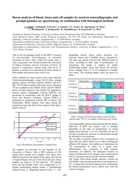
Netherlands. Blood samples were taken during the<br />
operations to provide data for the analysis of the drug’s<br />
kinetics (Fig.2).<br />
B-10 conc (ppm)<br />
25.00<br />
20.00<br />
15.00<br />
10.00<br />
5.00<br />
0.00<br />
0 20 40 60 80 100 120 140<br />
time (min)<br />
Patient 1<br />
Patient 2<br />
Patient 3<br />
Fig.2: Blood-Boron concentration vs infusion time<br />
All samples are provided by the hospital of the<br />
University of Mainz. After a BPA infusion of 200<br />
mg/kg body weight, liver samples were taken from<br />
different positions and depths of the organ, then frozen<br />
in liquid nitrogen and cut to thin slices (20 µm), blood<br />
samples are taken in 30 min intervals. The tumour<br />
areas and the healthy tissue were categorised as T+<br />
(highly proliferating tumour tissue), T (proliferating<br />
tumour tissue), T- (degrading tumour tissue), T- / Nec<br />
- B23 -<br />
(degrading tumour tissue, partly necrotic), Nec<br />
(necrotic tissue) and N (healthy tissue), respectively.<br />
The data vary greatly between the different kinds of<br />
tissue, according to their state of proliferation. As<br />
anticipated, the uptake is highest for highly<br />
proliferating tumour tissue and lowest for necrotic<br />
tissue, ranging from 1.5 to 4.0 compared to healthy<br />
liver tissue. The resulting uptake ratios are shown in<br />
table 1.<br />
Patient 1 before perf. after perfusion<br />
Tissue cat. T+ /T T- T-/Nec Nec N<br />
Con.(µg/g) --- --- n.f. 17.95 12.90 3.22 7.86<br />
Patient 2 before perf. after perfusion<br />
Tissue cat. T N T+ T T- Nec N<br />
Con.(µg/g) 19.91 10.26 24.33 20.23 n.f. n.f. 10.58<br />
Patient 3 before perf. after perfusion<br />
Tissue cat. Nec N T+ T T- Nec N<br />
Con.(µg/g) 4.70 9.06 25.15 n.f. 10.88 4.70 6.37<br />
Table 1: Results from the tissue samples of the first three<br />
patients (ppm 10 B, n.f.: no samples of such kind were found).<br />
Autoradiographic analysis demonstrated that the uptake<br />
behaviour of BPA into tumour cells is strongly<br />
dependent on the state of proliferation; we found<br />
uptake ratios between 1.5 to 4.0. Therefore, we are stil<br />
not able to predict the outcome of a therapeutic attempt<br />
for all kinds of cell structures.<br />
We are able to perform analysis with a resolution of<br />
about 500 µm, which means we are able to distinguish<br />
different cell clusters; this enables us to determine the<br />
10 B concentration in tissue in a much more<br />
differentiated way compared to bulk analysis<br />
procedures. In addition, the biological features of<br />
tumour proliferation in the samples were categorised in<br />
order to correlate tumour growth with boron uptake.<br />
The results from blood sample analysis, carried out<br />
with PGRA, showed maximum boron concentrations of<br />
20 – 25 ppm after two hours infusion time, furthermore<br />
the blood-concentration curves reflect very well the<br />
course of surgery.