
Create successful ePaper yourself
Turn your PDF publications into a flip-book with our unique Google optimized e-Paper software.
J O U R N A L O F L A S E R D E N T I S T R Y | 2 011 V O L . 19 , N O . 3<br />
286<br />
C L I N I C A L C A S E<br />
Gingival Depigmentation with an Er:YAG <strong>Laser</strong>:<br />
A Clinical Case with Three-Year Follow-Up<br />
Grace Sun, DDS, Los Angeles, California<br />
J <strong>Laser</strong> Dent 2011;19(3):286-288<br />
S Y N O P S I S<br />
This clinical case study describes the removal <strong>of</strong> gingival hyperpig-<br />
mentation using an Er:YAG laser. This benign condition was an<br />
esthetic concern for the patient, and the laser procedure produced<br />
good results. While the prognosis is good, the patient’s smoking can<br />
stimulate melanin production and the coloration can reappear.<br />
P R E T R E AT M E N T<br />
A. Case Outline<br />
A 43-year-old African American male<br />
presented with normal medical, oral,<br />
and dental health. The patient<br />
reported that his four upper incisors<br />
had a history <strong>of</strong> trauma, but he<br />
would not supply any details. A clinical<br />
examination revealed that those<br />
four teeth had received endodontic<br />
therapy and were then restored with<br />
porcelain-fused-to-metal crowns. A<br />
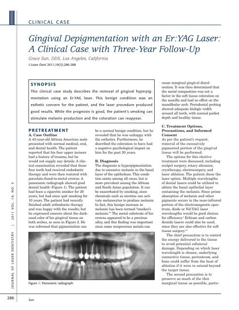
panoramic radiograph showed good<br />
dental health (Figure 1). The patient<br />
had been a cigarette smoker for 20<br />
years, but had since quit smoking for<br />
10 years. The patient had recently<br />
finished adult orthodontic therapy<br />
and was happy with the results; but<br />
he expressed concern about the darkened<br />
color <strong>of</strong> his gingival tissue on<br />
both arches, as seen in Figure 2. He<br />
was informed that pigmentation can<br />
Figure 1: Panoramic radiograph<br />
Sun<br />
be a normal benign condition, but he<br />
revealed that he was unhappy with<br />
the esthetics. Furthermore, he<br />
described the coloration to have had<br />
a negative psychological impact on<br />
him for the past 20 years.<br />
B. Diagnosis<br />
The diagnosis is hyperpigmentation<br />
due to excessive melanin in the basal<br />
layer <strong>of</strong> the epithelium. This condition<br />
exists among all races, but is<br />
more prevalent among the African<br />
and South Asian population. It can<br />
be exacerbated by smoking, since<br />
chemicals such as nicotine can activate<br />
melanocytes to produce melanin.<br />
In fact, this benign increase in<br />
melanin has been termed “smoker’s<br />
melanin.” 1 The metal substrate <strong>of</strong> his<br />
crowns appeared to be a precious<br />
alloy, and this finding was important<br />
since some nonprecious metals can<br />
cause marginal gingival discoloration.<br />
It was then determined that<br />
the metal composition was not a<br />
factor in the s<strong>of</strong>t tissue coloration on<br />
the maxilla and had no effect on the<br />
mandibular arch. Periodontal probing<br />
showed adequate biologic width<br />
around all teeth, with normal pocket<br />
depth and healthy tissue.<br />
C. Treatment Options,<br />
Precautions, and Informed<br />
Consent<br />
As per the patient’s request,<br />
removal <strong>of</strong> the excessively<br />
pigmented portion <strong>of</strong> the gingival<br />
tissue will be performed.<br />
The options for this elective<br />
treatment were discussed, including<br />
scalpel surgery, rotary abrasion,<br />
cryotherapy, electrosurgery, and<br />
laser ablation. The patient chose the<br />
laser option. Multiple wavelengths<br />
<strong>of</strong> dental lasers could be utilized to<br />
ablate the basal epithelial layer<br />
containing the melanin. Since prime<br />
absorption <strong>of</strong> melanin and other<br />
pigments occurs in the near-infrared<br />
portion <strong>of</strong> the electromagnetic spectrum,<br />
diode or Nd:YAG laser<br />
wavelengths would be good choices<br />
for efficiency. 2 Erbium and carbon<br />
dioxide lasers could also be used,<br />
since they are also effective for s<strong>of</strong>t<br />
tissue surgery. 3-5<br />
The chief precaution is to control<br />
the energy delivered to the tissue<br />
to avoid potential collateral<br />
damage. Depending on which laser<br />
wavelength is chosen, underlying<br />
connective tissue, periosteum, and<br />
bone could suffer from the heat <strong>of</strong><br />
ablation if it were to extend beyond<br />
the target tissue.<br />
The second precaution is to<br />
preserve as much <strong>of</strong> the thin<br />
marginal tissue as possible, partic-