
You also want an ePaper? Increase the reach of your titles
YUMPU automatically turns print PDFs into web optimized ePapers that Google loves.
Gallbladder #20 as soon as he was<br />
seated. This enabled us to perform<br />
the scheduled treatments (extraction,<br />
endodontics, and partial<br />
denture fabrication) during separate<br />
appointments.<br />
Among LLLT’s reported beneficial<br />
effects are: (1) Stimulating cell<br />
energy production, which aids in<br />
the restoration <strong>of</strong> normal cell<br />
morphology and function; (2)<br />
Increasing lymphatic flow, which<br />
decreases edema and swelling; (3)<br />
Increasing endorphin release,<br />
reducing conduction <strong>of</strong> C-Fibers,<br />
and decreasing release <strong>of</strong> histamine,<br />
bradykinins, and<br />
acetylcholine to reduce the sensation<br />
<strong>of</strong> postoperative pain; (4)<br />
Stimulating osteoblasts, odontoblasts,<br />
and fibroblasts to promote<br />
the growth <strong>of</strong> bone, dentin, and s<strong>of</strong>t<br />
tissue; (5) Stimulating nerve regeneration;<br />
(6) Increasing neutrophil<br />
and macrophage activity. The<br />
regime that I use takes advantage<br />
<strong>of</strong> all <strong>of</strong> these benefits. I predose or<br />
preload the area to be worked on to<br />
stimulate these wanted effects, and<br />
then use photobiomodulation again<br />
postoperatively to enhance these<br />
effects.<br />
The regime I use for extractions<br />
is as follows: For a maxillary tooth<br />
I place the LED instrument (total<br />
maximum power <strong>of</strong> 42 mW) for a<br />
full 3-minute cycle on the outside <strong>of</strong><br />
face by the tooth to be extracted.<br />
For a mandibular tooth I place it<br />
for half a cycle outside the face and<br />
half a cycle submandibular <strong>of</strong> the<br />
tooth to be extracted. I repeat this<br />
application postoperatively,<br />
followed by a full cycle using my<br />
660-nm probe on top <strong>of</strong> the extraction<br />
socket.<br />
The protocol I follow for<br />
endodontic treatment is as follows:<br />
For a tooth without a crown, I<br />
apply the LED device for a full<br />
cycle <strong>of</strong> 3 minutes outside the face,<br />
and then the 660-nm probe for 1<br />
minute on the buccal and lingual<br />
apex and on the occlusal aspect<br />
prior to beginning the endodontic<br />
treatment. I then apply the 660-nm<br />
Figure 3: Postoperative radiograph after<br />
root canal treatment and extraction<br />
probe to the cleaned and shaped<br />
canal for a full cycle just prior to<br />
placing the fillers. If there is a<br />
large radiolucency I will also use<br />
the 808-nm probe, 300 mW power<br />
output, for 1 minute at the apex <strong>of</strong><br />
the tooth after the filler has been<br />
placed. If the tooth has a crown,<br />
after application <strong>of</strong> the LED cycle I<br />
use the 660-nm probe for 1-1/2<br />
minutes at the buccal and lingual<br />
apices. Since I have been using this<br />
regime, I have had to provide fewer<br />
injections due to the analgesia<br />
induced from the LLLT. I do almost<br />
all my endodontics in one visit and<br />
my patients report having no postoperative<br />
pain.<br />
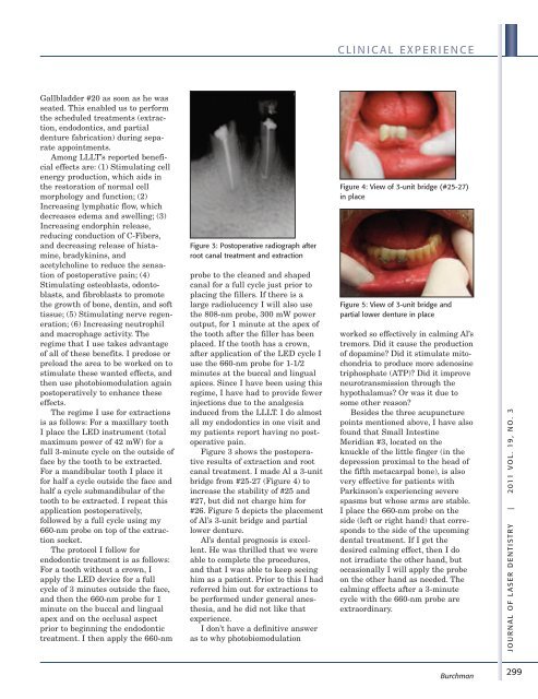
Figure 3 shows the postoperative<br />
results <strong>of</strong> extraction and root<br />
canal treatment. I made Al a 3-unit<br />
bridge from #25-27 (Figure 4) to<br />
increase the stability <strong>of</strong> #25 and<br />
#27, but did not charge him for<br />
#26. Figure 5 depicts the placement<br />
<strong>of</strong> Al’s 3-unit bridge and partial<br />
lower denture.<br />
Al’s dental prognosis is excellent.<br />
He was thrilled that we were<br />
able to complete the procedures,<br />
and that I was able to keep seeing<br />
him as a patient. Prior to this I had<br />
referred him out for extractions to<br />
be performed under general anesthesia,<br />
and he did not like that<br />
experience.<br />
I don’t have a definitive answer<br />
as to why photobiomodulation<br />
C L I N I C A L E X P E R I E N C E<br />
Figure 4: View <strong>of</strong> 3-unit bridge (#25-27)<br />
in place<br />
Figure 5: View <strong>of</strong> 3-unit bridge and<br />
partial lower denture in place<br />
worked so effectively in calming Al’s<br />
tremors. Did it cause the production<br />
<strong>of</strong> dopamine? Did it stimulate mitochondria<br />
to produce more adenosine<br />
triphosphate (ATP)? Did it improve<br />
neurotransmission through the<br />
hypothalamus? Or was it due to<br />
some other reason?<br />
Besides the three acupuncture<br />
points mentioned above, I have also<br />
found that Small Intestine<br />
Meridian #3, located on the<br />
knuckle <strong>of</strong> the little finger (in the<br />
depression proximal to the head <strong>of</strong><br />
the fifth metacarpal bone), is also<br />
very effective for patients with<br />
Parkinson’s experiencing severe<br />
spasms but whose arms are stable.<br />
I place the 660-nm probe on the<br />
side (left or right hand) that corresponds<br />
to the side <strong>of</strong> the upcoming<br />
dental treatment. If I get the<br />
desired calming effect, then I do<br />
not irradiate the other hand, but<br />
occasionally I will apply the probe<br />
on the other hand as needed. The<br />
calming effects after a 3-minute<br />
cycle with the 660-nm probe are<br />
extraordinary.<br />
Burchman<br />
J O U R N A L O F L A S E R D E N T I S T R Y | 2 011 V O L . 19 , N O . 3<br />
299