152 153 Intestinal Disease Meeting Berlin 2006 - Dr. Falk Pharma ...
152 153 Intestinal Disease Meeting Berlin 2006 - Dr. Falk Pharma ...
152 153 Intestinal Disease Meeting Berlin 2006 - Dr. Falk Pharma ...

Create successful ePaper yourself
Turn your PDF publications into a flip-book with our unique Google optimized e-Paper software.
Session 2<br />
Role of Endoscopy<br />
Chair:<br />
J. Devière, Brussels<br />
H. Lochs, <strong>Berlin</strong><br />
Gastroesophageal reflux disease –<br />
Will tomorrow’s therapy be<br />
endoscopic?<br />
According to J. Mössner (Leipzig), endoscopy<br />
plays a central role in the diagnosis of gastroesophageal<br />
reflux disease (GERD), although only<br />
about 50% of GERD patients actually exhibit<br />
endoscopically recognizable lesions in the esophagus.<br />
Obtaining a baseline examination early in<br />
the disease is important and provides a basis for<br />
assessing the subsequent disease course, as well<br />
as for work-up of a Barrett’s esophagus. In addition,<br />
an emergent endoscopic examination is indicated<br />
in any patient reporting alarm systems<br />
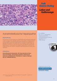
Fig. 11<br />
I Barrett’s esophagus (J. Mössner, Leipzig)<br />
Congress Short Report <strong>Falk</strong> Symposium <strong>152</strong><br />
J. Mössner<br />
such as dysphagia, weight loss or hemorrhage.<br />
Finally, patients with reflux symptoms who do<br />
not respond promptly to standard therapy with<br />
a proton pump inhibitor (PPI) represent a further<br />
indication for endoscopy.<br />
According to J. Mössner, the role of the newer<br />
techniques, such as chromoendoscopy, zoom<br />
endoscopy and narrow band imaging for the diagnosis<br />
of GERD cannot be definitively assessed<br />
at this time. However, he does expect significant<br />
progress from the application of high-resolution<br />
endoscopy in the identification of high-grade<br />
dysplasias and early carcinomas in patients with<br />
Barrett’s esophagus (figure 11). In this area, he<br />
13