JADC - Canadian Dental Association
JADC - Canadian Dental Association
JADC - Canadian Dental Association
Create successful ePaper yourself
Turn your PDF publications into a flip-book with our unique Google optimized e-Paper software.
Pratique C L I N I Q U E<br />
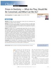
Chondrosarcoma of the Mandible: A Case Report<br />
Rajan Saini, BDS, MDS; Noor Hayati Abd Razak, BDS, M Clin Dent;<br />
Shiafulizan Ab Rahman, BDS, M Clin Dent; Abdul Rani Samsudin, BDS, FDSRCS, AM<br />
SOMMAIRE<br />
Les chondrosarcomes sont des tumeurs malignes d’origine cartilagineuse, qui varient de<br />
tumeurs bien différenciées ressemblant à une tumeur bénigne du cartilage à des tumeurs<br />
de haut grade de malignité affi chant un comportement local agressif et un potentiel<br />
métastatique. De 5 % à 10 % seulement des chondrosarcomes se manifestent dans la<br />
région de la tête et du cou. Nous présentons le cas d’un chondrosarcome siégeant dans la<br />
partie antérieure de la mandibule, et passons en revue la littérature pertinente.<br />
Mots clés MeSH : chondrosarcoma/pathology; female; mandibular neoplasms/pathology<br />
Pour les citations, la version définitive de cet article est la version électronique : www.cda-adc.ca/jcda/vol-73/issue-2/175.html<br />
Chondrosarcomas are malignant mesenchymal<br />
tumours with cartilaginous differentiation<br />
that only rarely affect the<br />
maxillofacial region. 1 Most chondrosarcomas of<br />
the head and neck region arise from the maxilla,<br />
with relatively few arising from the mandible. 2<br />
Although chondrosarcoma occurs in patients<br />
of all ages, most of those a ected are over 50<br />
years of age. 3 In most cases, the tumour presents<br />
as a painless mass or swelling associated with<br />
loosening of the associated teeth. is article<br />
describes a patient with chondrosarcoma of the<br />
anterior mandibular region. e clinical, radiographic,<br />
surgical and pathological aspects of this<br />
lesion are presented, and the relevant literature<br />
is reviewed.<br />
Case Report<br />
A 43-year-old woman was referred to a university-based<br />
dental clinic with swelling over<br />
the lingual aspect of the anterior mandible. e<br />
swelling had been present for 2 years and had<br />
increased gradually in size over that period. e<br />
patient denied any trauma or pain but reported<br />
di culty with swallowing solid foods. She had<br />
Auteur-ressource<br />
Dr Saini<br />
Courriel : rajan@<br />
kb.usm.my<br />
asthma but was not taking any medication for<br />
this condition.<br />
Extraoral examination did not reveal any<br />
obvious facial swelling or asymmetry. ere was<br />
no cervical lymphadenopathy, and all of the cranial<br />
nerves were intact. Intraoral examination<br />
revealed an indurated, painless, discoid swelling<br />
about 2.5 cm × 2 cm in the midline of the anterior<br />
mandible between the lower incisors and the<br />
opening of the sublingual ducts. e overlying<br />
mucosa was pink and appeared normal. e anterior<br />
margin of the swelling was con uent with<br />
the lower alveolus.<br />
Intraoral, occlusal (Fig. 1) and panoramic<br />
radiographs revealed a radiolucent lesion with<br />
di use margins, which had displaced the roots<br />
of the central incisors. Computed tomography<br />
showed an expansile lytic lesion involving the<br />
symphysis menti and the body of the mandible<br />
(Fig. 2). inning of the cortex was observed,<br />
and there was evidence of a cortical break.<br />
ree-dimensional imaging showed destruction<br />
of bone at the mid-mandibular region (Fig. 3).<br />
Histopathological examination revealed<br />
chondrocytes in lacunae that were arranged<br />
<strong>JADC</strong> • www.cda-adc.ca/jadc • Mars 2007, Vol. 73, N o 2 • 175


