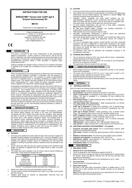
INSTRUCTIONS FOR USE BINDAZYME Human Anti ... - inova
INSTRUCTIONS FOR USE BINDAZYME Human Anti ... - inova
INSTRUCTIONS FOR USE BINDAZYME Human Anti ... - inova
You also want an ePaper? Increase the reach of your titles
YUMPU automatically turns print PDFs into web optimized ePapers that Google loves.
<strong>INSTRUCTIONS</strong> <strong>FOR</strong> <strong>USE</strong><br />
BINDAZYME <strong>Human</strong> <strong>Anti</strong>- ß 2 GP1 IgA S<br />
Enzyme Immunoassay Kit<br />
MK141<br />
This kit is for in-vitro diagnostic use<br />
Product manufactured by:<br />
The Binding Site Ltd., PO Box 11712, Birmingham B14 4ZB, U.K.<br />
www.bindingsite.co.uk<br />
Telephone: +44 (0)121 436 1000<br />
Fax: +44 (0)121 430 7061<br />
e-mail: info@bindingsite.co.uk<br />
1 INTENDED <strong>USE</strong><br />
This assay is intended for the in-vitro measurement of IgA autoantibodies<br />
against β 2 -glycoprotein 1 (β 2 GP1) present in human serum. This kit may be<br />
used in conjunction with other serological assays and clinical information to aid<br />
the diagnosis of thrombosis in at-risk patients, for instance those with primary<br />
antiphospholipid syndrome (APS) or APS secondary to systemic lupus<br />
erythematosus (SLE).<br />
Sufficient materials are supplied to allow a maximum of 41 samples to be tested<br />
in duplicate or 89 in single, with a calibration curve and a positive and negative<br />
control.<br />
2 BACKGROUND<br />
APS is characterised by the clinical syndrome of arterial and venous thrombosis<br />
and/or recurrent pregnancy morbidity in association with the presence of<br />
antiphospholipid antibodies (aPL) 1 . aPL comprise of a range of autoantibodies<br />
that bind to cardiolipin alone, to cardiolipin complexed to cofactor and to<br />
cofactor alone. This cofactor has been identified as the 50kD plasma protein<br />
β 2 GP1 (apolipoprotein H) 2,3 . β 2 GP1 inhibits the intrinsic coagulation pathway 4 ,<br />
binds to platelets 5 and inhibits ADP mediated platelet aggregation 6 .<br />
<strong>Anti</strong>cardiolipin autoantibodies from patients with SLE and primary APS bind to<br />
an altered form of β 2 GP1 produced by the interaction with negatively charged<br />
phospholipid 7 . This modified β 2 GP1 can be reproduced by binding β 2 GP1<br />
directly to polystyrene plates 7 , where clustering or the high density of antigen<br />
allows bi- or multivalent autoantibody binding and subsequent detection by<br />
ELISA methods. Eliminating the phospholipid and using only β 2 GP1 in this<br />
format produces an assay with an enhanced specificity for APS in view of their<br />
predominance as the major autoantibody in the sera of APS patients 8 .<br />
Recently IgG and IgM anti-β 2 GP1 have been included as laboratory criteria for<br />
APS when present at sufficiently high level, and the same committee concluded<br />
that IgA anti-β 2 GP1 are the most frequently detected antibodies in patients in<br />
specific ethnic groups 1 .<br />
3 PRINCIPLE OF THE ASSAY<br />
Microwells are pre-coated with human β 2 GP1 antigen. The calibrators, controls<br />
and diluted patient samples are added to the wells and autoantibodies<br />
recognising the β 2 GP1 antigen bind during the first incubation. After washing<br />
the wells to remove all unbound proteins, purified peroxidase labelled rabbit<br />
anti-human IgA (α chain specific) conjugate is added. The conjugate binds to<br />
the captured human autoantibody and the excess unbound conjugate is<br />
removed by a further wash step. Bound conjugate is visualised with 3,3’,5,5’<br />
tetramethylbenzidine (TMB) substrate which gives a blue reaction product, the<br />
intensity of which is proportional to the concentration of autoantibody in the<br />
sample. Phosphoric acid is added to each well to stop the reaction. This<br />
produces a yellow end-point colour, which is read at 450nm.<br />
4 PRECAUTIONS<br />
4.1 WARNING<br />
• All human sera supplied have been tested at donor level and found negative for<br />
Hepatitis B surface antigens and antibodies to HIV 1 and 2 and Hepatitis C virus.<br />
However, these tests cannot guarantee the absence of infectious agents. Proper<br />
handling and disposal methods should be established and only personnel<br />
qualified in handling of potentially infectious materials should be permitted to use<br />
this kit.<br />
• Sodium azide may react with lead and copper plumbing to form explosive metal<br />
azides. On disposal of reagents flush with a large volume of water, to prevent<br />
azide build-up.<br />
• The buffers and serum supplied in this kit contain various enzyme inhibitors as<br />
listed below. These should be handled with care.<br />
INHIBITOR<br />
CONCENTRATION<br />
Kathon<br />
0.02%<br />
Sodium azide<br />
0.099%<br />
ProClin 300<br />
0.045%<br />
Bromonitrodioxane<br />
0.002%<br />
Methylisothiazone<br />
0.002%<br />
ProClin is a trademark of Rohm and Haas Corp. Philadelphia, PA<br />
• Kathon is an irritant and may cause sensitisation by skin contact.<br />
• The stop solution contains 3M phosphoric acid, which is an irritant. Avoid contact<br />
with skin and eyes.<br />
• Reagent spills should be cleaned up appropriately, observing local and<br />
environmental regulations.<br />
4.2 CAUTION<br />
• This product should only be used by appropriately trained personnel.<br />
• Strict adherence to the protocol is recommended. Any deviation may affect assay<br />
performance, and the results obtained. Pay attention to specific ‘Notes’ and<br />
warnings throughout these Instructions for Use.<br />
• Calibrator, control, conjugate and plate batch numbers are not<br />
interchangeable. Substitution of such components with batch numbers that<br />
differ from those that are provided in the kit could lead to inconsistent and<br />
inaccurate results. All strips must be taken from the same foil pouch.<br />
• To avoid reagent contamination, only use new or clean plastic / glassware.<br />
Never return unused reagents to the bottles.<br />
• Do not leave reagent bottles uncapped; any resulting evaporation or<br />
contamination will lead to inconsistent results.<br />
• TMB substrate must not be exposed to light or water.<br />
• Microbially contaminated, haemolysed or lipaemic serum and specimens<br />
containing particulate matter should not be used.<br />
• Inaccurate sample dilution cannot be checked, as kit controls are ready to use.<br />
The use of calibrated pipettes and appropriate internal QC samples is<br />
recommended.<br />
• The use of automated assay systems, sample dilutors and other automated<br />
equipment may lead to differences in results when compared to the manual<br />
procedure. It is the responsibility of any laboratory to fully validate the system,<br />
and ensure the results fall within the limits as defined in this insert and<br />
associated QC certificate.<br />
• All equipment used must be calibrated and maintained according to the<br />
manufacturer’s instruction.<br />
4.3 STORAGE AND STABILITY<br />
• The kit should be stored at 2-8°C and should not be frozen. Inappropriate<br />
storage temperatures will affect the results.<br />
• Wash buffer diluted into a clean container can be stored at room temperature<br />
(20-24°C) for a maximum of 4 weeks.<br />
• The expiry date of the kit is shown on the outer label.<br />
5 SAMPLE COLLECTION AND STORAGE<br />
• Blood samples should be collected by venepuncture allowed to clot naturally<br />
and the serum separated.<br />
• The serum may be stored at 2-8°C for up to 7 days prior to assay 9 , or for<br />
prolonged storage, aliquoted and stored at -20°C or below.<br />
• Repeated thawing and freezing should be avoided.<br />
• Serum samples should not be heat-inactivated, as this may give false positive<br />
results.<br />
6 MATERIALS<br />
6.1 MATERIALS SUPPLIED<br />
Each kit contains the following product-specific reagents:<br />
• Instruction leaflet: Giving full assay details.<br />
• QC Certificate: Indicating the expected performance of the batch.<br />
• β 2 GP1 S Coated Wells: 12 breakapart 8-well strips coated with human β 2 GP1<br />
antigen. The plate is packaged in a re-sealable foil bag containing two<br />
desiccant pouches.<br />
• APS Sample Diluent: 2 bottles containing 50mL of buffer for sample dilution.<br />
Coloured yellow, ready to use.<br />
• APS Wash Buffer (20x Concentrate): 1 bottle containing 50mL of a 20-fold<br />
concentrated buffer for washing the wells.<br />
• β 2 GP1 IgA S Calibrators: 5 bottles each containing 1.2mL of diluted human<br />
serum, with the following concentrations of anti-β 2 GP1 autoantibody: 300, 100,<br />
33.3, 11.1, 3.7 U/mL. Ready to use.<br />
• β 2 GP1 IgA S Positive Control: 1 bottle containing 1.2mL of diluted human<br />
serum. The expected value is given on the QC certificate. Ready to use.<br />
• β 2 GP1 S Negative Control: 1 bottle containing 1.2mL of diluted human serum.<br />
The expected value is given on the QC certificate. Ready to use.<br />
• β 2 GP1 IgA S Conjugate: 1 bottle containing 12mL of purified peroxidase<br />
labelled antibody. Coloured green. Ready to use.<br />
• TMB Substrate: 1 bottle containing 14mL TMB substrate. Ready to use.<br />
• Stop Solution: 1 bottle containing 14mL of 3M phosphoric acid. Ready to use.<br />
6.2 ADDITIONAL MATERIALS AND EQUIPMENT - not supplied<br />
• Automatic microplate plate washer: This is recommended, however, plate<br />
washing can be performed manually.<br />
• Plate reader: Capable of measuring optical densities at 450nm referenced on<br />
air.<br />
• Distilled or deionised water: This should be of the highest quality available.<br />
• Calibrated micropipettes: For dispensing 1000, 100 & 10µL.<br />
• Multichannel pipette: Recommended for dispensing 100µL volumes of<br />
conjugate, substrate and stop solution.<br />
• Glass/plastic tubes: For sample dilution.<br />
7 ASSAY METHOD<br />
7.1 PRE-ASSAY STEPS<br />
1. Bring the kit to room temperature<br />
• The kit is designed for room temperature operation (20-24°C).<br />
• Remove the kit from storage and stand at room temperature for approximately<br />
60 minutes. Wells must not be removed from the foil bag until they have<br />
reached room temperature.<br />
Note: The kits may be maintained at room temperature for up to 1 week.<br />
2. Kit components<br />
Gently mix each kit component before use.<br />
3. Wash buffer dilution<br />
Add 50mL of the wash buffer concentrate to 950mL of distilled water (1 in 20<br />
dilution) into a clean container and mix. Smaller volumes can be diluted as<br />
appropriate.<br />
Note: Diluted wash buffer can be stored at room temperature for up to 4 weeks,<br />
therefore only dilute the appropriate amount.<br />
Insert Code: E141, Version: 17 January 2008, Page 1 of 13
4. Sample dilution<br />
Dilute 10µL of each sample with 1000µL of sample diluent (1:100) and mix well.<br />
Note: Diluted sample must be used within 8 hours.<br />
5. Strip and frame handling<br />
Place the required number of wells in the strip holder. Position from well A1,<br />
filling columns from left to right across the plate. When handling the plate,<br />
squeeze the long edges of the frame to prevent the wells falling out.<br />
Note: Return unused wells to the foil bag immediately with the two desiccant<br />
pouches and re-seal tightly to minimise exposure to moisture. Take care not to<br />
puncture or tear the foil bag, see below.<br />
WARNING: Exposure of wells to moisture or contamination by dust or<br />
other particulate matter will result in antigen degradation, leading to poor<br />
assay precision and potentially false results.<br />
7.2 ASSAY METHOD<br />
Maintain the same dispensing sequence throughout the assay.<br />
1. Sample addition<br />
Dispense 100µL of each calibrator, control and diluted (1:100) sample into the<br />
appropriate wells of the plate provided.<br />
Note: Samples should be added as quickly as possible to the plate to minimise<br />
assay drift, and the timer started after the addition of the last sample.<br />
Incubate at room temperature for 30 minutes.<br />
2. Washing<br />
The washing procedure is critical and requires special attention. An improperly<br />
washed plate will give inaccurate results, with poor precision and high<br />
backgrounds.<br />
After incubation remove the plate and wash wells 3 times with 250-350µL wash<br />
buffer per well. Wash the plate either by using an automatic plate washer or<br />
manually as indicated below. After the final automated wash, invert the plate<br />
and tap the wells dry on absorbent paper.<br />
Plates can be washed manually as follows:<br />
a. Flick out the contents of the plate into a sink.<br />
b. Tap the wells dry on absorbent paper.<br />
c. Fill each well with 250-350µL of wash buffer using a multichannel pipette.<br />
d. Gently shake the plate on a flat surface.<br />
e. Repeat a-d twice.<br />
f. Repeat a and b.<br />
3. Conjugate addition<br />
Dispense 100µL of conjugate into each well, blot the top of the wells with a<br />
tissue to remove any splashes.<br />
Note: To avoid contamination, never return excess conjugate to the reagent<br />
bottle.<br />
Incubate at room temperature for 30 minutes.<br />
4. Washing<br />
Repeat step 2.<br />
5. Substrate (TMB) addition<br />
Dispense 100µL of TMB substrate into each well, blot the top of the wells with a<br />
tissue to remove any splashes.<br />
Note: To avoid contamination, never return excess TMB to the reagent bottle.<br />
Incubate at room temperature in the dark for 30 minutes.<br />
6. Stopping<br />
Dispense 100µL of stop solution into each well. This causes a change in colour<br />
from blue to yellow.<br />
7. Optical density measurement<br />
Read the optical density (OD) of each well at 450nm on a microplate reader,<br />
within 30 minutes of stopping the reaction.<br />
8 RESULTS AND QUALITY CONTROL<br />
1. Quality control<br />
In order for an assay to be valid, all the following criteria must be met:<br />
• Calibrators and positive and negative controls must be included in each run.<br />
• The values obtained for the positive and negative controls should be in the<br />
ranges specified on the QC Certificate.<br />
• The curve shape should be similar to the calibration curve, shown on the QC<br />
Certificate.<br />
If the above criteria are not met, the assay is invalid and the test should<br />
be repeated.<br />
2. Calculate mean optical densities (for assays run in duplicate only)<br />
For each calibrator, control and sample calculate the mean OD of the duplicate<br />
readings. The percentage coefficient of variation (% CV) for each duplicate OD<br />
should be less than 15%.<br />
3. Plot calibration curve<br />
The calibration curve can be plotted either automatically or manually by plotting<br />
the anti-β 2 GP1 autoantibody concentration on the log scale against the OD on<br />
the linear scale for each calibrator:<br />
• Automatic - use appropriately validated software, and the curve fit that best fits<br />
the data.<br />
• Manual - using log/linear graph paper, draw a smooth curve through the points<br />
(not a straight line or point to point).<br />
4. Treatment of anomalous points<br />
If any one point does not lie on the curve, it can be removed. If the absence of<br />
this point means that the curve has a shape dissimilar to that of the sample<br />
calibration curve, or more than one point appears to be anomalous, then the<br />
assay should be repeated.<br />
5. Calculation of autoantibody levels in controls and samples<br />
Read the level of the anti-β 2 GP1 autoantibody in the controls and diluted<br />
samples directly from the calibration curve. The control values should fall within<br />
the ranges given on the QC Certificate.<br />
Note: The calibrator values have been adjusted by a factor of 100 to account<br />
for a 1:100 sample dilution. No further correction is required.<br />
6. Assay calibration<br />
The assay is calibrated in U/mL against an internal arbitrary reference<br />
calibrator, as no internationally recognised standard is currently available.<br />
7. Limitations<br />
• The results obtained from this assay are not diagnostic proof of the presence or<br />
absence of disease.<br />
• Where results produce a negative serum anti-β 2 GP1, in the presence of clinical<br />
indications, an anti-cardiolipin, lupus anti-coagulant or additional test is<br />
required.<br />
• Patient treatment must not begin on the basis of a positive anti-β 2 GP1 result<br />
alone. Clinical indications must also be present.<br />
9 PER<strong>FOR</strong>MANCE CHARACTERISTICS<br />
9.1 PRECISION<br />
The intra-assay precision was measured using six samples tested 20 times<br />
within one assay across the range of the calibration curve. The concentration<br />
and % C.V. for each sample are given below:<br />
INTRA-ASSAY PRECISION<br />
n=20 Concentration (U/mL) % CV<br />
Sample 1 6.4 5.8<br />
Sample 2 16.0 2.9<br />
Sample 3 20.5 2.9<br />
Sample 4 37.4 4.6<br />
Sample 5 48.7 3.6<br />
Sample 6 66.4 4.1<br />
The inter-assay precision was measured using six samples tested in duplicate<br />
six times over three days. The concentration and % C.V. for each sample are<br />
given below.<br />
INTER-ASSAY PRECISION<br />
n=6 Concentration (U/mL) % CV<br />
Sample 1 6.1 10.7<br />
Sample 2 15.8 3.6<br />
Sample 3 19.5 5.1<br />
Sample 4 37.9 4.4<br />
Sample 5 49.3 3.7<br />
Sample 6 65.4 2.6<br />
9.2 NORMAL RANGE<br />
<strong>Anti</strong>-β 2 GP1 autoantibody levels were measured in serum from 100 male and<br />
100 female normal blood donors. The results are displayed in the graph below.<br />
Based on a positive cut-off of 20.0U/mL, the two positive samples obtained<br />
were confirmed positive using an alternative manufacturer’s anti-β 2 GP1 IgA kit.<br />
Number of samples<br />
120<br />
100<br />
80<br />
60<br />
40<br />
20<br />
This normal range is provided as a guide only. It is recommended that each<br />
laboratory determine its own normal range.<br />
9.3 RELATIVE SPECIFICITY, SENSITIVITY, AGREEMENT<br />
The relative specificity, sensitivity and agreement have been determined<br />
against an alternative anti-β 2 GP1 IgA kit using 114 clinical and normal samples.<br />
The samples were from 46 SLE patients who had previously tested positive for<br />
anti-phospholipid antibodies, 8 from patients with primary anti-phospholipid<br />
syndrome, 20 RA patients, 20 SLE patients with unknown APS status and 20<br />
normal sera from healthy blood donors. The status of the samples was<br />
determined applying a positive cut-off of 20.0U/mL with a borderline region of<br />
15-20U/mL for the BINDAZYME IgA S assay and that recommended in the<br />
alternative kit.<br />
EIA<br />
BINDAZYME<br />
<strong>Anti</strong>-β 2 GP1 IgA S EIA<br />
Alternative EIA<br />
+ *Borderline -<br />
+ 18 2 1<br />
Borderline 3 3 3<br />
- 1^ 5 78<br />
Relative sensitivity 94.7%<br />
Relative specificity 98.7%<br />
Relative agreement 98.0%<br />
*Samples in the borderline range were excluded from the analysis<br />
^The discrepant sample tested negative in an alternate kit<br />
9.4 ANALYTICAL SENSITIVITY<br />
Confirmation that the anti-β 2 GP1 IgA S assay can distinguish between two<br />
samples with values close to the bottom of the measuring range (173 and 199%<br />
of the lowest calibrator) was obtained by statistical analysis (Student’s t-test) of<br />
the results obtained from testing multiple replicates of each sample.<br />
9.5 MEASURING RANGE<br />
0<br />
20.0<br />
IgA <strong>Anti</strong>-B2GP1 U/mL<br />
The measuring range of the assay is 3.7-300U/mL.<br />
Insert Code: E141, Version: 17 January 2008, Page 2 of 13
9.6 INTERFERING SUBSTANCES<br />
A range of interfering substances was spiked into anti-β 2 GP1 IgA positive and<br />
negative samples, which were subsequently assayed. The method used to<br />
check the effect of these substances used the Interference Check A plus kit<br />
from Kokusai, Shiyaku, Japan.<br />
Substance<br />
Concentration<br />
Bilirubin F (free)<br />
18.0mg/dL<br />
Bilirubin C (conjugate)<br />
19.3mg/dL<br />
Haemolysed haemoglobin<br />
482mg/dL<br />
Chyle<br />
1430FT Units<br />
Rheumatoid Factor<br />
50.0 IU/mL<br />
No interference was observed in samples tested.<br />
10 EXPECTED VALUES<br />
The positive cut-off based on the assay of 100 male and 100 female normal<br />
serum samples has been determined for the anti-β 2 GP1 IgA assay to be<br />
20.0U/mL, with a borderline region of 15.0-20.0U/mL. The ranges are provided<br />
as a guide only. ELISA assays are very sensitive and capable of detecting small<br />
differences in sample populations<br />
11 REFERENCES<br />
IgA anti-β 2 GP1<br />
< 15.0 U/mL Negative result<br />
15.0-20.0 U/mL Borderline<br />
>20.0 U/mL Positive result<br />
1. Miyakis S, Lockshin MD, Atsumi T, Branch DW, Brey RL, et al. (2006).<br />
International concensus statement on an update of the classification criteria for<br />
definite antiphospholipid syndrome. J Thromb Haemost; 4: 295-306.<br />
2. Galli M, Comfurius P, Maassen C, Hemker HC, de Baets MH, et al. (1990).<br />
<strong>Anti</strong>cardiolipin antibodies (ACA) directed not to cardiolipin but to a plasma<br />
protein cofactor. Lancet; 335: 1544-7.<br />
3. McNeil HP, Simpson RJ, Chesterman CN & Krilis SA. (1990). <strong>Anti</strong>-phospholipid<br />
antibodies are directed against a complex antigen that includes a lipid-binding<br />
inhibitor of coagulation: beta 2-glycoprotein I (apolipoprotein H). Proc Natl Acad<br />
Sci USA; 87:4120-4124.<br />
4. Schousboe I. (1985). β 2 -Glycoprotein I: A plasma inhibitor of the contact<br />
activation of the intrinsic blood coagulation pathway. Blood; 66: 1086-1091.<br />
5. Nimpf J, Bevers EM, Bomans PH, Till U, Wurm H, et al. (1986). Prothrombinase<br />
activity of human platelets is inhibited by β 2 -glycoprotein-I. Biochim. Biophys.<br />
Acta; 884: 142-149.<br />
6. Nimpf J, Wurm H & Kostner GM. (1987). β 2 -Glycoprotein-I (apo-H) inhibits the<br />
release reaction of human platelets during ADP-induced aggregation.<br />
Atherosclerosis; 63: 109-114.<br />
7. Matsuura E, Igarashi Y Yasuda T, et al (1994). <strong>Anti</strong>cardiolipin antibodies<br />
recognize beta 2-glycoprotein I structure altered by binding with an oxygenated<br />
modified solid phase surface. J Exp Med; 179:457-462.<br />
8. Matsuura E, & Koike T. (1996) in: Autoantibodies, pp 109-114, Peter JB &<br />
Shoenfeld Y (eds), Elsevier Science, Amsterdam.<br />
9. Protein Reference Unit Handbook of Autoimmunity (3rd Edition) 2004 Ed A<br />
Milford Ward. J. Sheldon, GD Wild. Publ. PRU Publications, Sheffield. 14.<br />
12 PLATE TEMPLATE<br />
Please see back of insert.<br />
Summary of procedure<br />
1. Add 100µL of each calibrator, control and 1:100 diluted sample to<br />
the appropriate wells.<br />
Incubate for 30 minutes.<br />
Wash.<br />
2. Add 100µL of conjugate to each well. Incubate for 30 minutes.<br />
Wash.<br />
3. Add 100µL of substrate to each well.<br />
Incubate for 30 minutes.<br />
4. Add 100µL of stop solution to each well.<br />
Measure the absorbance at 450nm.<br />
BINDAZYME is a trademark of<br />
The Binding Site Ltd.<br />
P.O. Box 11712, Birmingham<br />
B14 4ZB England<br />
Insert Code: E141, Version: 17 January 2008, Page 3 of 13
ARBEITSANLEITUNG<br />
BINDAZYME TM <strong>Human</strong> <strong>Anti</strong>-ß 2 GP1 IgA S<br />
Enzym-Immunoassay-Kit<br />
MK141<br />
Nur zur in-vitro Diagnostik<br />
Hergestellt von:<br />
The Binding Site Ltd, PO Box 11712, Birmingham, B14 4ZB, UK.<br />
www.bindingsite.co .uk<br />
Vertrieb in Deutschland und Österreich durch:<br />
The Binding Site GmbH, Robert-Bosch-Straβe 2A<br />
D-68723 Schwetzingen, Deutschland<br />
Telefon: +49 (0) 6202 92 62 0<br />
Fax: +49 (0) 6202 92 62 222<br />
e-mail: office@bindingsite.de<br />
1 VERWENDUNGSZWECK<br />
Dieser Kit dient der in-vitro Bestimmung von IgA-Autoantikörpern gegen<br />
ß 2 -Glykoprotein 1 (ß 2 GP1) in <strong>Human</strong>serum. Der Test kann zusammen mit<br />
anderen serologischen und klinischen Befunden bei der Diagnosestellung von<br />
Thrombosen bei Risikopatienten mit z.B. primärem <strong>Anti</strong>phospholipid-Syndrom<br />
(APS) oder sekundärem APS, bei dem in der Regel ein systemischer Lupus<br />
erythematodes (SLE) zugrunde liegt, helfen.<br />
Der Kit enthält ausreichend Material, um maximal 41 Proben in Doppelbestimmung<br />
oder 89 Proben in Einzelbestimmung, eine Kalibrationskurve und<br />
je eine Positiv- und eine Negativkontrolle messen zu können.<br />
2 EINFÜHRUNG<br />
APS ist klinisch durch arterielle und venöse Thrombosen und/oder<br />
wiederkehrende habituelle Aborte gekennzeichnet, wobei sich jeweils<br />
<strong>Anti</strong>phospholipid-<strong>Anti</strong>körpern (aPL) nachweisen lassen 1 . Zu diesen <strong>Anti</strong>phospholipid-<strong>Anti</strong>körpern<br />
gehören eine Reihe verschiedener Autoantikörper, die<br />
entweder nur an Cardiolipin, an den Cardiolipin-Cofaktor-Komplex oder nur an<br />
den Cofaktor binden. Als Cofaktor wurde das 50kD Plasmaprotein β 2 GP1<br />
(Apolipoprotein H) identifiziert 2,3 . β 2 GP1 inhibiert die intrinsische Gerinnungskaskade<br />
4 , bindet an Thrombozyten 5 und inhibiert die ADP-vermittelte<br />
Thrombozyten-Aggregation 6 .<br />
<strong>Anti</strong>-Cardiolipin-Autoantikörper von SLE- und APS-Patienten binden an eine durch<br />
die Wechselwirkung mit negativ geladenem Phospholipid veränderte Form von<br />
β 2 GP1 7 . Dieses modifizierte β 2 GP1 kann durch direkte Bindung von β 2 GP1 an<br />
Polystyrolplättchen 7 reproduziert werden, an denen die Clusterbildung oder hohe<br />
Dichte des <strong>Anti</strong>gens eine bi- oder multivalente Bindung des Autoantikörpers<br />
erlaubt und demzufolge im ELISA nachweisbar ist. Durch Eliminierung des<br />
Phospholipids und ausschließliche Verwendung dieser Form von β 2 GP1 erhält<br />
man einen Test, der in Hinblick auf das vorherrschende Auftreten dieses<br />
Autoantikörpers in den Seren von APS-Patienten eine erhöhte Spezifität für APS<br />
aufweist 8 .<br />
Unlängst wurden anti-ß 2 GP1-<strong>Anti</strong>körper vom IgG- und IgM-Typ unter der<br />
Vorausssetzung, dass sie einen entsprechend hohen Titer aufweisen, in die<br />
Laborkriterien zur Diagnose eines definitiven APS neu aufgenommen; hinsichtlich<br />
der anti-ß 2 GP1-<strong>Anti</strong>körper vom IgA-Typ wurde von demselben Komitee<br />
abschließend festgehalten, dass sie am häufigsten bei spezifischen ethnischen<br />
Gruppen nachweisbar sind 1 .<br />
3 TESTPRINZIP<br />
Die Vertiefungen (Wells) der Mikrotiterplatten sind mit humanem β 2 GP1<br />
beschichtet. Die Kalibratoren, Kontrollen und verdünnten Patientenproben<br />
werden in den Vertiefungen inkubiert, so dass in diesem ersten<br />
Inkubationsschritt Autoantikörper an das immobilisierte ß 2 GP1-<strong>Anti</strong>gen binden<br />
können. Nach dem Waschen der Vertiefungen - um ungebundenes Protein zu<br />
entfernen - wird Peroxidase-markiertes Kaninchen anti-<strong>Human</strong>-IgA-Konjugat<br />
(spezifisch für die α-Kette) zugegeben. Das Konjugat bindet an die gebundenen<br />
humanen Autoantikörper. Ungebundenes Konjugat wird durch einen weiteren<br />
Waschschritt entfernt. Das gebundene Konjugat wird mit Hilfe von 3,3’,5,5’<br />
Tetramethylbenzidin (TMB) sichtbar gemacht. TMB ergibt ein blaues Reaktionsprodukt,<br />
dessen Intensität proportional zur Autoantikörper-Konzentration der<br />
Probe ist. Um die Reaktion zu stoppen, wird in jede Vertiefung Phosphorsäure<br />
pipettiert. Das gelbe Reaktionsprodukt wird bei 450nm gemessen.<br />
4 WARNUNGEN UND VORSICHTSMAßNAHMEN<br />
4.1 WARNUNGEN<br />
• Jede Einzelspende wurde bezüglich <strong>Anti</strong>körpern gegen <strong>Human</strong>-<br />
Immunschwäche-Virus (HIV 1 & 2), Hepatitis C-Virus und Hepatitis-B-Oberflächenantigen<br />
(HBsAg) untersucht und als negativ befunden.<br />
• Es gibt aber zur Zeit keine absolut sicheren Testmethoden zum Ausschluss von<br />
Infektionsträgern. Deshalb sollten die Reagenzien als potenziell infektiös<br />
behandelt werden. Umgangs- und Entsorgungsmethoden sollten denen für<br />
potenziell infektiösem Material entsprechen und der Test nur von entsprechend<br />
geschultem Personal durchgeführt werden.<br />
• Natriumazid kann mit Blei- oder Kupferrohren explosive Metallazide bilden.<br />
Nach der Entsorgung mit ausreichender Menge Wasser nachspülen, um<br />
Azidablagerungen zu vermeiden.<br />
• Alle Puffer und Seren im Kit enthalten die unten aufgelisteten Enzym-Inhibitoren<br />
als Konservierungsmittel. Diese sollten mit der entsprechenden Vorsicht<br />
behandelt werden.<br />
INHIBITOR<br />
KONZENTRATION<br />
Kathon<br />
Natriumazid<br />
ProClin 300<br />
Bromonitrodioxan<br />
Methylisothiazon<br />
0,02%<br />
0,099%<br />
0,045%<br />
0,002%<br />
0,002%<br />
ProClin ist ein eingetragenes Warenzeichen der Rohm and Haas Corp. Philadelphia, PA<br />
• Kathon wirkt reizend und kann bei Hautkontakt zu einer Sensibilisierung führen.<br />
• Die Stopp-Lösung enthält 3M Phosphorsäure. Sie ist reizend, deshalb Kontakt<br />
mit Haut und Augen vermeiden.<br />
• Verschüttete Reagenzien entsprechend beseitigen, dabei sind ökologische und<br />
gesetzliche Bestimmung zu beachten.<br />
4.2 VORSICHTSMAßNAHMEN<br />
• Dieser Test sollte nur von entsprechend geschultem Laborpersonal<br />
durchgeführt werden.<br />
• Testdurchführung strikt nach Arbeitsanleitung. Jegliche Abweichung kann die<br />
Testqualität und Ergebnisse beeinflussen. Beachten Sie die spezifischen<br />
‚Hinweise‘ und Warnungen in dieser Arbeitsanleitung.<br />
• Die Chargen von Kalibratoren, Kontrollen, Konjugat und Mikrotiterstreifen sind<br />
nicht austauschbar. Wird eine dieser Komponenten durch eine andere Charge,<br />
die nicht in dem gelieferten Kit enthalten ist, ersetzt, kann es zu inkonsistenten<br />
und inakkuraten Ergebnissen kommen. Alle Mikrotiterstreifen müssen aus<br />
demselben Folienbeutel entnommen werden.<br />
• Zur Vermeidung von Kontaminationen der Reagenzien immer saubere Plastikoder<br />
Glasgefäße verwenden. Niemals unbenutztes Reagenz in die Flaschen<br />
zurückgeben.<br />
• Reagenzienflaschen nicht für längere Zeit offen stehen lassen. Die daraus<br />
resultierende Verdunstung oder Verunreinigung führt zu widersprüchlichen<br />
Ergebnissen.<br />
• TMB darf weder mit Licht noch mit Wasser in Berührung kommen!<br />
• Die Verwendung von mikrobiell oder mit Partikeln verunreinigter, hämolytischer<br />
oder lipämischer Seren vermeiden.<br />
• Falsche Probenverdünnungen können nicht erkannt werden, da die Kit-<br />
Kontrollen gebrauchsfertig sind. Es wird empfohlen, kalibrierte Pipetten und<br />
geeignete interne Kontrollen zu verwenden.<br />
• Die Verwendung von ELISA-Automaten, Pipettierautomaten oder anderen<br />
automatisierten Geräten kann im Vergleich zur manuellen Testdurchführung zu<br />
abweichenden Ergebnissen führen. Jedes Labor muss das System vollständig<br />
validieren und sicherstellen, dass die Ergebnisse innerhalb der im QC-Zertifikat<br />
angegebenen Spezifikationen liegen. Das QC-Zertifikat ist dieser<br />
Arbeitsanleitung beigefügt.<br />
• Die verwendeten Geräte und Pipetten müssen gemäß der Herstellerangaben<br />
kalibriert und gewartet werden.<br />
4.3 LAGERUNG UND STABILITÄT<br />
• Den Kit bei 2-8°C lagern, nicht einfrieren. Lagerung bei falscher Temperatur<br />
beeinflusst die Ergebnisse.<br />
• Der verdünnte Waschpuffer ist in einem sauberen Gefäß bei Raumtemperatur<br />
(20-24°C) bis maximal 4 Wochen haltbar.<br />
• Die Haltbarkeit des Kits wird auf dem Außenetikett angegeben.<br />
5 PROBENENTNAHME UND -VORBEREITUNG<br />
• Blutproben über Venenpunktur sammeln und auf natürliche Weise gerinnen<br />
lassen. Serum vom Gerinnsel trennen.<br />
• Die Seren können bei 2-8°C bis zu 7 Tage vor dem Test gelagert werden 9 . Für<br />
eine längere Lagerung empfiehlt es sich, die Seren unverdünnt und aliquotiert<br />
bei mindestens -20°C einzufrieren.<br />
• Wiederholtes Einfrieren und Auftauen der Proben vermeiden.<br />
• Serumproben nicht hitzeinaktivieren, dies kann zu falsch-positiven Ergebnissen<br />
führen.<br />
6 MATERIALIEN<br />
6.1 GELIEFERTE MATERIALIEN<br />
• Arbeitsanleitung: mit genauen Angaben zur Testdurchführung.<br />
• QC-Zertifikat: mit Angabe der Leistungsdaten der Charge.<br />
• ß 2 GP1 S Coated Wells (mit ß 2 GP1 S beschichtete Wells): 12 Mikrotiterstreifen<br />
mit 8 vereinzelbaren Vertiefungen, die mit humanem ß 2 GP1 beschichtet sind.<br />
Die Streifen sind in einem wiederverschließbaren Folienbeutel, der ein<br />
Trockenmittel enthält, verpackt.<br />
• APS Sample Diluent (APS-Probendiluens): 2 Flaschen mit je 50mL<br />
gebrauchsfertigem Puffer zur Probenverdünnung. Farbcodierung: gelb.<br />
• APS Wash Buffer (20x Concentrate) (APS-Waschpuffer, 20fach-Konzentrat):<br />
1 Flasche mit 50mL eines 20-fach konzentrierten Puffers zum Waschen der<br />
Vertiefungen der Mikrotiterstreifen.<br />
• ß 2 GP1 IgA S Calibrators (ß 2 GP1 IgA S Kalibratoren): 5 Flaschen mit jeweils<br />
1,2mL vorverdünntem <strong>Human</strong>serum, das folgende Konzentrationen an <strong>Anti</strong>ß<br />
2 GP1-Autoantikörpern enthält: 300; 100; 33,3; 11,1 und 3,7 U/mL.<br />
Gebrauchsfertig.<br />
• ß 2 GP1 IgA S Positive Control (ß 2 GP1 IgA S Positivkontrolle): 1 Flasche mit<br />
1,2mL vorverdünntem <strong>Human</strong>serum. Der Sollwert ist auf dem QC-Zertifikat<br />
angegegen. Gebrauchsfertig.<br />
• ß 2 GP1 S Negative Control (ß 2 GP1 S Negativkontrolle): 1 Flasche mit 1,2mL<br />
vorverdünntem <strong>Human</strong>serum. Der Sollwert ist auf dem QC-Zertifikat<br />
angegegen. Gebrauchsfertig.<br />
• ß 2 GP1 IgA S Conjugate (ß 2 GP1 IgA S-Konjugat): 1 Flasche mit 12mL<br />
gereinigtem, Peroxidase-markiertem <strong>Anti</strong>körper. Farbcodierung: grün.<br />
Gebrauchsfertig.<br />
• TMB Substrate (TMB-Substrat): 1 Flasche mit 14mL TMB-Lösung. Gebrauchsfertig.<br />
• Stop Solution (Stopp-Lösung): 1 Flasche mit 14mL 3M Phosphorsäure.<br />
Gebrauchsfertig.<br />
6.2 BENÖTIGTE, NICHT IM KIT ENTHALTENE MATERIALIEN<br />
• Automatisches Mikrotiterplatten-Waschgerät: ist empfehlenswert, aber die<br />
Platten können auch manuell gewaschen werden.<br />
• Mikrotiterplatten-Reader: Messung der optischen Dichte (OD) in den<br />
Vertiefungen bei 450nm gegen Luft<br />
• Destilliertes Wasser: Es sollte von höchster Qualität und frei von Metallionen<br />
sein.<br />
Insert Code: E141, Version: 17 January 2008, Page 4 of 13
• Kalibrierte Mikropipetten: zum Pipettieren von 1000, 100 & 10µL.<br />
• Mehrkanal-Pipette: zum Pipettieren eines Volumens von 100µL von Konjugat,<br />
Substrat und Stopp-Lösung.<br />
• Glas- oder Plastikgefäße: zur Probenverdünnung.<br />
7 TESTDURCHFÜHRUNG<br />
7.1 TESTVORBEREITUNG<br />
1. Kit auf Raumtemperatur erwärmen:<br />
• Die optimale Temperatur für die Testdurchführung liegt bei 20-24°C.<br />
• Den Kit nach Entnahme aus dem Kühlschrank ca. 60 Minuten bei Raumtemperatur<br />
stehen lassen. Die Wells dürfen erst nach Erreichen der<br />
Raumtemperatur aus dem Folienbeutel entnommen werden.<br />
Hinweis: Der Kit kann bis zu 1 Woche bei Raumtemperatur gelagert werden.<br />
2. Kit-Komponenten<br />
Jede Kit-Komponente vor Gebrauch kurz schütteln.<br />
3. Verdünnung des Waschpuffers (20fach-Konzentrat):<br />
50mL Waschpuffer-Konzentrat und 950mL destilliertes Wasser (1/20-<br />
Verdünnung) in ein sauberes Gefäß geben und mischen. Es können auch<br />
entsprechend kleinere Volumina verdünnt werden.<br />
Hinweis: Der verdünnte Puffer ist bei Raumtemperatur bis zu 4 Wochen<br />
haltbar; deshalb nur die benötigte Menge verdünnen.<br />
4. Verdünnung der Proben<br />
10µL jeder Probe mit 1000µL Probendiluens verdünnen (1:100) und gut mischen.<br />
Hinweis: Die verdünnten Proben müssen innerhalb von 8 h gemessen werden.<br />
5. Umgang mit Streifen und Rahmen<br />
Die benötigte Anzahl Wells aus dem Folienbeutel entnehmen und in den<br />
Rahmen setzen. Dabei von Position A1 ausgehend die Spalten von links nach<br />
rechts auffüllen. Achten Sie darauf, die langen Seiten des Rahmens<br />
zusammenzudrücken, damit die Wells nicht herausfallen können.<br />
Hinweis: Um die Kontaktzeit mit der Luftfeuchtigkeit zu minimieren, die nicht<br />
verwendeten Wells sofort in den wiederverschließbaren Folienbeutel mit dem<br />
Trockenmittel geben und gut verschließen. Darauf achten, dass der<br />
Folienbeutel nicht beschädigt wird.<br />
ACHTUNG: Kontakt mit Feuchtigkeit oder Verunreinigung durch Staub<br />
oder anderen Partikeln kann einen <strong>Anti</strong>gen-Abbau verursachen. Daraus<br />
resultieren eine schlechte Präzision und potenziell falsche Ergebnisse.<br />
7.2 TESTDURCHFÜHRUNG<br />
Alle Reagenzien in der gleichen Reihenfolge pipettieren.<br />
1. Pipettieren der Kontrollen und Proben<br />
100µL von jeder Kontrolle und verdünnten (1:100) Proben in das dafür laut<br />
Plattenschema vorgesehene Well pipettieren.<br />
Hinweis: Die Proben müssen so schnell wie möglich pipettiert werden, um eine<br />
‚Assay-Drift’ zu vermeiden; die Inkubationszeit läuft ab Zugabe der letzten<br />
Probe.<br />
30 Minuten bei Raumtemperatur inkubieren.<br />
2. Waschen der Platte<br />
Sorgfältiges Waschen der Wells ist wichtig für eine hohe Präzision und genaue<br />
Testergebnisse. Bei nicht korrekt gewaschenen Wells können ungenaue<br />
Ergebnisse mit schlechter Präzision und hohem Hintergrund auftreten.<br />
Nach der Inkubation die Wells dreimal mit 250-350µL Waschpuffer waschen.<br />
Das Waschen kann mit einem automatischen Platten-Waschgerät oder<br />
manuell, wie nachfolgend beschrieben, erfolgen. Nach dem letzten Waschschritt<br />
die Wells durch leichtes Klopfen auf eine absorbierende Unterlage (z.B.<br />
Papiertücher) trocknen.<br />
Manuelles Waschen:<br />
a. Den Inhalt der Wells in den Ausguss schütten, dabei die<br />
langen Seiten des Rahmens zusammendrücken.<br />
b. Die Wells auf einer absorbierenden Unterlage (z.B.<br />
Papiertücher) leicht ausklopfen.<br />
c. 250-350µL Waschpuffer in der Arbeitskonzentration mit<br />
einer Mehrkanalpipette in die Wells pipettieren.<br />
d. Die Mikrotiterplatte auf einer flachen Unterlage stehend<br />
leicht schütteln.<br />
e. Zweimaliges Wiederholen der Schritte a bis d.<br />
f. Die Wells wie unter a und b entleeren.<br />
3. Pipettieren des Konjugats<br />
100µL Konjugat in jedes Well pipettieren. Die Ränder der Wells mit Filterpapier<br />
abtrocknen, um Spritzer zu entfernen.<br />
Hinweis: Um Verunreinigungen zu vermeiden, niemals überschüssiges<br />
Konjugat in die Konjugat-Flasche zurückgeben.<br />
30 Minuten bei Raumtemperatur inkubieren.<br />
4. Waschen der Platte<br />
Siehe Abschnitt 2.<br />
5. Pipettieren von Substrat (TMB)<br />
Nach dem Waschen 100µL TMB in jedes Well pipettieren. Die Ränder der<br />
Wells mit Filterpapier abtrocknen, um Spritzer zu entfernen.<br />
Hinweis: Um Verunreinigungen zu vermeiden, niemals überschüssiges TMB in<br />
die TMB-Flasche zurückgeben.<br />
30 Minuten bei Raumtemperatur im Dunkeln inkubieren.<br />
6. Reaktionsende<br />
100µL Stopp-Lösung in jedes Well pipettieren. Die Farbe schlägt von blau nach<br />
gelb um.<br />
7. Farbmessung<br />
Die optische Dichte (OD) bei 450nm in jedem Well mit einem Mikrotiterplatten-<br />
Reader innerhalb von 30 Minuten nach Zugabe der Stopp-Lösung bestimmen.<br />
8 ERGEBNISSE UND QUALITÄTSKONTROLLE<br />
1. Qualitätskontrolle<br />
Nur, wenn alle nachfolgend aufgeführten Kriterien erfüllt sind, ist der Test gültig.<br />
• Alle angegebenen Kalibratoren sowie die positiven und negativen Kontrollen<br />
müssen bei jedem Testansatz mitgeführt werden.<br />
• Alle für die Kontrollen erzielten Ergebnisse müssen in dem angegebenen<br />
Wertebereich (siehe QC-Zertifikat) liegen.<br />
• Der Kurvenverlauf der erzielten Kalibrationskurve sollte in etwa dem auf dem<br />
QC-Zertifikat gezeigen entsprechen.<br />
Sind die oben aufgeführten Kriterien nicht erfüllt, ist der Test ungültig und<br />
muss wiederholt werden.<br />
2. Berechnung der OD-Mittelwerte (Nur bei Doppelbestimmungen)<br />
Berechnen Sie für jeden Kalibrator, jede Kontrolle und jede Probe den<br />
Mittelwert aus den beiden OD-Messergebnissen. Der prozentuale Variationskoeffizient<br />
(VK%) der beiden OD-Messwerte sollte dabei jeweils unter 15%<br />
liegen.<br />
3. Erstellung der Kalbrationskurve<br />
Die Kalibrationskurve kann automatisch oder manuell erstellt werden, indem<br />
man die <strong>Anti</strong>-Cardiolipin-Autoantikörper-Konzentrationen (logarithmische Skala)<br />
halblogarithmisch gegen die zugehörigen OD-Werte der Kalibratoren (lineare<br />
Skala) aufträgt.<br />
• Automatisch – verwenden Sie eine geeignete und validierte Software und die<br />
die Daten am besten widerspiegelnde Kurvenanpassung.<br />
• Manuell – verwenden Sie halblogarithmisches Millimeterpapier und zeichen Sie<br />
eine glatte Kurve durch die Punkte (keine Gerade oder Punkt-zu-Punkt-<br />
Verbindung).<br />
4. Handhabung abweichender Kalibrationspunkte<br />
Sollte irgendein Messpunkt nicht auf der Kurve liegen, kann er gestrichen<br />
werden. Hat diese Streichung allerdings zur Folge, dass der Kurvenverlauf von<br />
dem im QC-Zertifikat vorgegebenen abweicht, oder müssten mehrere Punkte<br />
der Kalibration gestrichen werden, sollte die gesamte Kalibration wiederholt<br />
werden.<br />
5. Berechnung der Autoantikörper-Konzentrationen in den Kontrollen und<br />
Proben<br />
Lesen Sie die <strong>Anti</strong>-ß2GP1-Autoantikörper-Konzentrationen in den Kontrollen<br />
und verdünnten Patientenproben direkt aus der Kalibrationskurve ab. Die<br />
Kontrollwerte sollten in den im QC-Zertifikat vorgegebenen Konzentrationsbereich<br />
fallen.<br />
Hinweis: Die Standardprobenverdünnung von 1:100 ist in der Kalibrationskurve<br />
bereits berücksichtigt. Es sind keine weiteren Korrekturen nötig.<br />
6. Kalibration des Tests<br />
Der Test ist gegen einen intenternen Standard kalibriert (in U/mL), da kein<br />
anerkannter internationaler Standard verfügbar ist.<br />
7. Grenzen des Tests<br />
• Die Testergebnisse können nicht als diagnostischer Beweis für das<br />
Vorhandensein bzw. Nichtvorhandensein einer Krankheit verwendet werden.<br />
• Falls der Nachweis von ß 2 GP1-Autoantikörpern im Serum trotz klinischer<br />
Indikationen negativ ausfällt, ist eine Testung auf <strong>Anti</strong>-Cardiolipin-<strong>Anti</strong>körper,<br />
Lupus <strong>Anti</strong>koagulans oder einen weiteren Test erforderlich.<br />
• Die Behandlung eines Patienten darf nicht allein aufgrund eines positiven<br />
ß 2 GP1-Ergebnisses begonnen werden; sie muss auch klinisch indiziert sein.<br />
9 LEISTUNGSDATEN<br />
9.1 PRÄZISION<br />
Die Intra-Assay-Präzision wurde durch eine 20-fache Messung von 6 innerhalb<br />
der Kalibrationskurve liegenden Proben in einem Assay ermittelt. Die<br />
Konzentrationen und Variationskoeffizienten (VK%) jeder einzelnen Probe sind<br />
nachfolgend aufgelistet:<br />
INTRA-ASSAY PRÄZISION<br />
n=20 Konzentration (U/mL) VK %<br />
Probe 1 6,4 5,8<br />
Probe 2 16,0 2,9<br />
Probe 3 20,5 2,9<br />
Probe 4 37,4 4,6<br />
Probe 5 48,7 3,6<br />
Probe 6 66,4 4,1<br />
Für die Bestimmung der Inter-Assay-Präzision wurden sechs Proben in<br />
Doppelbestimmung in 6 verschiedenen Testansätzen in einem Zeitraum von 3<br />
Tagen gemessen. Die Konzentrationen und Variationskoeffizienten (VK%) jeder<br />
einzelnen Probe sind nachfolgend aufgelistet.<br />
INTER-ASSAY PRÄZISION<br />
n=6 Konzentration (U/mL VK %<br />
Probe 1 6,1 10,7<br />
Probe 2 15,8 3,6<br />
Probe 3 19,5 5,1<br />
Probe 4 37,9 4,4<br />
Probe 5 49,3 3,7<br />
Probe 6 65,4 2,6<br />
9.2 NORMALBEREICH<br />
Die Serumwerte an ß 2 GP1 IgA-Autoantikörpern wurden bei je 100 männlichen und<br />
100 weiblichen normalen Blutspendern bestimmt; die Ergebnisse sind in der<br />
nachfolgenden Graphik dargestellt. Basierend auf einem positiven Cut-off-Wert von<br />
20,0 U/mL waren zwei Proben positiv; dieses positive Ergebnis wurde mit einem<br />
alternativen anti-ß 2 GP1-IgA-Kit jeweils bestätigt.<br />
Anzahl an Proben<br />
120<br />
100<br />
80<br />
60<br />
40<br />
20<br />
0<br />
20,0<br />
IgA <strong>Anti</strong>-ß 2 GP1 U/mL<br />
Dieser Normalbereich dient nur zur Orientierung. Jedes Labor sollte seine eigenen<br />
Normalbereiche festlegen.<br />
Insert Code: E141, Version: 17 January 2008, Page 5 of 13
9.3 SPEZIFITÄT, SENSITIVITÄT UND ÜBEREINSTIMMUNG<br />
Die relative Spezifität, Sensitivität und Übereinstimmung wurde im Vergleich zu<br />
einem alternativen <strong>Anti</strong>-ß 2 GP1 IgA-Kit anhand von 114 klinischen und normalen<br />
Proben bestimmt. Diese Proben stammten von 46 SLE-Patienten, die zuvor auf<br />
<strong>Anti</strong>phospholipid-<strong>Anti</strong>körper positiv getestet worden waren, 8 Patienten mit<br />
primärem <strong>Anti</strong>phospholipid-Syndrom (APS), 20 RA-Patienten, 20 SLE-<br />
Patienten mit unbekanntem APS-Status und 20 gesunden Blutspendern. Die<br />
Bewertung der Proben erfolgte beim BINDAZYME IgA S Assay bei einem<br />
grenzwertigen „Grauzonenbereich“ von 15-20 U/mL und eines positiven Cut-off-<br />
Wertes von 20,0 U/mL bzw. dem im Alternativtest angegebenen Cut-off.<br />
EIA<br />
BINDAZYME<br />
<strong>Anti</strong>-β 2 GP1 IgA S EIA<br />
Alternativer EIA<br />
+ *Grenzwertig -<br />
+ 18 2 1<br />
Grenzwertig 3 3 3<br />
- 1^ 5 78<br />
Relative Sensitivität 94,7%<br />
Relative Spezifität 98,7%<br />
Relative Übereinstimmung 98,0%<br />
*Die im grenzwertigen Konzentrationsbereich liegenden Proben wurden bei der<br />
Auswertung nicht berücksichtigt<br />
^Die eine unterschiedlich befundete Probe war bei Testung mit einem weiteren<br />
Kit negativ.<br />
9.4 ANALYTISCHE SENSITIVITÄT<br />
Der Beweis, dass der <strong>Anti</strong>-ß 2 GP1 IgA S auch noch zwischen zwei Proben mit<br />
Werten im unteren Messbereich (173 und 199% des niedrigsten Kalibrators)<br />
unterscheiden kann, wurde durch statistische Analyse (t-Test, Vergleich von<br />
Mittelwerten aus unabhängigen Stichproben) der Ergebnisse mehrfacher<br />
Messungen jeder einzelnen Probe erbracht.<br />
9.5 MESSBEREICH<br />
Der Messbereich des Kits liegt zwischen 3,7 und 300 U/mL.<br />
9.6 INTERFERIERENDE SUBSTANZEN<br />
<strong>Anti</strong>-ß 2 GP1 IgA negative und positive Seren wurden mit verschiedenen<br />
interferierenden Substanzen versetzt und anschließend mit diesem Kit<br />
gemessen. Hierzu wurde ein Interference Check A Plus TM (Kokusai Shiyaku,<br />
Japan) verwendet.<br />
Substanz<br />
Bilirubin F (frei)<br />
Bilirubin C (konjugiert)<br />
Hämolysiertes Hämoglobin<br />
Chylus (Milchsaft)<br />
Rheumafaktor<br />
Konzentration<br />
18,0 mg/dL<br />
19,3 mg/dL<br />
482 mg/dL<br />
1430 FT Units<br />
50,0 IU/mL<br />
12 PLATTENSCHEMA<br />
Siehe Ende der Arbeitsanleitung.<br />
KURZARBEITSANLEITUNG<br />
1. 100µL von jeder Kontrolle und 1:100 verdünnten<br />
Probe in das entsprechende Well pipettieren.<br />
30 Minuten inkubieren. Waschen.<br />
2. 100µL Konjugat in jedes Well geben.<br />
30 Minuten inkubieren. Waschen.<br />
3. 100µL Substrat in jedes Well geben.<br />
30 Minuten im Dunkeln inkubieren.<br />
4. 100µL Stopp-Lösung in jedes Well pipettieren.<br />
Absorption bei 450nm messen.<br />
BINDAZYME<br />
ist ein Warenzeichen von<br />
The Binding Site Ltd., UK.<br />
P.O. Box 11712, Birmingham<br />
B14 4ZB. England<br />
Bei keiner der eingesetzten Substanzen wurde ein interferierender Einfluss in<br />
den getesteten Proben beobachtet.<br />
10 ERWARTETE WERTE<br />
Der positive Cut-off-Wert für den <strong>Anti</strong>-ß 2 GP1 IgA S Assay wurde basierend auf<br />
den Messergebnissen der Serumproben von 100 männlichen und 100<br />
weiblichen Blutspendern bei einem grenzwertigen „Grauzonenbereich“ von 15,0<br />
- 20,0 U/mL mit 20,0 U/mL festgelegt. Diese Bereiche dienen nur zur<br />
Orientierung. ELISAs sind sehr sensitive Tests, die kleinste Unterschiede in<br />
Probenpopulationen erkennen können.<br />
11 REFERENZEN<br />
IgA <strong>Anti</strong>-ß 2 GP1<br />
< 15,0 U/mL Negativ<br />
15,0 - 20,0 U/mL Grenzwertig<br />
> 20,0 U/mL Positiv<br />
1. Miyakis S, Lockshin MD, Atsumi T, Branch DW, Brey RL, et al. (2006).<br />
International concensus statement on an update of the classification criteria for<br />
definite antiphospholipid syndrome. J Thromb Haemost; 4: 295-306.<br />
2. Galli M, Comfurius P, Maassen C, Hemker HC, de Baets MH, et al. (1990).<br />
<strong>Anti</strong>cardiolipin antibodies (ACA) directed not to cardiolipin but to a plasma<br />
protein cofactor. Lancet; 335: 1544-7.<br />
3. McNeil HP, Simpson RJ, Chesterman CN & Krilis SA. (1990). <strong>Anti</strong>-phospholipid<br />
antibodies are directed against a complex antigen that includes a lipid-binding<br />
inhibitor of coagulation: beta 2-glycoprotein I (apolipoprotein H). Proc Natl Acad<br />
Sci USA; 87:4120-4124.<br />
4. Schousboe I. (1985). β 2 -Glycoprotein I: A plasma inhibitor of the contact<br />
activation of the intrinsic blood coagulation pathway. Blood; 66: 1086-1091.<br />
5. Nimpf J, Bevers EM, Bomans PH, Till U, Wurm H, et al. (1986). Prothrombinase<br />
activity of human platelets is inhibited by β 2 -glycoprotein-I. Biochim. Biophys.<br />
Acta; 884: 142-149.<br />
6. Nimpf J, Wurm H & Kostner GM. (1987). β 2 -Glycoprotein-I (apo-H) inhibits the<br />
release reaction of human platelets during ADP-induced aggregation.<br />
Atherosclerosis; 63: 109-114.<br />
7. Matsuura E, Igarashi Y Yasuda T, et al. (1994). <strong>Anti</strong>cardiolipin antibodies<br />
recognize beta 2-glycoprotein I structure altered by binding with an oxygenated<br />
modified solid phase surface. J Exp Med; 179:457-462.<br />
8. Matsuura E, & Koike T. (1996) in: Autoantibodies, pp 109-114, Peter JB &<br />
Shoenfeld Y (eds), Elsevier Science, Amsterdam.<br />
9. Protein Reference Unit Handbook of Autoimmunity (3rd Edition) 2004 Ed A<br />
Milford Ward. J. Sheldon, GD Wild. Publ. PRU Publications, Sheffield. 14.<br />
Insert Code: E141, Version: 17 January 2008, Page 6 of 13
<strong>INSTRUCTIONS</strong> D’UTILISATION<br />
Coffret BINDAZYME S de dosage des<br />
anticorps humains <strong>Anti</strong> β 2 GP1 IgA S<br />
MK141<br />
Pour un usage en diagnostic in-vitro<br />
Produits fabriqués par:<br />
The Binding Site Ltd, PO Box 11712, Birmingham B14 4ZB, UK<br />
www.bindingsite.co.uk<br />
Distribués en France par la société :<br />
The Binding Site, 14 rue des Glairaux, BP226, 38522 Saint-Egrève Cedex.<br />
Téléphone : 04.38.02.19.19<br />
Fax : 04.38.02.19.20<br />
e-mail: info@bindingsite.fr<br />
1 INDICATIONS<br />
Ce coffret permet de mesurer in vitro les autoanticorps IgA spécifiques dirigés<br />
contre la ß 2 -glycoproteine 1 (ß 2 GP1) du sérum humain. Ce coffret doit être<br />
utilisé en conjonction avec d’autres tests sérologiques et informations cliniques<br />
pour aider au diagnostic chez les patients présentant par exemple un risque<br />
thrombotique; syndrome primaires des antiphospholipides (SAPL) ou consécutif<br />
à un lupus érythémateux disséminé (LED).<br />
Ce coffret permet de tester au maximum 41 échantillons en double ou 89 en<br />
simple, avec une courbe de calibration, un contrôle positif, un contrôle négatif.<br />
2 PRESENTATION GENERALE<br />
Le SAPL est caractérisé par des symptômes tels que des thromboses<br />
artérielles et veineuses et/ou des morts fœtales récurrentes en association<br />
avec la présence d’anticorps antiphospholipides (aPL) 1 . Les aPL comprennent<br />
toute une gamme d’autoanticorps qui se fixent aux cardiolipides seuls, aux<br />
cardiolipides complexés à un cofacteur et au cofacteur seul. Ce cofacteur a été<br />
identifié comme étant une protéine plasmatique de 50kD, la β 2 GP1<br />
(apolipoprotéine H) 2,3 . La β 2 GP1 inhibe la voie intrinsèque de la coagulation 4 , se<br />
fixe aux plaquettes 5 et inhibe l’agrégation des plaquettes médiées par les ADP 6 .<br />
Les autoanticorps anticardiolipides des patients atteints de LED et de SAPL<br />
primaire se fixent à une forme altérée de la β 2 GP1 produite par l’interaction<br />
avec des phospholipides 7 chargés négativement. Cette β 2 GP1 modifiée peut<br />
être reproduite en fixant de la β 2 GP1 directement dans les plaques de<br />
polystyrène 7 , lorsque le regroupement ou la forte densité de l’antigène permet<br />
un accrochage des autoanticorps bi ou polyvalent et la détection par des<br />
méthodes ELISA. Eliminer les phospholipides et utiliser uniquement la β 2 GP1<br />
sous ce format permet d’avoir un test avec une spécificité accrue pour le SAPL<br />
du fait de leur prédominance chez les patients atteints de le SAPL 8 .<br />
Récemment, les IgG et les IgM anti-β 2 GP1 ont été inclus dans les critères<br />
d’identification de le SAPL, quand ceux-ci sont présents à un taux suffisamment<br />
élevé 1 . Il a aussi été admis que les IgA anti-β 2 GP1 sont les anticorps spécifiques<br />
les plus fréquemment detéctés chez les patients de certains groupes ethniques 1 .<br />
3 PRINCIPE DU TEST<br />
Les micropuits sont recouverts de l’antigène ß 2 GP1. Les contrôles et les<br />
échantillons dilués sont déposés dans les puits permettant ainsi la liaison<br />
spécifique de l’anticorps à l’antigène fixé ß 2 GP1 pendant la première<br />
incubation. Après avoir rincé les puits pour éliminer toutes traces de protéines<br />
non accrochées, un anticorps de lapin anti IgG (spécifique de la chaîne γ)<br />
humain purifié par affinité et conjugué à la péroxydase est déposé. Le conjugué<br />
se fixe aux autoanticorps humains capturés puis l’excès de conjugué non fixé<br />
est éliminé par une étape de lavage supplémentaire. Le conjugué fixé sera<br />
visualisé lors de l’ajout du substrat 3,3’,5,5’ tetramethylbenzidine (TMB) qui<br />
donne une coloration bleue après réaction, l’intensité de celle-ci est<br />
proportionnelle à la concentration en autoanticorps de l’échantillon. De l’acide<br />
phosphorique est ajouté dans chaque puits pour stopper la réaction. Cela<br />
produit une couleur finale jaune, qui sera lue à 450nm.<br />
4 PRECAUTIONS<br />
4.1 AVERTISSEMENTS<br />
• Tous les sérums des donneurs humains ont subi un dépistage négatif pour les<br />
anticorps anti-VIH 1 et 2, les anticorps anti-VHC et l’Ag HBs. Toutefois, ces tests<br />
ne peuvent garantir une absence totale d’agents infectieux. De bonnes méthodes<br />
de manipulation et d’élimination doivent être établies et seul un personnel qualifié<br />
dans la manipulation d’échantillons potentiellement infectieux devrait être<br />
autorisé à utiliser ce coffret.<br />
• L’azide de sodium peut réagir avec les tuyauteries en plomb ou cuivre et former<br />
des azides de métaux explosifs. Il est nécessaire d’ajouter de grands volumes<br />
d’eau pour éviter la formation d’azides.<br />
• Les tampons et sérums fournis dans ce coffret contiennent plusieurs inhibiteurs<br />
d’enzymes listés ci-dessous. Les manipuler avec précautions.<br />
INHIBITEURS<br />
CONCENTRATION<br />
Kathon<br />
Sodium Azide<br />
ProClin 300<br />
Bromonitrodioxane<br />
Methylisothiazone<br />
0.02%<br />
0.099%<br />
0.045%<br />
0.002%<br />
0.002%<br />
ProClin est une marque déposée de Rohm and Haas Corp. Philadelphia, PA<br />
• Le kathon est un irritant et peut induire une sensibilisation en cas de contact<br />
avec la peau.<br />
• La solution d’arrêt contient de l’acide phosphorique 3M qui est un irritant. Eviter<br />
le contact avec la peau et les yeux.<br />
• Les éclaboussures de réactifs doivent être nettoyées en respectant la<br />
réglementation légale et environnementale.<br />
4.2 PRECAUTIONS<br />
• Ce coffret doit être utilisé par un personnel formé.<br />
• Il est recommandé de suivre scrupuleusement le protocole. Toute déviation par<br />
rapport à celui-ci peut affecter les performances et les résultats obtenus. Lire<br />
attentivement les ‘Notes’ et les avertissements dans cette fiche technique.<br />
• Les réactifs et les plaques des coffrets ayant des numéros différents ne sont<br />
pas interchangeables. La substitution d’un des réactifs peut induire des<br />
résultats incorrects. Toutes les barrettes doivent être issues du même sachet.<br />
• Afin d’éviter la contamination des réactifs, utiliser uniquement de nouveaux<br />
récipients ou des récipients propres en verre ou en plastique. Ne jamais<br />
remettre les réactifs non utilisés dans leur flacon d’origine.<br />
• Eviter de laisser les flacons ouverts; l’évaporation ou la contamination des<br />
réactifs peut induire des résultats incorrects.<br />
• Le substrat TMB ne doit pas être exposé à la lumière ou mis en contact avec de<br />
l’eau.<br />
• Les échantillons hémolysés, lipidiques, contaminés par des bactéries ou<br />
contenant des particules de matières ne doivent pas être utilisés.<br />
• Les dilutions des échantillons ne peuvent pas être vérifiées car les contrôles du<br />
coffret sont prêts à l’emploi. L’utilisation de pipettes calibrées et de contrôles<br />
qualité internes est recommandée.<br />
• L’utilisation d’automates, de diluteurs et d’autres équipements automatiques peut<br />
induire des différences de résultats par rapport à la technique manuelle. Il est de<br />
la responsabilité de chaque laboratoire de valider complètement le système et de<br />
s’assurer que les résultats sont conformes à la fiche technique et au certificat de<br />
contrôle qualité.<br />
• Tous les équipements doivent être calibrés et suivis selon les recommandations<br />
du fournisseur.<br />
4.3 STOCKAGE ET STABILITE<br />
• Ce coffret doit être conservé à 2-8°C et ne doit pas être congelé. Des<br />
températures de conservation inappropriées peuvent affecter les résultats.<br />
• Le tampon de lavage dilué dans un récipient propre peut être conservé à<br />
température ambiante (20-24°C) pendant 4 semaines.<br />
• La date de péremption du coffret se trouve sur l’étiquette extérieure<br />
5 ECHANTILLONS<br />
• Les échantillons doivent être prélevés par ponction veineuse et le sérum doit<br />
être séparé après coagulation.<br />
• Les sérums peuvent être conservés à 2-8°C jusqu’à 7 jours avant les tests 9 , ou<br />
aliquotés et congelés à -20°C pour une conservation prolongée.<br />
• Des congélations et décongélations répétées doivent être évitées.<br />
• Les sérums ne doivent pas être inactivés par la chaleur, ceci peut induire de<br />
faux positifs.<br />
6 MATERIEL<br />
6.3 MATERIEL FOURNI<br />
Chaque coffret contient les produits spécifiques ci-dessous:<br />
• Une fiche technique : donnant tous les détails de la technique.<br />
• Un certificat de contrôle qualité : indiquant les performances attendues du<br />
lot.<br />
• ß 2 GP1 S Coated Wells (puits) : 12 barrettes de 8 puits sécables coatés avec<br />
de l’antigène β 2 GP1. Chaque plaque est emballée dans un étui contenant deux<br />
dessiccateurs.<br />
• APS Sample Diluent (diluant échantillon pour SAPL) : 2 flacons contenant<br />
50mL de tampon pour diluer les échantillons. Coloré en jaune et prêt à l’emploi.<br />
• APS Wash Buffer (Tampon de lavage pour SAPL) : 1 flacon contenant 50mL<br />
de tampon concentré 20 fois pour le lavage des puits.<br />
• β 2 GP1 IgA S Calibrators (Calibrateurs β 2 GP1 IgG S) : 5 flacons, chacun<br />
contenant 1.2mL de sérum humain dilué avec les concentrations en autoanticorps<br />
anti-β 2 GP1 suivantes: 300; 100; 33,3; 11,1; 3,7 U/mL. Prêts à<br />
l’emploi.<br />
• β 2 GP1 IgA S Positive Control (Contrôle Positif β 2 GP1 IgA S) : 1 flacon<br />
contenant 1.2mL de sérum humain dilué. La valeur attendue est inscrite sur le<br />
certificat de contrôle qualité. Prêt à l’emploi.<br />
• β 2 GP1 S Negative Control (Contrôle Négatif): 1 flacon de contenant 1.2ml de<br />
sérum humain dilué. La valeur attendue est inscrite sur le certificat de contrôle<br />
qualité. Prêt à l’emploi.<br />
• β 2 GP1 IgA S Conjugate (Conjugué β 2 GP1 IgA S) : 1 flacon contenant 12mL<br />
d’anticorps marqué à la péroxidase purifié. Coloré en vert. Prêt à l’emploi.<br />
• TMB Substrate (Substrat TMB) : 1 flacon contenant 14mL de substrat TMB.<br />
Prêt à l’emploi.<br />
• Stop Solution (Solution d’arrêt) : 1 flacon contenant 14mL d’acide phosphorique<br />
3M. Prêt à l’emploi.<br />
6.2 MATERIEL NECESSAIRE - non fourni<br />
• Laveur automatique de plaque : recommandé, cependant les lavages<br />
manuels sont également possibles.<br />
• Lecteur de microplaques : capable de mesurer la densité optique à 450nm.<br />
• Eau distillée ou eau désionisée : elle doit être de très bonne qualité.<br />
• Micropipettes calibrées : pour la distribution de volumes de 1000, 100 et 10µL.<br />
• Pipette multicanaux : recommandée pour la distribution de volumes de 100µL<br />
de conjugué, de substrat et de solution d’arrêt.<br />
• Tubes en verre ou plastique : pour la dilution des échantillons.<br />
7 PROCEDURE<br />
7.1 ETAPES PREALABLES<br />
1. Ramener le coffret à température ambiante<br />
• Le coffret est opérationnel à température ambiante (20-24°C).<br />
• Sortir le coffret et le ramener à température ambiante pendant approximativement<br />
60 minutes. Ne pas retirer les puits de leur sachet aluminium durant cette<br />
période. Attendre que les barrettes soient à température ambiante.<br />
Note : Le coffret peut être maintenu à température ambiante au plus 1<br />
semaine.<br />
2. Composants du coffret<br />
Mélanger doucement chaque composant du coffret avant utilisation.<br />
3. Tampon de lavage<br />
Ajouter 50mL de tampon de lavage concentré à 950mL d’eau distillée (dilution<br />
Insert Code: E141, Version: 17 January 2008, Page 7 of 13
au 1/20) et mélanger. De plus petits volumes peuvent être dilués si nécessaire.<br />
Note: Le tampon de lavage dilué peut être conservé à température ambiante<br />
jusqu’à 4 semaines, cependant il est recommandé de diluer uniquement la<br />
quantité appropriée.<br />
4. Dilution de l’échantillon<br />
Diluer 10µL de chaque échantillon avec 1000µL de diluant échantillon (1/100)<br />
et mélanger bien.<br />
Note : Les échantillons dilués doivent être utilisés dans les huit heures.<br />
5. Manipulation des barrettes et du support<br />
Placer le nombre de puits requis sur le support de la position A1 en remplissant<br />
les colonnes de gauche à droite. Manipuler la plaque par les bords longs du<br />
support de plaque afin d’empêcher les puits de tomber.<br />
Note : Remettre immédiatement les puits non utilisés dans leur emballage<br />
aluminium avec les deux dessiccateurs et refermer soigneusement afin de<br />
minimiser l’exposition à l’humidité.<br />
Faire attention à ne pas percer ou déchirer le sachet, cf. ci-dessous.<br />
ATTENTION: L’exposition des puits à l’humidité, à la poussière ou à des<br />
particules de matière peut induire une dégradation de l’antigène<br />
conduisant à une mauvaise précision et à des résultats potentiellement<br />
faux.<br />
7.2 METHODE DE TEST<br />
Maintenir la même séquence tout au long du test.<br />
2. Dépôt des échantillons<br />
Déposer 100µL de chaque contrôle et échantillon dilué (1:100) dans les puits<br />
appropriés de la plaque fournie.<br />
Note : Les échantillons doivent être déposés sur la plaque aussi rapidement<br />
que possible afin de minimiser les écarts. Le décompte du temps doit<br />
commencer après l’addition du dernier échantillon.<br />
Incuber à température ambiante pendant 30 minutes.<br />
2. Lavage<br />
La procédure de lavage est très importante et requiert une attention spéciale.<br />
Une plaque mal lavée peut donner de mauvais résultats avec une mauvaise<br />
précision et du bruit de fond.<br />
Après l’incubation, vider la plaque et laver 3 fois les puits avec 250 à 350 µL de<br />
tampon de lavage par puits. Laver la plaque manuellement ou avec un lecteur<br />
de plaque automatique comme indiqué ci-dessous. Après le lavage final,<br />
renverser la plaque et taper les puits sur du papier absorbant.<br />
Les plaques peuvent être lavées manuellement comme indiqué cidessous<br />
:<br />
a. Vider le contenu de la plaque dans un réservoir.<br />
b. Taper les puits sur du papier absorbant.<br />
c. Remplir chaque puits avec 250 à 350µL de tampon de lavage en<br />
utilisant une pipette multicanaux.<br />
d. Agiter doucement la plaque.<br />
e. Répéter 2 fois les étapes a à d.<br />
f. Répéter les étapes a et b.<br />
3. Dépôt du conjugué<br />
Déposer 100µL de conjugué par puits. Enlever les éclaboussures autour des<br />
puits à l’aide d’un tissu.<br />
Note: Afin d’éviter les contaminations, ne jamais remettre l’excès de conjugué<br />
dans le flacon d’origine.<br />
Incuber à température ambiante pendant 30 minutes.<br />
4. Lavage<br />
Répéter l’étape 2.<br />
5. Dépôt du substrat (TMB)<br />
Déposer 100µL de substrat TMB dans chaque puits. Enlever les éclaboussures<br />
autour des puits à l’aide d’un tissu.<br />
Note: Afin d’éviter les contaminations, ne jamais remettre l’excès de TMB dans<br />
le flacon d’origine.<br />
Incuber à température ambiante dans l’obscurité pendant 30 minutes.<br />
6. Solution d’arrêt<br />
Déposer 100µL de solution d’arrêt dans chaque puits. Ceci induit un<br />
changement de couleur du bleu au jaune.<br />
7. Mesure de la densité optique<br />
Lire la densité optique (DO) de chaque puits à 450nm avec un lecteur de<br />
microplaques dans les 30 minutes suivant l’arrêt de la réaction.<br />
8 RESULTATS ET CONTRÔLE DE QUALITE<br />
1. Contrôle de Qualité<br />
Afin de valider le test, tous les critères suivants doivent être respectés :<br />
• Tous les calibrateurs, les contrôles positif et négatig spécifiques doivent être<br />
inclus dans chaque test.<br />
• Les valeurs obtenues pour les contrôles doivent être dans la gamme spécifiée<br />
sur le certificat de contrôle de qualité.<br />
• L’allure de la courbe doit être similaire à l’allure de la courbe de calibration se<br />
trouvant sur le certificat de contrôle qualité.<br />
Si les critères ci-dessus ne sont pas respectés, le test est invalide et doit<br />
être répété.<br />
2. Calcul de la moyenne des densités optiques (pour les tests lancés en<br />
duplicats)<br />
Pour chaque calibrateur, les contrôles et les échantillons, calculer la moyenne<br />
des DO obtenues. Le pourcentage du coefficient de variation (% C.V.) pour<br />
chaque duplicats doit être inférieur à 15%.<br />
3. Réalisation de la courbe de calibration<br />
La courbe de calibration peut être tracée automatiquement ou manuellement en<br />
reportant la concentration des autoanticorps anti-β 2 GP1 en abscisse avec une<br />
échelle logarithmique et la DO de chaque calibrateur en ordonnée:<br />
• Automatiquement - utiliser le logiciel approprié et l’allure de la courbe qui<br />
correspond le mieux aux données.<br />
• Manuellement - utiliser du papier grphique millimétré logarithmique, dessiner<br />
une courbe reliant les points (pas une ligne droite).<br />
4. Traitement des points anormaux<br />
Si un point ne se trouve pas sur la courbe, il peur être enlevé. Une fois ce point<br />
enlevé, le test devra être refait si l’aspect de la courbe obtenue est différent de<br />
celui de la courbe de calibration donnée sur le certificat de contrôle qualité, ou<br />
si plus d’un point deviennent anormaux.<br />
5. Calcul du taux d’autoanticorps dans les échantillons et les contôles<br />
Lire le taux d’autoanticorps anti-β 2 GP1 dans les échantillons dilués et les<br />
contrôles directement à partir de la courbe de calibration. Les valeurs des<br />
contrôles doivent être comprises dans les gammes données sur le certificat de<br />
contrôle qualité.<br />
Note : Les valeurs des calibrateurs doivent être ajustées par un facteur 100<br />
pour prendre en compte la dilution au 1 : 100 de l’échantillon.<br />
6. Calibration du test<br />
Ce test est calibré en U/Ml contre un calibratuer référence interne arbitraire,<br />
etétn donné qu’aucun standard internantional n’est disponible actuellement.<br />
7. Limites<br />
• Les résultats obtenus avec ces coffrets ne sont pas une preuve diagnostique de<br />
la présence ou de l’absence de maladies.<br />
• Lorsque les résultats sont négatifs et que des indications cliniques sont<br />
fournies, un test anti-cardiolipides, anticoagulant de type lupique ou un test<br />
suplémentaire devra être réalisé.<br />
• Le traitement d’un patient ne doit pas être réalisé sur la seule base d’un résultat<br />
anti-β 2 GP1 positif. D’autres données cliniques doivent être présentes.<br />
9 PER<strong>FOR</strong>MANCES ET CHARACTERISTIQUES<br />
9.1 PRECISION<br />
La précision intra-essai a été mesurée en utilisant 6 échantillons testés 20 fois<br />
dans le même test. La concentration et le pourcentage de Coefficient de<br />
Variation (C.V.) pour chaque échantillon sont donnés ci-dessous :<br />
PRECISION INTRA-ESSAI<br />
n=20 Concentration (U/mL) % CV<br />
Echantillon 1 6,4 5,8<br />
Echantillon 2 16,0 2,9<br />
Echantillon 3 20,5 2,9<br />
Echantillon 4 37,4 4,6<br />
Echantillon 5 48,7 3,6<br />
Echantillon 6 66,4 4,1<br />
La précision inter-essai a été mesurée en utilisant six échantillons testés en<br />
duplicats six fois de suite sur trois jours. La concentration et le %CV de chaque<br />
échantillon sont indiqués ci-dessous.<br />
PRECISION INTER-ESSAI<br />
n=6 Concentration (U/mL) % CV<br />
Echantillon 1 6,1 10,7<br />
Echantillon 2 15,8 3,6<br />
Echantillon 3 19,5 5,1<br />
Echantillon 4 37,9 4,4<br />
Echantillon 5 49,3 3,7<br />
Echantillon 6 65,4 2,6<br />
9.2 GAMME NORMALE<br />
Les taux d’auto-anticorps anti- ß 2 GP1 ont été mesurés dans le sérum de 100<br />
hommes et 100 femmes normaux. Les résultats obtenus sont montrés dans le<br />
graphique ci-dessous. En se basant sur un cut-off de 20,0 U/mL, des deux<br />
échantillons positifs ont été confirmés en utilisant un coffret anti-β 2 GP1 IgA<br />
alternatif d’une autre fabricant.<br />
Nombre d’échantillons<br />
120<br />
100<br />
80<br />
60<br />
40<br />
20<br />
0<br />
Cette gamme normale est fournie à titre indicatif. IL est fortement recommandé<br />
à chaque laboratoire d’établir sa propore gamme normale.<br />
9.3 SPECIFICITE, SENSIBILITE, CORRELATION<br />
La spécificité relative, la sensibilité et la correlation ont été déterminées contre<br />
un autre coffret de dosage d’IgA anti-β 2 GP1 en utilisant 114 échantillons<br />
cliniques et normaux. Ces échantillons provenaient de 46 patients atteints de<br />
LED auparavant testés positifs pour les anticorps anti-phospholipides; 8<br />
patients atteints du Syndrome primaire des antiphospholipides; 20 patients<br />
atteints d’Arthrite Rhumatoïde; 20 patients atteints de LED et de statut inconnu<br />
pour le SAPL et 20 sérums normaux issus d’adultes sains. Le statut des<br />
échantillons a été determiné en appliquant un cut-off positif de 20.0 U/mL avec<br />
une zone douteuse de 15-20 U/mL pour le coffret BINDAZYME IgA S et le<br />
coffret alternatif.<br />
Coffret EIA Alternatif<br />
EIA<br />
BINDAZYME<br />
<strong>Anti</strong>-β 2 GP1 IgA S EIA<br />
20.0<br />
IgA <strong>Anti</strong>-B2GP1 U/mL<br />
+ *Douteux -<br />
+ 18 2 1<br />
Douteux 3 3 3<br />
- 1^ 5 78<br />
Sensibilité Relative 94,7%<br />
Spécificité Relative 98,7%<br />
Corrélation Relative 98,0%<br />
* Les échantillons douteux ont été exclus de l’analyse.<br />
^ Ces échantillons ont tous été testés négatifs avec un autre coffret.<br />
Insert Code: E141, Version: 17 January 2008, Page 8 of 13
9.4 SENSIBILITE ANALYTIQUE<br />
La confirmation que le test anti-β 2 GP1 IgA S permet de faire la distinction entre<br />
deux échantillons avec des valeurs proches de la limite haute de la gamme de<br />
mesure (173 et 199% du calibrateur le plus faible) a été obtenue par analyse<br />
statistique (Student’s t-test) des résultats obtenus après le test de plusieurs<br />
replicats de chaque échantillon.<br />
9.5 GAMME DE MESURE<br />
La gamme de mesure de ce test est 3,7-300U/mL.<br />
9.6 SUBSTANCES INTERFERANTES<br />
Une gamme de substances interférentes a été mise en évidence dans le<br />
dépistage des anticorps anti-ß 2 GP IgA sur des échantillons positifs et négatifs,<br />
qui ont été testés les uns après les autres. La méthode utilisée pour vérifier<br />
l’effet de ces substances est le coffret Interference Check A plus de Kokusai,<br />
Shiyaku, Japon.<br />
Substance<br />
Concentration<br />
Bilirubine F (libre)<br />
18,0mg/dL<br />
Bilirubine C (conjuguée)<br />
19,3mg/dL<br />
Hémoglobine hémolysée<br />
482mg/dL<br />
Chyle<br />
1430FT Units<br />
Facteur Rhumatoïde<br />
50,0 IU/mL<br />
Aucune interférence n’a été observée dans les échantillons testés.<br />
10 RESULTATS ET INTERPRETATION<br />
Le cut-off positif du test de 100 sérums normaux d’homme et de 100 sérums<br />
normaux de femmes a été déterminé à 20,0 U/mL avec une zone douteuse de<br />
0,65-0,90µg/mL pour le test anti-β 2 GP1 IgA S. Ces gammes sont fournies à titre<br />
indicatif. Les tests ELISA sont très sensibles et permettent la détection de<br />
petites différences dans des populations de sérums<br />
11 REFERENCES<br />
IgA anti-β 2 GP1<br />
< 15,0 U/mL Résultat négatif<br />
15,0-20,0 U/mL Douteux<br />
>20,0 U/mL Résultat positif<br />
1. Miyakis S, Lockshin MD, Atsumi T, Branch DW, Brey RL, et al. (2006).<br />
International concensus statement on an update of the classification criteria for<br />
definite antiphospholipid syndrome. J Thromb Haemost; 4: 295-306.<br />
2. Galli M, Comfurius P, Maassen C, Hemker HC, de Baets MH, et al. (1990).<br />
<strong>Anti</strong>cardiolipin antibodies (ACA) directed not to cardiolipin but to a plasma<br />
protein cofactor. Lancet; 335: 1544-7.<br />
3. McNeil HP, Simpson RJ, Chesterman CN & Krilis SA. (1990). <strong>Anti</strong>-phospholipid<br />
antibodies are directed against a complex antigen that includes a lipid-binding<br />
inhibitor of coagulation: beta 2-glycoprotein I (apolipoprotein H). Proc Natl Acad<br />
Sci USA; 87:4120-4124.<br />
4. Schousboe I. (1985). β 2 -Glycoprotein I: A plasma inhibitor of the contact<br />
activation of the intrinsic blood coagulation pathway. Blood; 66: 1086-1091.<br />
5. Nimpf J, Bevers EM, Bomans PH, Till U, Wurm H, et al. (1986). Prothrombinase<br />
activity of human platelets is inhibited by β 2 -glycoprotein-I. Biochim. Biophys.<br />
Acta; 884: 142-149.<br />
6. Nimpf J, Wurm H & Kostner GM. (1987). β 2 -Glycoprotein-I (apo-H) inhibits the<br />
release reaction of human platelets during ADP-induced aggregation.<br />
Atherosclerosis; 63: 109-114.<br />
7. Matsuura E, Igarashi Y Yasuda T, et al. (1994). <strong>Anti</strong>cardiolipin antibodies<br />
recognize beta 2-glycoprotein I structure altered by binding with an oxygenated<br />
modified solid phase surface. J Exp Med; 179:457-462.<br />
8. Matsuura E, & Koike T. (1996) in: Autoantibodies, pp 109-114, Peter JB &<br />
Shoenfeld Y (eds), Elsevier Science, Amsterdam.<br />
9. Protein Reference Unit Handbook of Autoimmunity (3rd Edition) 2004 Ed A<br />
Milford Ward. J. Sheldon, GD Wild. Publ. PRU Publications, Sheffield. 14.<br />
12 PLAN DE PLAQUE<br />
Veuillez regarder au dos de la Fiche Technique<br />
Résumé de la procédure<br />
1. Déposer 100µL de chaque contrôle et de chaque échantillon dilué au<br />
1/100 dans le puits approprié.<br />
Incuber 30 minutes.<br />
Laver<br />
2. Ajouter 100µL de conjugué à chaque puits<br />
Incuber 30 minutes.<br />
Laver<br />
3. Ajouter 100µL de Substrat à chaque puits<br />
Incuber 30 minutes.<br />
4. Ajouter de solution stop à chaque puits.<br />
Lire l’absorbance à 450nm.<br />
BINDAZYME est une marque de<br />
The Binding Site Ltd.<br />
P.O. Box 11712, Birmingham<br />
B14 4ZB England<br />
Insert Code: E141, Version: 17 January 2008, Page 9 of 13
INSTRUCCIONES DE USO<br />
<strong>Anti</strong>-β 2 GP1 IgA S <strong>Human</strong>a BINDAZYME TM<br />
Kit Enzimoinmunoensayo<br />
MK141<br />
Kit para diagnóstico in vitro únicamente<br />
Producto fabricado por:<br />
The Binding Site Ltd, PO Box 11712, Birmingham B14 4ZB, U.K.<br />
www.bindingsite.co.uk<br />
1 UTILIZACIÓN<br />
The Binding Site sucursal en España,<br />
C/ Balmes 243 4º 3ª, 08006 Barcelona<br />
Teléfono 902027750<br />
Fax: 902027752<br />
e-mail: info@bindingsite.es<br />
web: www.bindingsite.es<br />
Spanish<br />
Este kit está preparado para la medida in vitro de anticuerpos anti-β 2 -<br />
glicoproteína 1 IgA (β 2 GP1) en suero humano. Puede utilizarse junto a otros<br />
ensayos de laboratorio e información clínica como ayuda en el diagnóstico<br />
pacientes en riesgo de trombosis, por ejemplo pacientes con síndrome antifosfolípido<br />
primario (SAF) o SAF secundario a lupus eritematoso sistémico<br />
(LES).<br />
Se suministra material suficiente para analizar un máximo de 41 muestras en<br />
duplicado o bien 89 en un único ensayo, con una curva de calibración y<br />
controles positivo y negativo.<br />
2 GENERALIDADES<br />
El SAF se caracteriza por el síndrome clínico de trombosis arterial y venosa y/o<br />
morbilidad neonatal recurrente en asociación con la presencia de anticuerpos<br />
anti-fosfolípido (aPL) 1 . aPL incluyen un rango de autoanticuerpos que se unen<br />
a cardiolipina sola, a cardiolipina unida a cofactor y a cofactor solo. Este<br />
cofactor ha sido identificado como la proteína plasmática de 50kD β 2 GP1<br />
(apolipoproteína H) 2,3 . La β 2 GP1 inhibe la vía de coagulación intrínseca 4 , se<br />
une a las plaquetas 5 e inhibe la agregación de plaquetas mediada por ADP 6 .<br />
Los autoanticuerpos anti-cardiolipina de pacientes con SLE y SAF primaria se<br />
unen a una forma alterada de β 2 GP1 producida por la interacción con<br />
fosfolípidos cargados negativamente 7 . Esta β 2 GP1 modificada puede<br />
reproducirse mediante la unión de β 2 GP1 directamente a microplacas de<br />
poliestireno 7 , donde el acumulo o alta densidad de antígeno permite la unión bio<br />
multivalente de autoanticuerpos y su detección posterior por métodos ELISA.<br />
La eliminación del fosfolípido y la utilización de únicamente β 2 GP1 hace de este<br />
kit un ensayo con una especificidad mejorada para SAF dada su<br />
predominancia como el autoanticuerpo principal en el suero de pacientes SAF 8 .<br />
Recientemente se han incluido los ensayos anti-β 2 GP1 IgG e IgM como<br />
criterios de laboratorio para SAF cuando se encuentran presentes en un nivel<br />
suficientemente elevado, y el mismo comité concluyó que los anticuerpos antiβ<br />
2 GP1 IgA son los anticuerpos que se detectan con mayor frecuencia en<br />
pacientes de grupos étnicos específicos 1 .<br />
3 PRINCIPIO DEL MÉTODO<br />
Los pocillos se encuentran sensibilizados con β 2 -glicoproteína 1 (β 2 GP1)<br />
humana. Los calibradores, controles y muestras diluidas de pacientes se<br />
añaden a los pocillos y los autoanticuerpos que reconocen la β 2 GP1 se unen<br />
durante la primera incubación. Tras el lavado de los pocillos para eliminar las<br />
proteínas no unidas, se añade un conjugado de conejo purificado anti-IgA<br />
(específico de cadena α) humana marcado con peroxidasa. El conjugado se<br />
une al anticuerpo humano capturado y el exceso de conjugado no unido se<br />
elimina mediante un lavado posterior. El conjugado unido se visualiza con el<br />
sustrato 3,3’,5,5’ tetrametilbenzidina (TMB) que da un producto de color azul<br />
cuya intensidad es proporcional a la concentración de anticuerpo en la<br />
muestra. Se añade ácido fosfórico a cada pocillo para parar la reacción, lo que<br />
produce un color amarillo final que se lee a 450nm.<br />
4 PRECAUCIONES<br />
4.1 AVISOS<br />
• Todos los donantes de suero humano suministrado en este kit han sido<br />
analizados para la presencia de HBsAg, HIV1 y HIV2 y HCV, con resultados<br />
negativos. De todos modos dichos ensayos no pueden asegurar la ausencia de<br />
dichos agentes infecciosos, por tanto deben establecerse procedimientos<br />
adecuados para la manipulación y eliminación de material potencialmente<br />
infeccioso, y únicamente personal con entrenamiento adecuado debe llevar a<br />
cabo dichos procedimientos.<br />
• La azida sódica puede provocar la formación de azidas explosivas en contacto<br />
con el cobre y plomo de los desagües. Al eliminar los reactivos dejar correr<br />
abundantes cantidades de agua para evitar la formación de dichos compuestos.<br />
• Los tampones y suero suministrado con este kit contienen diversos inhibidores<br />
enzimáticos, como se indica a continuación. Deben manipularse con precaución.<br />
INHIBIDOR<br />
CONCENTRACIÓN<br />
Kathon<br />
Azida sódica<br />
ProClin 300<br />
Bromonitrodioxano<br />
Metilisotiazona<br />
0.02%<br />
0.099%<br />
0.045%<br />
0.002%<br />
0.002%<br />
ProClin es marca registrada de Rohm and Haas Corp. Philadelphia, PA<br />
• Kathon es irritante y puede causar sensibilización por contacto con la piel.<br />
• La solución de parada contiene ácido fosfórico 3M, que es irritante. Evite el<br />
contacto con la piel y los ojos.<br />
• Derrames de reactivo deben limpiarse adecuadamente, según las normativas<br />
locales y medioambientales.<br />
4.2 ADVERTENCIAS<br />
• Este producto debe ser utilizado únicamente por personas correctamente<br />
entrenadas.<br />
• Se recomienda seguir el protocolo de forma estricta. Cualquier desviación puede<br />
afectar a la funcionalidad de este ensayo y a los resultados obtenidos. Ponga<br />
especial atención a los avisos y “Notas” que encontrará en estas instrucciones<br />
de uso.<br />
• No se pueden intercambiar calibradores, controles, conjugados y microplacas de<br />
distintos lotes. La sustitución de cualquier componente por otro con un número<br />
de lote distinto al incluido en el kit puede conducir a resultados erróneos. Deben<br />
tomarse todas las tiras del mismo envoltorio.<br />
• Para evitar la contaminación de los reactivos utilice únicamente material plástico<br />
o de vidrio limpio. Nunca devuelva reactivos no utilizados a los viales.<br />
• No deje viales de reactivo abiertos; cualquier posible contaminación o<br />
evaporación dará resultados inconsistentes.<br />
• El sustrato TMB no debe exponerse a la luz o al agua.<br />
• No deben ser utilizadas muestras hemolizadas, lipémicas, con material<br />
particulado o contaminación microbiana.<br />
• No se puede comprobar una posible dilución de muestra incorrecta ya que los<br />
controles están listos para su uso. Se recomienda la utilización de pipetas<br />
calibradas y muestras CQ internas apropiadas.<br />
• La utilización de sistemas automáticos, dilutores de muestra u otro equipo<br />
automatizado puede llevar a diferencias en los resultados cuando se compara<br />
con el procedimiento manual. Cada laboratorio es responsable de validar el<br />
sistema completo y asegurar que los resultados entran dentro de los límites<br />
indicados en esta hoja de instrucciones y el certificado QC asociado.<br />
• Todo el equipamiento ha de ser calibrado y mantenido según las instrucciones<br />
del fabricante.<br />
4.3 CONSERVACIÓN Y ESTABILIDAD<br />
• El kit debe conservarse a 2-8ºC y no debe congelarse. Temperaturas de<br />
conservación no adecuadas afectarán a los resultados.<br />
• El tampón de lavado diluido en una botella limpia puede conservarse a<br />
temperatura ambiente (20-24ºC) durante un máximo de 4 semanas.<br />
• La fecha de caducidad de este kit se muestra en la etiqueta exterior.<br />
5 OBTENCIÓN Y CONSERVACIÓN DE LAS MUESTRAS<br />
• Las muestras de sangre se han de obtener por punción en vena, dejar que<br />
coagule de modo natural y separar el suero.<br />
• El suero puede conservarse a 2-8ºC hasta 7 días antes del ensayo 9 , o para<br />
periodos superiores, ha de alicuotarse y conservarse a -20ºC.<br />
• Evitar congelaciones y descongelaciones repetidas.<br />
• Las muestras de suero no deben ser inactivadas por calor ya que esto puede<br />
ocasionar resultados falsos positivos.<br />
6 MATERIALES<br />
6.1 MATERIAL SUMINISTRADO<br />
• Hoja de instrucciones: Indica todos los detalles del ensayo.<br />
• Certificado QC: Indica la funcionalidad esperada del lote.<br />
• β 2 GP1 S Coated Wells (Pocillos sensibilizados con β2GP1 S): 12 tiras de 8<br />
pocillos separables, sensibilizados con β2GP1 humana. La placa se suministra<br />
en una bolsa que puede volver a cerrarse y que contiene dos bolsas de<br />
desecante.<br />
• APS Sample Diluent (Diluyente de muestra SAF): 2 botellas con 50mL de<br />
tampón para dilución de la muestra. Listo para uso. Coloreado en amarillo.<br />
• APS Wash Buffer (20x Concentrate) (Tampón de lavado SAF (concentrado<br />
20x): 1 botella con 50mL de tampón concentrado 20 veces para el lavado de<br />
los pocillos.<br />
• β 2 GP1 IgA S Calibrators (Calibradores de β2GP1 IgA S): 5 viales cada uno<br />
con 1.2mL de suero humano diluido, con las siguientes concentraciones de<br />
autoanticuerpo anti-β2GP1: 300, 100, 33.3, 11.1, 3.7 U/mL. Listo para uso.<br />
• β 2 GP1 IgA S Positive Control (Control positivo β2GP1 IgA S): 1 vial con<br />
1.2mL de suero humano diluido. El valor esperado se indica en el certificado<br />
QC. Listo para uso.<br />
• β 2 GP1 S Negative Control (Control negativo β2GP1 S): 1 vial con 1.2mL de<br />
suero humano diluido. El valor esperado se indica en el certificado QC. Listo<br />
para uso.<br />
• β 2 GP1 IgA S Conjugate (Conjugado β2GP1 IgA S): 1 vial con 12 mL de<br />
anticuerpo humano purificado marcado con peroxidasa. Color verde. Listo para<br />
uso.<br />
• TMB Substrate (SustratoTMB): 1 botella con 14mL de sustrato TMB. Listo<br />
para uso.<br />
• Stop Solution (Solución de parada): 1 botella con 14 mL de ácido fosfórico<br />
3M. Listo para uso.<br />
6.2 MATERIAL ADICIONAL Y EQUIPO NO SUMINISTRADO<br />
• Lavador automático de microplacas: Su uso es recomendable, aunque las<br />
placas pueden lavarse manualmente.<br />
• Lector de microplacas: Capaz de medir densidades ópticas a 450nm en<br />
referencia al aire.<br />
• Agua destilada o desionizada: Ha de ser de la calidad más alta disponible.<br />
• Micropipetas calibradas: Para dispensar 1000, 100 y 10µL.<br />
• Pipeta multicanal: Recomendada para dispensar volúmenes de 100µL de<br />
conjugado, solución sustrato y solución de parada.<br />
• Tubos de vidrio/plástico: Para dilución de la muestra.<br />
7 METODOLOGÍA DE ENSAYO<br />
7.1 PASOS PRE-ENSAYO<br />
1. Lleve el kit a temperatura ambiente<br />
• Los kits están diseñados para su uso a temperatura ambiente (20-24°C).<br />
• Retire el kit de la cámara frigorífica y déjelo a temperatura ambiente durante<br />
aproximadamente 60 minutos. Los pocillos no deben sacarse de su envoltorio<br />
hasta que hayan alcanzado la temperatura ambiente.<br />
Nota: los kits pueden mantenerse a temperatura ambiente hasta una semana.<br />
Insert Code: E141, Version: 17 January 2008, Page 10 of 13
2. Componentes del kit<br />
Mezcle cuidadosamente cada componente del kit antes de su uso.<br />
3. Dilución del tampón de lavado<br />
Añada 50mL del tampón de lavado concentrado a 950mL de agua destilada<br />
(dilución 1/20) en un recipiente limpio y mezcle. Pueden prepararse volúmenes<br />
más pequeños diluyendo adecuadamente.<br />
Nota: El tampón de lavado diluido puede conservarse a temperatura ambiente<br />
hasta 4 semanas, por tanto diluya únicamente la cantidad necesaria.<br />
4. Dilución de la muestra<br />
Diluya 10µL de cada muestra con 1000µL de diluyente de muestra (1:100) y<br />
mezcle bien.<br />
Nota: La muestra diluida debe utilizarse dentro de las 8 horas siguientes a la<br />
dilución.<br />
5. Manipulación de tira y marco<br />
Coloque el número de pocillos necesarios en el soporte de las tiras. Posicione<br />
a partir del pocillo A1, rellenando las columnas de izquierda a derecha a través<br />
de la placa. Cuando manipule la placa, apriete los largos bordes del marco<br />
para evitar que los pocillos caigan fuera.<br />
Nota: Vuelva a colocar inmediatamente los pocillos no utilizados en su<br />
envoltorio junto a las dos bolsitas de desecante, y séllelo fuertemente para<br />
minimizar su exposición a la humedad.<br />
Tenga cuidado de no perforar o rasgar el envoltorio, ver más abajo.<br />
ATENCIÓN: La exposición de los pocillos a la humedad o a<br />
contaminación por polvo u otro material particulado provocará la<br />
degradación del antígeno, dando como resultado una pobre precisión del<br />
método y potencialmente resultados falsos.<br />
7.2 METODOLOGÍA DE ENSAYO<br />
Mantenga la misma secuencia de dispensación durante todo el proceso.<br />
1. Adición de la muestra<br />
Dispense 100µL de cada calibrador, control y muestra diluida (1:100) en los<br />
pocillos adecuados de la placa.<br />
Nota: Las muestras deben añadirse lo más rápidamente posible a la placa para<br />
minimizar la deriva del ensayo, y conectar el timer tras la adición de la última<br />
muestra.<br />
Incube a temperatura ambiente durante 30 minutos.<br />
2. Lavado<br />
El procedimiento de lavado es crítico y requiere una atención especial. Un<br />
lavado inadecuado de la placa dará resultados inexactos, con una pobre<br />
precisión y fondos elevados.<br />
Tras la incubación retire la placa y lave 3 veces con 250-350µL de tampón de<br />
lavado por pocillo. Puede lavar la placa tanto con un lavador de placas<br />
automático o manualmente como se indica a continuación. Después del último<br />
lavado automático, invierta la placa y golpee los pocillos sobre papel secante.<br />
Las placas pueden lavarse de modo manual como sigue:<br />
a. Sacuda el contenido de la placa en un fregadero<br />
b. Golpee los pocillos sobre papel secante<br />
c. Pipetee 250-350µL de tampón de lavado en cada pocillo<br />
con una pipeta multicanal<br />
d. Agite la placa suavemente en una superficie plana<br />
e. Repita a-d dos veces.<br />
f. Repita a y b.<br />
3. Adición del conjugado<br />
Dispense 100µL de conjugado en cada pocillo, seque la parte superior de los<br />
pocillos con un pañuelo de papel para eliminar salpicaduras.<br />
Nota: No devuelva cantidades que sobren de conjugado al vial original del<br />
reactivo, con el fin de evitar su contaminación.<br />
Incube a temperatura ambiente durante 30 minutos.<br />
4. Lavado<br />
Repita el paso 2.<br />
5. Adición del sustrato (TMB)<br />
Dispense 100µL de sustrato TMB en cada pocillo, seque la parte superior de<br />
los pocillos con un pañuelo de papel para eliminar salpicaduras.<br />
Nota: Nunca devuelva cantidades sobrantes de TMB a la botella de reactivo,<br />
para evitar su contaminación.<br />
Incube a temperatura ambiente en la oscuridad durante 30 minutos.<br />
6. Parada<br />
Dispense 100µL de solución de parada en cada pocillo. De este modo se<br />
produce un cambio de color de azul a amarillo.<br />
7. Medición de la densidad óptica<br />
Lea la densidad óptica (OD) de cada pocillo a 450nm en un lector de<br />
microplacas, dentro de los 30 minutos siguientes a la parada de la reacción.<br />
8 RESULTADOS Y CONTROL DE CALIDAD<br />
1. Control de calidad<br />
Para que un ensayo sea válido se han de cumplir los siguientes requisitos:<br />
• Se han de incluir calibradores y controles positivo y negativo en cada ensayo.<br />
• El valor obtenido para el control positivo y negativo ha de entrar en el rango<br />
especificado en el certificado QC.<br />
• La forma de la curva ha de ser similar a la curva de calibración que se muestra<br />
en el certificado QC.<br />
Si estos requisitos no se cumplen el ensayo no es válido y debe repetirse.<br />
2. Cálculo de densidades ópticas medias (Solo para ensayos que se lleven a<br />
cabo en duplicado)<br />
Calcule la media de DO de las lecturas duplicadas para cada calibrador, control<br />
y muestra. El %CV para cada duplicado de DO ha de ser menor al 15%.<br />
3. Trazado de la curva de calibración<br />
Se puede trazar la curva de calibración de modo manual o automático, con los<br />
valores de concentración de autoanticuerpo anti- β 2 GP1 en la escala log frente<br />
a la DO en la escala lineal, para cada calibrador:<br />
• Automático – utilice un software adecuado y validado y el ajuste de curva que<br />
mejor se adapte a los datos.<br />
• Manual – utilice papel gráfico log/lineal y dibuje una suave curva a través de los<br />
puntos (no una línea recta o punto a punto).<br />
4. Tratamiento de puntos anómalos<br />
Si alguno de los puntos no entra en la curva, puede eliminarse. Si la ausencia<br />
de dicho punto implica que la forma de la curva sea distinta a la curva de<br />
calibración de muestra, o más de un punto parece ser anómalo, el ensayo debe<br />
repetirse.<br />
5. Cálculo del nivel de autoanticuerpo en controles y muestras<br />
Lea el nivel de autoanticuerpo anti- β 2 GP1 de los controles y muestras diluidas<br />
directamente de la curva de calibración. Los valores de control deben entrar<br />
dentro del rango indicado en el certificado QC.<br />
Nota: Los valores de calibrador han sido ajustados en un factor de 100 para<br />
tener en cuenta la dilución de la muestra 1:100. Por tanto, no hace falta<br />
ninguna corrección posterior.<br />
6. Calibración del ensayo<br />
El ensayo se encuentra calibrado en U/mL frente a un calibrador de referencia<br />
interno arbitrario, ya que actualmente no existe un patrón internacional<br />
reconocido.<br />
7. Limitaciones<br />
• Los resultados de este ensayo no son una prueba diagnóstica de la presencia<br />
o ausencia de enfermedad.<br />
• Cuando se obtiene un resultado negativo anti-β 2 GP1 en presencia de<br />
indicaciones clínicas, se requiere realizar un ensayo de anti-cardiolipina, anticoagulante<br />
lupus u otros ensayos adicionales.<br />
• No se debe iniciar el tratamiento de un paciente únicamente en base a un<br />
resultado positivo anti-β 2 GP1, se han de tener también en cuenta los datos<br />
clínicos.<br />
9 CARACTERÍSTICAS DEL ENSAYO<br />
9.1 PRECISIÓN<br />
Se midió la precisión intra-ensayo utilizando 6 muestras ensayadas 20 veces<br />
dentro del mismo ensayo en el rango de la curva de calibración. Se muestra a<br />
continuación la concentración y %CV para cada una:<br />
PRECISIÓN INTRA-ENSAYO<br />
n=20 Concentración (U/mL) % CV<br />
Muestra 1 6,4 5,8<br />
Muestra 2 16,0 2,9<br />
Muestra 3 20,5 2,9<br />
Muestra 4 37,4 4,6<br />
Muestra 5 48,7 3,6<br />
Muestra 6 66,4 4,1<br />
Se midió la precisión inter-ensayo utilizando 6 muestras ensayadas en<br />
duplicado seis veces durante 3 días. Se muestra a continuación la<br />
concentración y %CV para cada una:<br />
PRECISIÓN INTER-ENSAYO<br />
n=6 Concentración (U/mL) % CV<br />
Muestra 1 6,1 10,7<br />
Muestra 2 15,8 3,6<br />
Muestra 3 19,5 5,1<br />
Muestra 4 37,9 4,4<br />
Muestra 5 49,3 3,7<br />
Muestra 6 65,4 2,6<br />
9.2 RANGO NORMAL<br />
Se midieron los niveles de autoanticuerpos anti-β 2 GP1 en sueros de donantes<br />
de sangre normales (100 hombres y 100 mujeres) y los resultados se muestran<br />
en la siguiente gráfica. Tomando como referencia un cut-off positivo de 20,0 U/mL,<br />
las dos muestras positivas obtenidas se confirmaron como positivas utilizando<br />
un kit alternativo de anti-β 2 GP1 IgA.<br />
Número de muestras<br />
120<br />
100<br />
80<br />
60<br />
40<br />
20<br />
0<br />
20.0<br />
<strong>Anti</strong>-B2GP1 IgA U/mL<br />
Estos rangos de normalidad se indican únicamente como una guía. Es<br />
recomendable que cada laboratorio establezca sus propios rangos normales.<br />
9.3 ESPECIFICIDAD, SENSIBILIDAD Y CORRELACIÓN<br />
La especificidad, sensibilidad y correlación relativa se determinaron frente a un<br />
kit alternativo EIA anti- β 2 GP1 IgA, utilizando 114 muestras clínicas y normales.<br />
Las muestras eran de 46 pacientes LES que dieron un resultado positivo previo<br />
para anticuerpos anti-fosfolípido, 8 de pacientes con síndrome anti-fosfolípido,<br />
20 pacientes AR, 20 pacientes LES con un estado SAF desconocido y 20<br />
sueros normales de donantes de sangre sanos. El ensayo de las muestras se<br />
realizó aplicando un cut-off de 20.0U/mL para el kit BINDAZYME IgA S y el<br />
recomendado en el kit alternativo.<br />
Insert Code: E141, Version: 17 January 2008, Page 11 of 13
EIA alternativo<br />
EIA<br />
+ *Dudoso -<br />
+ 18 2 1<br />
BINDAZYME<br />
Dudoso 3 3 3<br />
<strong>Anti</strong>-β 2 GP1 IgA S EIA<br />
- 1^ 5 78<br />
Sensibilidad relativa 94,7%<br />
Especificidad relativa 98,7%<br />
Correlación relativa 98,0%<br />
*Las muestras en el rango dudoso se excluyeron del análisis.<br />
^La muestra discrepante fue negativa en el kit alternativo.<br />
9.4 SENSIBILIDAD ANALÍTICA<br />
Se ensayaron múltiples replicados de cada muestra y se estudiaron mediante<br />
el análisis estadístico t-Student. Dicho estudio confirmó que el ensayo antiβ<br />
2 GP1 IgA S puede diferenciar entre dos muestras con valores cercanos al<br />
valor inferior del rango de medida (173% y 199% del calibrador inferior).<br />
9.5 RANGO DE MEDIDA<br />
El rango de medida del ensayo es de 3,7 – 300 U/mL.<br />
9.6 SUSTANCIAS INTERFERENTES<br />
Se añadieron distintas sustancias interferentes a muestras anti-β 2 GP1 IgA<br />
positivas y negativas, que fueron posteriormente analizadas. El método<br />
utilizado para comprobar el efecto de dichas sustancias fue el kit Interference<br />
Check A plus TM de Kokusai, Shiyaku, Japón.<br />
Sustancia<br />
Bilirrubina L (libre)<br />
Bilirrubina C (conjugada)<br />
Hemoglobina hemolizada<br />
Quilo<br />
Factor reumatoide<br />
Concentración<br />
18.0mg/dL<br />
19,3mg/dL<br />
482mg/dL<br />
1430 Unidades FT<br />
50,0 UI/mL<br />
No se observaron interferencias en las muestras ensayadas.<br />
10 VALORES ESPERADOS<br />
Se determinó el cut-off positivo en base al ensayo de muestras normales (100<br />
hombres y 100 mujeres) para el ensayo anti-β 2 GP1 IgA S, siendo el resultado<br />
de 20,0U/mL, con una zona dudosa de 15,0 – 20,0U/mL. Los rangos indicados<br />
son únicamente orientativos. Los ensayos ELISA son muy sensibles y capaces<br />
de detectar pequeñas diferencias en grupos de población.<br />
11 REFERENCIAS<br />
<strong>Anti</strong>-β 2 GP1 IgA<br />
< 15,0 U/mL Resultado negativo<br />
15,0-20,0 U/mL Dudoso<br />
>20,0 U/mL Resultado positivo<br />
1. Miyakis S, Lockshin MD, Atsumi T, Branch DW, Brey RL, et al. (2006).<br />
International concensus statement on an update of the classification criteria for<br />
definite antiphospholipid syndrome. J Thromb Haemost; 4: 295-306.<br />
2. Galli M, Comfurius P, Maassen C, Hemker HC, de Baets MH, et al. (1990).<br />
<strong>Anti</strong>cardiolipin antibodies (ACA) directed not to cardiolipin but to a plasma<br />
protein cofactor. Lancet; 335: 1544-7.<br />
3. McNeil HP, Simpson RJ, Chesterman CN & Krilis SA. (1990). <strong>Anti</strong>-phospholipid<br />
antibodies are directed against a complex antigen that includes a lipid-binding<br />
inhibitor of coagulation: beta 2-glycoprotein I (apolipoprotein H). Proc Natl Acad<br />
Sci USA; 87:4120-4124<br />
4. Schousboe I. (1985). β 2 -Glycoprotein I: A plasma inhibitor of the contact<br />
activation of the intrinsic blood coagulation pathway. Blood; 66: 1086-1091.<br />
5. Nimpf J, Bevers EM, Bomans PH, Till U, Wurm H, et al. (1986). Prothrombinase<br />
activity of human platelets is inhibited by β 2 -glycoprotein-I. Biochim. Biophys.<br />
Acta; 884: 142-149<br />
6. Nimpf J, Wurm H & Kostner GM. (1987). β 2 -Glycoprotein-I (apo-H) inhibits the<br />
release reaction of human platelets during ADP-induced aggregation.<br />
Atherosclerosis; 63: 109-114.<br />
7. Matsuura E, Igarashi Y Yasuda T, et al. (1994). <strong>Anti</strong>cardiolipin antibodies<br />
recognize beta 2-glycoprotein I structure altered by binding with an oxygenated<br />
modified solid phase surface. J Exp Med; 179:457-462<br />
8. Matsuura E, & Koike T. (1996) in: Autoantibodies, pp 109-114, Peter JB &<br />
Shoenfeld Y (eds), Elsevier Science, Amsterdam.<br />
9. Protein Reference Unit Handbook of Autoimmunity (3rd Edition) 2004 Ed A<br />
Milford Ward. J. Sheldon, GD Wild. Publ. PRU Publications, Sheffield. 14<br />
12 PLANTILLA DE PLACA<br />
Vea la parte posterior de la hoja de instrucciones.<br />
Resumen del procedimiento<br />
1. Añada 100µL de cada calibrador, control y muestra diluida 1:100 en<br />
los pocillos correspondientes.<br />
Incube durante 30 minutos.<br />
Lave.<br />
2. Añada100µL de conjugado a cada pocillo.<br />
Incube durante 30 minutos.<br />
Lave.<br />
3. Añada 100µL de sustrato a cada pocillo.<br />
Incube durante 30 minutos.<br />
4. Añada 100µL de solución de parada a cada pocillo.<br />
Mida la absorbancia a 450 nm.<br />
BINDAZYME es una marca de<br />
The Binding Site Ltd.<br />
P.O. Box 11712, Birmingham<br />
B14 4ZB. England<br />
Insert Code: E141, Version: 17 January 2008, Page 12 of 13
Plate Template<br />
Plattenschema<br />
Plan de plaque<br />
Plantilla de placa<br />
A<br />
1 2 3 4 5 6 7 8 9 10 11 12<br />
B<br />
C<br />
D<br />
E<br />
F +<br />
G<br />
H<br />
Insert Code: E141, Version: 17 January 2008, Page 13 of 13


