

Monovision and cataract surgery Visual field and OCT correlation ...
Monovision and cataract surgery Visual field and OCT correlation ...
Monovision and cataract surgery Visual field and OCT correlation ...

Create successful ePaper yourself
Turn your PDF publications into a flip-book with our unique Google optimized e-Paper software.
MOURA FC, COSTA-CUNHA LVF, ET AL.<br />
Average thickness was calculated for the inferonasal, superonasal,<br />
inferotemporal <strong>and</strong> superotemporal quadrants by joining<br />
two half scans <strong>and</strong> averaging values from each test point<br />
within the respective quadrants. A total of 108 A-scan points<br />
per quadrant were averaged. To calculate the average thickness<br />
of the nasal <strong>and</strong> temporal hemiretinas, the horizontal scan<br />
was weighted-averaged with measures from the corresponding<br />
quadrants (inferonasal <strong>and</strong> superonsal quadrants for the<br />
nasal hemiretina; the superotemporal <strong>and</strong> inferotemporal<br />
quadrants for temporal hemiretina).<br />
Macular thickness parameters measurements from patients<br />
with BA <strong>and</strong> normal controls were compared using<br />
Student’s t test. Pearson’s <strong>correlation</strong> coefficient (r) was used<br />
to evaluate the strength of relationship between the macular<br />
thickness parameters <strong>and</strong> corresponding VFS loss. Subsequently,<br />
the relationship between <strong>OCT</strong>-measured macular thickness<br />
<strong>and</strong> VF sensitivity loss was described with linear regression<br />
analysis. VF data were treated as dependent variables <strong>and</strong><br />
FD-<strong>OCT</strong> measurements as independent variables in all regressions.<br />
To evaluate the dependence between variables from a<br />
same patient <strong>and</strong> to provide valid statistical inferences, intraclass<br />
<strong>correlation</strong> coefficient (ICC) was performed (16) . Values of ICC lower<br />
than 0.3 were considered as a weak association (17) . P values of less<br />
than 0.05 were considered statistically significant.<br />
RESULTS<br />
A total of 40 eyes with temporal hemianopia <strong>and</strong> 40<br />
control eyes were studied. Thirty-three patients had pituitary<br />
adenoma, four had craniopharyngioma <strong>and</strong> three had parasellar<br />
meningioma. Table 1 shows the deviation from normal<br />
values in each quadrant <strong>and</strong> hemi<strong>field</strong> of the 16 central VF<br />
points both in dB <strong>and</strong> 1/L scale in eyes with BA. The greatest<br />
loss of sensitivity measured in dB was observed in the superotemporal<br />
quadrant. When expressed in the 1/L scale, the<br />
inferotemporal quadrant was the most severely depressed.<br />
The mean (SD) macular thickness in the nasal hemiretina<br />
was 231.23 (12.98) μm <strong>and</strong> 260.91 (19.34) μm in eyes with BA<br />
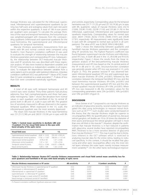
Table 1. Central mean sensitivity in decibels (dB) <strong>and</strong><br />
1/Lambert (1/L) for visual <strong>field</strong> zones in the eyes with<br />
b<strong>and</strong> atrophy of the optic nerve<br />
<strong>Visual</strong> <strong>field</strong> zone dB 1/L<br />
Superotemporal central quadrant -20.81 ± 12.03 765.28 ± 993.24<br />
Inferotemporal central quadrant -17.65 ± 13.87 786.68 ± 1137.45<br />
Superonasal central quadrant -0.78 ±1.94 1.27 ± 0.66<br />
Inferonasal central quadrant -1.01 ± 1.61 1.29 ± 0.54<br />
Temporal central 8 points -19.22 ± 12.46 616.91 ± 946.45<br />
Nasal central 8 points -0.89 ± 1.68 1.26 ± 0.58<br />
<strong>and</strong> controls, respectively. Corresponding values for the temporal<br />
hemiretina was 231.71 (11.25) μm <strong>and</strong> 257.18 (19.36) μm. In eyes<br />
with BA, quadrantic values of macular thickness were 232.16<br />
(13.69); 233.96 (13.43); 232.99 (11.92) <strong>and</strong> 231.68 (11.76) in the<br />
inferonasal, superonasal, inferotemporal <strong>and</strong> superotemporal<br />
quadrants respectively. Corresponding values for normal eyes<br />
were: 260.41 (19.02); 262.62 (19.33); 258.86 (19.91) <strong>and</strong> 256.49<br />
(17.81), respectively. All measurements were significantly lower<br />
in eyes with BA compared to normal eyes (p


