

Monovision and cataract surgery Visual field and OCT correlation ...
Monovision and cataract surgery Visual field and OCT correlation ...
Monovision and cataract surgery Visual field and OCT correlation ...

Create successful ePaper yourself
Turn your PDF publications into a flip-book with our unique Google optimized e-Paper software.
GENE TRANSFER TO PRIMARY CORNEAL EPITHELIAL CELLS WITH AN INTEGRATING LENTIVIRAL VECTOR<br />
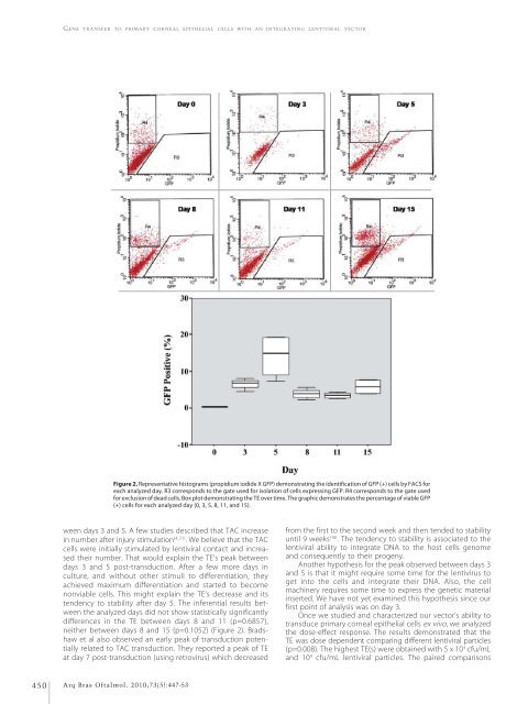
Figure 2. Representative histograms (propidium iodide X GFP) demonstrating the identification of GFP (+) cells by FACS for<br />
each analyzed day. R3 corresponds to the gate used for isolation of cells expressing GFP. R4 corresponds to the gate used<br />
for exclusion of dead cells. Box plot demonstrating the TE over time. The graphic demonstrates the percentage of viable GFP<br />
(+) cells for each analyzed day (0, 3, 5, 8, 11, <strong>and</strong> 15).<br />
ween days 3 <strong>and</strong> 5. A few studies described that TAC increase<br />
in number after injury stimulation (4, 21) . We believe that the TAC<br />
cells were initially stimulated by lentiviral contact <strong>and</strong> increased<br />
their number. That would explain the TE’s peak between<br />
days 3 <strong>and</strong> 5 post-transduction. After a few more days in<br />
culture, <strong>and</strong> without other stimuli to differentiation, they<br />
achieved maximum differentiation <strong>and</strong> started to become<br />
nonviable cells. This might explain the TE’s decrease <strong>and</strong> its<br />
tendency to stability after day 5. The inferential results between<br />
the analyzed days did not show statistically significantly<br />
differences in the TE between days 8 <strong>and</strong> 11 (p=0.6857),<br />
neither between days 8 <strong>and</strong> 15 (p=0.1052) (Figure 2). Bradshaw<br />
et al also observed an early peak of transduction potentially<br />
related to TAC transduction. They reported a peak of TE<br />
at day 7 post-transduction (using retrovirus) which decreased<br />
from the first to the second week <strong>and</strong> then tended to stability<br />
until 9 weeks (16) . The tendency to stability is associated to the<br />
lentiviral ability to integrate DNA to the host cells genome<br />
<strong>and</strong> consequently to their progeny.<br />
Another hypothesis for the peak observed between days 3<br />
<strong>and</strong> 5 is that it might require some time for the lentivirus to<br />
get into the cells <strong>and</strong> integrate their DNA. Also, the cell<br />
machinery requires some time to express the genetic material<br />
inserted. We have not yet examined this hypothesis since our<br />
first point of analysis was on day 3.<br />
Once we studied <strong>and</strong> characterized our vector’s ability to<br />
transduce primary corneal epithelial cells ex vivo, we analyzed<br />
the dose-effect response. The results demonstrated that the<br />
TE was dose dependent comparing different lentiviral particles<br />
(p=0.008). The highest TE(s) were obtained with 5 x 10 3 cfu/mL<br />
<strong>and</strong> 10 4 cfu/mL lentiviral particles. The paired comparisons<br />
450<br />
Arq Bras Oftalmol. 2010;73(5):447-53


