

Monovision and cataract surgery Visual field and OCT correlation ...
Monovision and cataract surgery Visual field and OCT correlation ...
Monovision and cataract surgery Visual field and OCT correlation ...

You also want an ePaper? Increase the reach of your titles
YUMPU automatically turns print PDFs into web optimized ePapers that Google loves.
CUNHA LP, COSTA-CUNHA LVF, ET AL.<br />
A<br />
B<br />
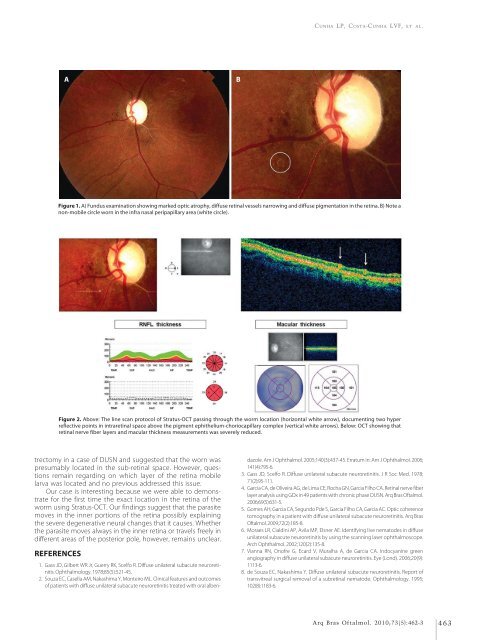
Figure 1. A) Fundus examination showing marked optic atrophy, diffuse retinal vessels narrowing <strong>and</strong> diffuse pigmentation in the retina. B) Note a<br />
non-mobile circle worn in the infra nasal peripapillary area (white circle).<br />
Figure 2. Above: The line scan protocol of Stratus-<strong>OCT</strong> passing through the worm location (horizontal white arrow), documenting two hyper<br />
reflective points in intraretinal space above the pigment ephithelium-choriocapillary complex (vertical white arrows). Below: <strong>OCT</strong> showing that<br />
retinal nerve fiber layers <strong>and</strong> macular thickness measurements was severely reduced.<br />
trectomy in a case of DUSN <strong>and</strong> suggested that the worn was<br />
presumably located in the sub-retinal space. However, questions<br />
remain regarding on which layer of the retina mobile<br />
larva was located <strong>and</strong> no previous addressed this issue.<br />
Our case is interesting because we were able to demonstrate<br />
for the first time the exact location in the retina of the<br />
worm using Stratus-<strong>OCT</strong>. Our findings suggest that the parasite<br />
moves in the inner portions of the retina possibly explaining<br />
the severe degenerative neural changes that it causes. Whether<br />
the parasite moves always in the inner retina or travels freely in<br />
different areas of the posterior pole, however, remains unclear.<br />
REFERENCES<br />
1. Gass JD, Gilbert WR Jr, Guerry RK, Scelfo R. Diffuse unilateral subacute neuroretinitis.<br />
Ophthalmology. 1978;85(5):521-45.<br />
2. Souza EC, Casella AM, Nakashima Y, Monteiro ML. Clinical features <strong>and</strong> outcomes<br />
of patients with diffuse unilateral subacute neuroretinitis treated with oral albendazole.<br />
Am J Ophthalmol. 2005;140(3):437-45. Erratum in: Am J Ophthalmol. 2006;<br />
141(4):795-6.<br />
3. Gass JD, Scelfo R. Diffuse unilateral subacute neuroretinitis. J R Soc Med. 1978;<br />
71(2):95-111.<br />
4. Garcia CA, de Oliveira AG, de Lima CE, Rocha GN, Garcia Filho CA. Retinal nerve fiber<br />
layer analysis using GDx in 49 patients with chronic phase DUSN. Arq Bras Oftalmol.<br />
2006;69(5):631-5.<br />
5. Gomes AH, Garcia CA, Segundo Pde S, Garcia Filho CA, Garcia AC. Optic coherence<br />
tomography in a patient with diffuse unilateral subacute neuroretinitis. Arq Bras<br />
Oftalmol. 2009;72(2):185-8.<br />
6. Moraes LR, Cialdini AP, Avila MP, Elsner AE. Identifying live nematodes in diffuse<br />
unilateral subacute neuroretinitis by using the scanning laser ophthalmoscope.<br />
Arch Ophthalmol. 2002;120(2):135-8.<br />
7. Vianna RN, Onofre G, Ecard V, Muralha A, de Garcia CA. Indocyanine green<br />
angiography in diffuse unilateral subacute neuroretinitis. Eye (Lond). 2006;20(9):<br />
1113-6.<br />
8. de Souza EC, Nakashima Y. Diffuse unilateral subacute neuroretinitis. Report of<br />
transvitreal surgical removal of a subretinal nematode. Ophthalmology. 1995;<br />
102(8):1183-6.<br />
Arq Bras Oftalmol. 2010;73(5):462-3 463


