

Monovision and cataract surgery Visual field and OCT correlation ...
Monovision and cataract surgery Visual field and OCT correlation ...
Monovision and cataract surgery Visual field and OCT correlation ...

Create successful ePaper yourself
Turn your PDF publications into a flip-book with our unique Google optimized e-Paper software.
FORTES FILHO JB, MAGNANI AC, ET AL.<br />
large size of the lesion coupled with the absence of any visual<br />
potential, enucleation was indicated. The eye containing the<br />
malignant tumor was enucleated with a good surgical outcome.<br />
After <strong>surgery</strong>, the macroscopic assessment of the enucleated<br />
globe revealed a 10 mm thick melanocytic mass associated<br />
with a serous retinal detachment. The basal size of the<br />
lesion was 17 mm by 16 mm. Tumor dimension findings were<br />
similar to the previous imaging study. Microscopic evaluation<br />
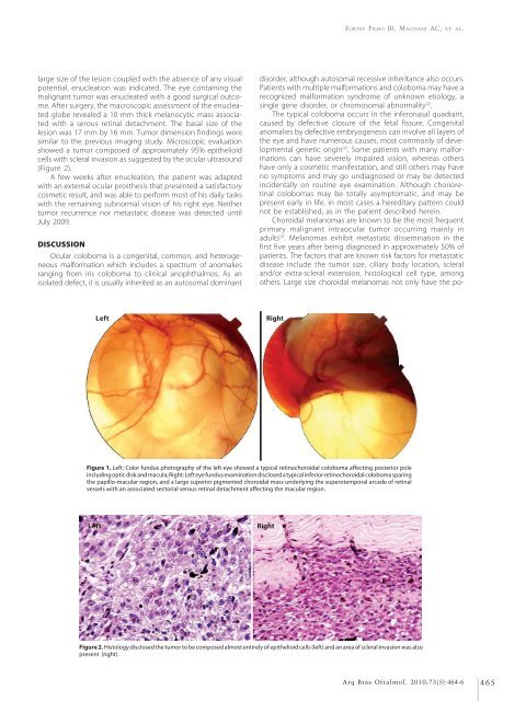
showed a tumor composed of approximately 95% epithelioid<br />
cells with scleral invasion as suggested by the ocular ultrasound<br />
(Figure 2).<br />
A few weeks after enucleation, the patient was adapted<br />
with an external ocular prosthesis that presented a satisfactory<br />
cosmetic result, <strong>and</strong> was able to perform most of his daily tasks<br />
with the remaining subnormal vision of his right eye. Neither<br />
tumor recurrence nor metastatic disease was detected until<br />
July 2009.<br />
DISCUSSION<br />
Ocular coloboma is a congenital, common, <strong>and</strong> heterogeneous<br />
malformation which includes a spectrum of anomalies<br />
ranging from iris coloboma to clinical anophthalmos. As an<br />
isolated defect, it is usually inherited as an autosomal dominant<br />
disorder, although autosomal recessive inheritance also occurs.<br />
Patients with multiple malformations <strong>and</strong> coloboma may have a<br />
recognized malformation syndrome of unknown etiology, a<br />
single gene disorder, or chromosomal abnormality (2) .<br />
The typical coloboma occurs in the inferonasal quadrant,<br />
caused by defective closure of the fetal fissure. Congenital<br />
anomalies by defective embryogenesis can involve all layers of<br />
the eye <strong>and</strong> have numerous causes, most commonly of developmental<br />
genetic origin (4) . Some patients with many malformations<br />
can have severely impaired vision, whereas others<br />
have only a cosmetic manifestation, <strong>and</strong> still others may have<br />
no symptoms <strong>and</strong> may go undiagnosed or may be detected<br />
incidentally on routine eye examination. Although chorioretinal<br />
colobomas may be totally asymptomatic, <strong>and</strong> may be<br />
present early in life, in most cases a hereditary pattern could<br />
not be established, as in the patient described herein.<br />
Choroidal melanomas are known to be the most frequent<br />
primary malignant intraocular tumor occurring mainly in<br />
adults (3) . Melanomas exhibit metastatic dissemination in the<br />
first five years after being diagnosed in approximately 50% of<br />
patients. The factors that are known risk factors for metastatic<br />
disease include the tumor size, ciliary body location, scleral<br />
<strong>and</strong>/or extra-scleral extension, histological cell type, among<br />
others. Large size choroidal melanomas not only have the po-<br />
Left<br />
Right<br />
Figure 1. Left: Color fundus photography of the left eye showed a typical retinochoroidal coloboma affecting posterior pole<br />
including optic disk <strong>and</strong> macula; Right: Left eye fundus examination disclosed a typical inferior retinochoroidal coloboma sparing<br />
the papillo-macular region, <strong>and</strong> a large superior pigmented choroidal mass underlying the superotemporal arcade of retinal<br />
vessels with an associated sectorial serous retinal detachment affecting the macular region.<br />
Left<br />
Right<br />
Figure 2. Histology disclosed the tumor to be composed almost entirely of epithelioid cells (left) <strong>and</strong> an area of scleral invasion was also<br />
present (right).<br />
Arq Bras Oftalmol. 2010;73(5):464-6 465


