

Monovision and cataract surgery Visual field and OCT correlation ...
Monovision and cataract surgery Visual field and OCT correlation ...
Monovision and cataract surgery Visual field and OCT correlation ...

Create successful ePaper yourself
Turn your PDF publications into a flip-book with our unique Google optimized e-Paper software.
SIQUEIRA RC, VOLTARELLI JC, ET AL.<br />
that HSCs isolated from BM are plastic <strong>and</strong> are able to “transdifferentiate”<br />
into tissue-committed stem cells (TCSCs) for<br />
other organs (e.g., heart, liver, or brain). Unfortunately, the<br />
concept of SC plasticity was not confirmed in recent studies<br />
<strong>and</strong> previously encouraging data demonstrating this phenomenon<br />
in vitro could be explained by a phenomenon of cell<br />
fusion or, as postulated by our group, by the presence, of<br />
heterogeneous populations of SCs in BM (11-12) . The identification<br />
of VSELSCs (primitive, very small, embryonic-like) in BM<br />
supports the notion that this tissue contains a population of<br />
primitive stem cell, which, if transplanted together with<br />
HSCs, was able to regenerate damaged tissues in certain<br />
experimental settings. Cells from BM could be easily <strong>and</strong> safely<br />
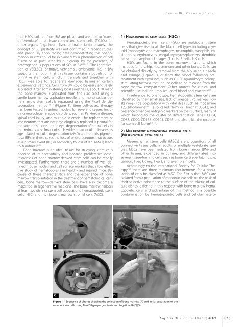
aspirated. After administering local anesthesia, about 10 ml of<br />
the bone marrow is aspirated from the iliac crest using a<br />
sterile bone marrow aspiration needle, <strong>and</strong> mononuclear bone<br />
marrow stem cells is separated using the Ficoll density<br />
separation method (14-17) (Figure 1). Stem cell–based therapy<br />
has been tested in animal models for several diseases, including<br />
neurodegenerative disorders, such as Parkinson disease,<br />
spinal cord injury, <strong>and</strong> multiple sclerosis. The replacement of<br />
lost neurons that are not physiologically replaced is pivotal for<br />
therapeutic success. In the eye, degeneration of neural cells in<br />
the retina is a hallmark of such widespread ocular diseases as<br />
age-related macular degeneration (AMD) <strong>and</strong> retinitis pigmentosa<br />
(RP). In these cases the loss of photoreceptors that occurs<br />
as a primary event (RP) or secondary to loss of RPE (AMD) leads<br />
to blindness (8-9) .<br />
Bone marrow is an ideal tissue for studying stem cells<br />
because of its accessibility <strong>and</strong> because proliferative doseresponses<br />
of bone marrow-derived stem cells can be readily<br />
investigated. Furthermore, there are a number of well-defined<br />
mouse models <strong>and</strong> cell surface markers that allow effective<br />
study of hematopoiesis in healthy <strong>and</strong> injured mice. Because<br />
of these characteristics <strong>and</strong> the experience of bone<br />
marrow transplantation in the treatment of hematological cancers,<br />
bone marrow–derived stem cells have also become a<br />
major tool in regenerative medicine. The bone marrow harbors<br />
at least two distinct stem cell populations: hematopoietic stem<br />
cells (HSC) <strong>and</strong> multipotent marrow stromal cells (MSC).<br />
1) HEMATOPOIETIC STEM CELLS (HSCS)<br />
Hematopoietic stem cells (HSCs) are multipotent stem<br />
cells that give rise to all the blood cell types including myeloid<br />
(monocytes <strong>and</strong> macrophages, neutrophils, basophils, eosinophils,<br />
erythrocytes, megakaryocytes/platelets, dendritic<br />
cells), <strong>and</strong> lymphoid lineages (T-cells, B-cells, NK-cells).<br />
HSCs are found in the bone marrow of adults, which<br />
includes femurs, hip, ribs, sternum, <strong>and</strong> other bones. Cells can<br />
be obtained directly by removal from the hip using a needle<br />
<strong>and</strong> syringe (Figure 1), or from the blood following pretreatment<br />
with cytokines, such as G-CSF (granulocyte colonystimulating<br />
factors), that induce cells to be released from the<br />
bone marrow compartment. Other sources for clinical <strong>and</strong><br />
scientific use include umbilical cord blood <strong>and</strong> placenta (10-11) .<br />
In reference to phenotype, hematopoeitic stem cells are<br />
identified by their small size, lack of lineage (lin) markers, low<br />
staining (side population) with vital dyes such as rhodamine<br />
123 (rhodamine DULL , also called rho lo ) or Hoechst 33342, <strong>and</strong><br />
presence of various antigenic markers on their surface, many of<br />
which belong to the cluster of differentiation series: CD34,<br />
CD38, CD90, CD133, CD105, CD45 <strong>and</strong> also c-kit, the receptor<br />
for stem cell factor (12-17) .<br />
2) MULTIPOTENT MESENCHYMAL STROMAL CELLS<br />
(MESENCHYMAL STEM CELLS)<br />
Mesenchymal stem cells (MSCs) are progenitors of all<br />
connective tissue cells. In adults of multiple vertebrate species,<br />
MSCs have been isolated from bone marrow (BM) <strong>and</strong><br />
other tissues, exp<strong>and</strong>ed in culture, <strong>and</strong> differentiated into<br />
several tissue-forming cells such as bone, cartilage, fat, muscle,<br />
tendon, liver, kidney, heart, <strong>and</strong> even brain cells.<br />
Accordingly to the International Society for Cellular Therapy<br />
(18) there are three minimum requirements for a population<br />
of cells be classified as MSC. The first is that MSCs are<br />
isolated from a population of mononuclear cells on the basis of<br />
their selective adherence to the surface of the plastic of culture<br />
dishes, differing in this respect with bone marrow hematopoietic<br />
cells, a disadvantage of this method is a possible<br />
contamination by hematopoietic cells <strong>and</strong> cellular hetero-<br />
A<br />
B<br />
C<br />
D<br />
Figure 1. Sequence of photos showing the collection of bone marrow (A) <strong>and</strong> initial separation of the<br />
mononuclear cells using Ficoll’Hypaque gradient centrifugation (B)(C)(D).<br />
C<br />
Arq Bras Oftalmol. 2010;73(5):474-9 475


