

Monovision and cataract surgery Visual field and OCT correlation ...
Monovision and cataract surgery Visual field and OCT correlation ...
Monovision and cataract surgery Visual field and OCT correlation ...

You also want an ePaper? Increase the reach of your titles
YUMPU automatically turns print PDFs into web optimized ePapers that Google loves.
USE OF CD25 AS AN IMMUNOHISTOCHEMICAL MARKER FOR ACQUIRED OCULAR TOXOPLASMOSIS<br />
from patients with probable congenital toxoplasmosis was decreased,<br />
compared to patients with presumed acquired infection<br />
(7) .<br />
CD25 is a transmembrane protein which forms the alpha<br />
chain of the IL-2 receptor (19-20) . It plays a crucial role in IL-2 homeostasis<br />
(20) . The goal of this study was to examine the immunohistochemistry<br />
expression of CD25 in enucleated eyes of patients<br />
with toxoplasmosis, considering the possibility of evaluating<br />
histopathologic specimens (Yamamoto et al. studied blood samples<br />
by ELISA (7) . In addition we aimed to determine whether<br />
CD25, as a marker for the expression of IL-2, could differentiate<br />
acquired from congenital ocular toxoplasmosis.<br />
METHODS<br />
Ten formalin-fixed paraffin-embedded enucleated globes<br />
from ten immunocompetent patients with clinical diagnosis<br />
of toxoplasmosis were evaluated. The globes were obtained<br />
from the Henry C. Witelson Ocular Pathology Laboratory. All<br />
the subjects had the titers of antibody (IgM or IgG) to T. gondii<br />
determined. Age, gender <strong>and</strong> number of recurrences was also<br />
retrieved.<br />
Histopathological slides were reviewed by one experienced<br />
ocular pathologist (MNB) for the extension of the retinal<br />
necrosis, number of toxo cysts, the granulomatous inflammatory<br />
reaction <strong>and</strong> the presence of T <strong>and</strong> B cells within the choroid,<br />
as well as the IL-2 expression.<br />
Expression of CD4, CD8, CD20, CD25 <strong>and</strong> CD68 was also<br />
evaluated by the avidin-biotin complex (ABC) immunohistochemistry<br />
method, using monoclonal antibodies (CD4, CD8,<br />
CD 20, CD 25 <strong>and</strong> CD68) from Dako Laboratory (SPA-830; Stressgen,<br />
Victoria, BC, Canada).<br />
The study protocol followed the precepts of the Declaration<br />
of Helsinki <strong>and</strong> was approved by the Research Institute of<br />
the McGill University Hospital Centre.<br />
RESULTS<br />
Clinical information for all ten patients included in the<br />
study is summarized in table 1. Histopathological examination<br />
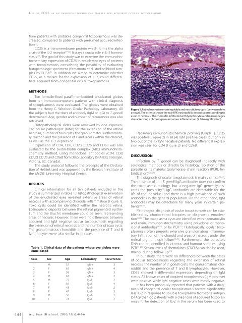
of the enucleated eyes revealed extensive areas of retinal<br />
necrosis with accompanying choroidal inflammation (Figure 1).<br />
Toxo cysts could be identified within the necrotic retina.<br />
Eosinophilic deposits between the retinal pigmented epithelium<br />
<strong>and</strong> the Bruch’s membrane could be seen, representing<br />
areas of necrosis. However, there were no differences between<br />
acquired <strong>and</strong> IgM negative ocular toxoplasmosis regarding<br />
the extension of retinal necrosis <strong>and</strong> the number of toxo cysts.<br />
The granulomatous choroiditis <strong>and</strong> the presence of T <strong>and</strong> B<br />
lymphocytes were also similar in all cases.<br />
Table 1. Clinical data of the patients whose eye globes were<br />
enucleated<br />
Case Sex Age Laboratory Recurrence<br />
1 M 37 IgM+ 3<br />
2 F 61 IgM+ 4<br />
3 F 58 IgM+ 3<br />
4 M 63 IgM+ 2<br />
5 M 72 IgM- 2<br />
6 F 56 IgM- 4<br />
7 F 43 IgM- 2<br />
8 F 47 IgM- 3<br />
9 M 56 IgM- 2<br />
10 F 74 IgM- 2<br />
Figure 1. Retinal necrosis containing viable <strong>and</strong> necrotic toxo cysts (between white<br />
arrows). The asterisk shows the sub-RPE eosinophilic deposits corresponding to<br />
areas of necrosis. The choroid is infiltrated with lymphocytes <strong>and</strong> macrophages<br />
characterizing a chronic granulomatous inflammation (X 50 magnification).<br />
Regarding immunohistochemical profiling (Graph 1), CD25<br />
was positive (Figure 2) in all (4) IgM positive cases, but only in<br />
two out of the six IgM negative patients. No differential expression<br />
was seen for CD4 (Figure 3) <strong>and</strong> CD68.<br />
DISCUSSION<br />
Infection by T. gondii can be diagnosed indirectly with<br />
serological methods or directly by histology, isolation of the<br />
parasite or its material (polymerase chain reaction (PCR), hybridization)<br />
(2,6,21) .<br />
The diagnosis of ocular toxoplasmosis is mainly clinical (3,22) .<br />
The presence of anti T. gondii IgG antibodies does not confirm<br />
the toxoplasmic etiology, but a negative IgG generally discards<br />
the possibility (3) . IgG antibodies are detectable for the<br />
life of the individual <strong>and</strong> there is a high prevalence of such<br />
antibodies in the general population. On the other h<strong>and</strong>, IgM<br />
antibodies may be detectable for many years in certain patients<br />
(22-25) .<br />
Pathological diagnosis of ocular toxoplasmosis can be established<br />
by chorioretinal biopsies or diagnostic enucleation<br />
(3,26) . The toxoplasma cysts are identified with haematoxylin<br />
<strong>and</strong> eosin, immunohistochemistry using polyclonal or monoclonal<br />
antibodies (3,27) , or by PCR (17) . Histologically, ocular toxoplasmosis<br />
often presents extensive granulomatous inflammatory<br />
infiltration of the choroid <strong>and</strong> areas of necrosis under the<br />
retinal pigment epithelium (3,26) . Furthermore, the parasite’s<br />
DNA can be identified in vitreous <strong>and</strong> humour samples using<br />
PCR (21,28) . Serum levels of chemokines (CXCL8) can also be used,<br />
mainly during follow-up (29) .<br />
In our study, there were no differences between the cases<br />
of ocular toxoplasmosis regarding the extension of retinal<br />
necrosis, the number of T. gondii cysts, the granulomatous choroiditis<br />
<strong>and</strong> the presence of T <strong>and</strong> B lymphocytes. However,<br />
CD25 showed a differential expression, depending on IgM<br />
status. All known cases of acquired toxoplamosis (IgM positive)<br />
were positive, while IgM negative cases were mostly negative.<br />
It has been previously reported that patients with a diagnosis<br />
of congenital ocular toxoplasmosis secrete significantly<br />
less IL-2 in response to soluble toxoplasma tachyzoite antigen<br />
(STAg) than do patients with a diagnosis of acquired toxoplasmosis<br />
(7) . The detection of IL-2 in the serum has been used to<br />
444<br />
Arq Bras Oftalmol. 2010;73(5):443-6


