

Monovision and cataract surgery Visual field and OCT correlation ...
Monovision and cataract surgery Visual field and OCT correlation ...
Monovision and cataract surgery Visual field and OCT correlation ...

You also want an ePaper? Increase the reach of your titles
YUMPU automatically turns print PDFs into web optimized ePapers that Google loves.
SANTOS TS, MELO AR, ET AL.<br />
of Recife (Pernambuco, Brazil), presenting to the emergency<br />
unit with impacted foreign bodies in the orbital region<br />
between January, 2008 <strong>and</strong> January, 2010 were evaluated<br />
retrospectively. These patients had been submitted to the<br />
surgical removal of foreign bodies with different degrees of<br />
severity. Following general anesthesia by orotracheal or nasotracheal<br />
intubation, the foreign body was removed back<br />
along its path of insertion, with care being taken with hemostasis.<br />
The patients received tetanus prophylaxis <strong>and</strong> antibiotic<br />
therapy. The following data were collected: age,<br />
gender, etiology of injury, occurrence of fracture, anatomical<br />
location of fracture, type of object, signs <strong>and</strong> symptoms, type<br />
of imaging exam used, approach <strong>and</strong> transoperative complications.<br />
RESULTS<br />
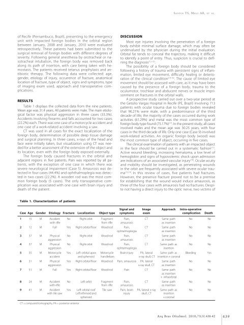
Table 1 displays the collected data from the nine patients.<br />
Mean age was 31.4 years. All patients were male. The main etiological<br />
factor was physical aggression in three cases (33.3%).<br />
Accidents involving firearms <strong>and</strong> falls accounted for two cases<br />
(22.2%) each. There was one case of a motorcycle accident <strong>and</strong><br />
one case of a work-related accident involving a saw.<br />
CT was used in all cases for the exact localization of the<br />
foreign body, determination of possible deep tissue damage<br />
<strong>and</strong> surgical planning. In three cases, x-rays of the head <strong>and</strong><br />
face were initially taken, but visualization using CT was needed<br />
for a better assessment of the extension of the object <strong>and</strong><br />
its location, even with the foreign body exposed externally.<br />
The foreign body caused fractures in the orbital <strong>and</strong><br />
adjacent regions in five patients. Pain was reported by all patients,<br />
with the exception of one case in which there was<br />
severe neurological impairment (Case 5). Amaurosis was detected<br />
in four cases (44.4%) <strong>and</strong> ophthalmoplegia was detected<br />
in two cases (22.2%). A wooden rod was the most common<br />
foreign body (5 cases). The only transoperative complication<br />
was associated with one case with brain injury <strong>and</strong><br />
death of the patient.<br />
DISCUSSION<br />
Most eye injuries involving the penetration of a foreign<br />
body exhibit minimal surface damage, which may often be<br />
undervalued by the physician during the initial evaluation.<br />
Orbital fat tends to conceal the trajectory, making it difficult<br />
to identify a point of entry. Thus, suspicion is crucial to defining<br />
the diagnosis (12-13) .<br />
The possibility of a foreign body should be considered<br />
following a history of trauma with persistent signs of inflammation,<br />
limited eye movement, difficulty healing or deterioration<br />
of the clinical condition (14-16) . The cause of limited eye<br />
movement should be assessed with care, as it may have been<br />
caused by the presence of a foreign body, trauma to the<br />
oculumotor, trochlear <strong>and</strong> abducent nerves or muscle imprisonment<br />
on fractures in the orbital walls.<br />
A prospective study carried out over a two-year period at<br />
the Getúlio Vargas Hospital in Recife (PE, Brazil) involving 713<br />
patients with ocular trauma due to foreign bodies revealed<br />
that 96.21% were male, with a predominance of the third<br />
decade of life; the majority of the cases occurred during work<br />
activities (61.29%) <strong>and</strong> metal was the most common type of<br />
foreign body type found (75.17%) (17) . In the present study, all cases<br />
involved males <strong>and</strong> the mean age was 30.25 years, with four<br />
cases in the third decade of life. Only one case (Case 9) involved<br />
work-related activities. An organic foreign body (wood) was<br />
the most common type of object, occurring in five cases.<br />
The clinical examination of patients with an impacted object<br />
in the face should be carried out in a systematic fashion (11) .<br />
Active wound bleeding, increasing hematoma, a low level of<br />
hemoglobin <strong>and</strong> signs of hypovolemic shock upon admission<br />
are indications of an associated vascular injury (18) . Ocular acuity<br />
<strong>and</strong> mobility should be investigated, as penetrating wounds<br />
in the orbit are frequently associated with severe ocular trauma<br />
(10,19) . In this review of cases, five patients had fractures.<br />
However, the presence fracture proved not to be a premise<br />
for establishing that the wound would induce amaurosis, as<br />
three of the four cases with amaurosis had no fractures. Despite<br />
not having a direct injury to the optic nerve, two victims of<br />
Table 1. Characterization of patients<br />
Signal <strong>and</strong> Image Approach Intra-operative<br />
Case Age Gender Etiology Fracture Localization Object type symptoms exam complication Death<br />
1 15 M Accident No Right orbit Fragment Pain, CT Same path No No<br />
with rifle from rifle amaurosis as insertion<br />
2 12 M Fall Yes Right orbital floor Wood rod Pain, CT Same path No No<br />
ophtalmoplegia<br />
as insertion<br />
3 37 M Physical No Right orbit Wood rod Pain, CT Same path No No<br />
aggression amaurosis as insertion<br />
4 37 M Physical No Right orbit Wood rod Pain, CT Same path as No No<br />
aggression ophtalmoplegia insertion<br />
5 35 M Motorcycle Yes Left orbital apex Motorcycle Brain injury PA, lateral Same path as Bleeding Yes<br />
accident <strong>and</strong> sphenoid h<strong>and</strong>lebars x-ray skull, CT insertion + coronal<br />
6 31 M Physical Yes Right orbital floor Wood rod Pain, amaurosis PA, lateral Same path No No<br />
aggression x-ray skull, CT as insertion<br />
7 51 M Fall Yes Right orbital floor Wood rod Pain CT Same path No No<br />
as insertion<br />
+ infraorbital<br />
8 24 M Accident No Left orbit Fragment Pain, CT Same path No No<br />
with rifle from rifle amaurosis as insertion<br />
9 41 M Accident Yes Left orbital roof Tile saw Pain, brain PA, lateral x-ray Same path as No No<br />
with tile saw Left ethmoid <strong>and</strong> injury skull, CT insertion<br />
sphenoid<br />
+ coronal<br />
CT = computed tomography, PA = posterior-anterior<br />
Arq Bras Oftalmol. 2010;73(5):438-42 439


