

Monovision and cataract surgery Visual field and OCT correlation ...
Monovision and cataract surgery Visual field and OCT correlation ...
Monovision and cataract surgery Visual field and OCT correlation ...

You also want an ePaper? Increase the reach of your titles
YUMPU automatically turns print PDFs into web optimized ePapers that Google loves.
GENE TRANSFER TO PRIMARY CORNEAL EPITHELIAL CELLS WITH AN INTEGRATING LENTIVIRAL VECTOR<br />
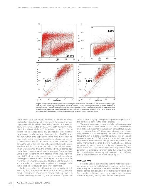
A<br />
B<br />
C<br />
Figure 4. Representative histograms demonstrating the identification of transduced side population phenotypic<br />
cells by FACS. A) Histogram (propidium iodide X forward scatter) isolating viable cells (gate R1: 43.80%). B)<br />
Histogram (GFP X forward scatter) isolating GFP(+) cells (gate R4: 4.61%). C) Histogram (Hoechst blue X Hoechst red)<br />
isolating side population phenotypic cells (gate R3: 1.67%). D) Histogram (Hoechst blue X Hoechst red) after<br />
incubation with verapamil isolating the subpopulation “side population” (gate R3: 0.47%).<br />
D<br />
thelial stem cells continues, however, a number of investigators<br />
have isolated putative stem cells functionally as side<br />
population cells based on their ability to efflux the Hoechst<br />
33342 dye when sorted by FACS (10,23) . Both human (24-25) <strong>and</strong><br />
rabbit limbal epithelial cells (26) have been sorted in order to<br />
isolate the side population (SP) phenotypic cells. Additionally,<br />
strong <strong>correlation</strong>s with the expression of ABCG2 markers<br />
for human side population limbal cells have been reported<br />
reinforcing the characterization of phenotypic corneal<br />
epithelial stem cells (24-25) . Our results are similar to others comparing<br />
the size of the side-population phenotypic cells found.<br />
We detected that 0.47% of the cells in our cell suspension,<br />
which was obtained from the limbal <strong>and</strong> whole cornea epithelial<br />
layer, demonstrated that phenotype. Some authors,<br />
using a cell suspension from rabbit’s limbal area, reported<br />
that 0.40% of the cells demonstrated the side-population<br />
phenotype (27) . When double sorted by FACS using two different<br />
channels simultaneously, one to isolate GFP positive cells<br />
<strong>and</strong> the other to isolate side population phenotypic cells,<br />
transduced side population cells were identified.<br />
While gene delivery to differentiated corneal epithelial cells<br />
will likely have utility for a number of ocular surface diseases,<br />
genetic modification of presumed corneal epithelial stem cells<br />
may be promising by enabling the production of gene products<br />
in their progeny or by providing bioactive proteins to<br />
the epithelium early in the repair process.<br />
The use of transduced corneal epithelial cells may augment<br />
our ability to treat severe ocular surface disease. Depletion of<br />
stem cells leads to cornea vascularization, fibrous tissue growth,<br />
<strong>and</strong> cornea opacification (28) . Current techniques for reconstructing<br />
the ocular surface damaged by stem cell deficiency involve<br />
transplantation of exp<strong>and</strong>ed corneal epithelial cells ex vivo, as<br />
previously reported (8-9) . Cell therapy is a typical example of<br />
regenerative medicine. Gene therapy makes regenerative medicine<br />
more attractive, since it allows modification of cellular<br />
properties by gene introduction before transplanting the<br />
tissue to the host. This combination allows exploration of the<br />
potential to genetically transform cells that would promote<br />
biochemical signals influencing cell proliferation <strong>and</strong> differentiation,<br />
relevant steps in functional tissue repair.<br />
CONCLUSIONS<br />
Lentiviral vectors can effectively transfer heterologous genes<br />
to primary corneal epithelial cells exp<strong>and</strong>ed ex vivo. Genes<br />
were stably expressed over time <strong>and</strong> could be transferred to<br />
mature corneal cells as well as presumable putative stem cells.<br />
Transduction efficiency was dose-dependent, however<br />
concerns regarding toxicity need further investigation.<br />
452<br />
Arq Bras Oftalmol. 2010;73(5):447-53


