

Monovision and cataract surgery Visual field and OCT correlation ...
Monovision and cataract surgery Visual field and OCT correlation ...
Monovision and cataract surgery Visual field and OCT correlation ...

Create successful ePaper yourself
Turn your PDF publications into a flip-book with our unique Google optimized e-Paper software.
IMPACTED FOREIGN BODIES IN ORBITAL REGION: REVIEW OF NINE CASES<br />
A<br />
B<br />
C<br />
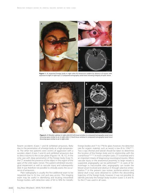
Figure 1. A) Impacted foreign body in right orbit; B) Amaurosis evident by absence of myosis with<br />
exposure to light in right eye; C) Computed tomography (axial view) showing integrity of optic nerve.<br />
A<br />
B<br />
C<br />
Figure 2. A) Wooden splinter in right orbit; B) Soft tissue window in computed tomography (axial view)<br />
showing space similar to air in right orbit; C) Hard tissue window in computed tomography (axial view)<br />
showing space similar to air in right orbit.<br />
firearm accidents (Cases 1 <strong>and</strong> 8) exhibited amaurosis, likely<br />
due to the penetration of a foreign body at a high temperature.<br />
The other two patients were victims of aggression with a<br />
wooden object (Cases 3 <strong>and</strong> 6) <strong>and</strong> exhibited amaurosis due<br />
to direct trauma to the ocular globe (Figures 1A, 1B, 1C). In the<br />
only case with deep penetration of the foreign body (Case 5),<br />
the CT revealed the presence of the object in the region of the<br />
apex of the orbit (optic nerve). This patient exhibited neurological<br />
impairment as well as vascular injury <strong>and</strong> subsequently<br />
died. It was therefore not possible to clinically assess the occurrence<br />
of amaurosis.<br />
Plain radiography is usually the first additional exam to be<br />
requested due to its low cost <strong>and</strong> easy access. This imaging<br />
exam may be useful in identifying <strong>and</strong> locating intraorbital<br />
foreign bodies, with detection rates of 69 to 90% for metallic<br />
foreign bodies <strong>and</strong> 71 to 77% for glass; however, the detection<br />
rate for organic material, such as wood, is low (0 to 15%) (20-21) .<br />
Two x-rays (frontal <strong>and</strong> lateral) should be taken to determine<br />
the location of the foreign body <strong>and</strong> its relationship with the<br />
cranial fossa (10,22-23) . In more complex cases, CT is essential <strong>and</strong> is<br />
an important means of diagnosing neurological injuries. When<br />
vascular injury or the anatomical proximity to large vessels is<br />
suspected, angiography can be performed (18,22) . In cases of hemorrhage<br />
in inaccessible sites, angiography can locate the<br />
blood vessel involved <strong>and</strong> obstruct it through selective embolization.<br />
(24) In the present case review, posterior-anterior <strong>and</strong><br />
lateral skull x-rays were obtained to confirm the descending<br />
trajectory of the foreign body, however, it was not possible to<br />
identify precisely the foreign body location (cases 5, 6 <strong>and</strong> 9).<br />
So the CT was used in all cases.<br />
440<br />
Arq Bras Oftalmol. 2010;73(5):438-42


