Diagnosticul imagistic al tumorilor de unghi ponto-cerebelos
Diagnosticul imagistic al tumorilor de unghi ponto-cerebelos
Diagnosticul imagistic al tumorilor de unghi ponto-cerebelos

Create successful ePaper yourself
Turn your PDF publications into a flip-book with our unique Google optimized e-Paper software.
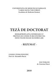
DIAGNOSTICUL IMAGISTIC AL TUMORILOR DE UNGHI<br />
PONTO-CEREBELOS<br />
TEZĂ DE DOCTORAT – REZUMAT<br />
COORDONATOR DOCTORAND<br />
PROF.DR.EMANOIL POPESCU DR.DANA PAULA GEORGESCU<br />
-2007-
2<br />
Rezumat <strong>Diagnosticul</strong> <strong>imagistic</strong> <strong>al</strong> <strong>tumorilor</strong> <strong>de</strong> <strong>unghi</strong> <strong>ponto</strong>-<strong>cerebelos</strong><br />
CUPRINS<br />
Introducere<br />
Partea I: Partea gener<strong>al</strong>ă<br />
1.Anatomia <strong>unghi</strong>ului <strong>ponto</strong>-<strong>cerebelos</strong><br />
1.1.Noţiuni <strong>de</strong> anatomie a fosei cerebr<strong>al</strong>e posterioare<br />
1.2. Delimitarea <strong>unghi</strong>ului <strong>ponto</strong>-<strong>cerebelos</strong><br />
1.3. Meningele<br />
1.4. Conţinutul spaţiului <strong>ponto</strong>-<strong>cerebelos</strong><br />
1.5. Conductul auditiv intern<br />
2. Tumorile <strong>de</strong> <strong>unghi</strong> <strong>ponto</strong>-<strong>cerebelos</strong><br />
2.1. Clasificarea <strong>tumorilor</strong> <strong>de</strong> <strong>unghi</strong> <strong>ponto</strong>-<strong>cerebelos</strong><br />
2.2. Date etiopatogenice şi histologice<br />
2.3. Date clinice<br />
3. Tehnici <strong>de</strong> explorare <strong>imagistic</strong>ă în tumorile <strong>de</strong> <strong>unghi</strong> <strong>ponto</strong><strong>cerebelos</strong><br />
3.1. Tomografia Computerizată<br />
3.2. Imagistica prin Rezonanţă Magnetică<br />
3.3. Angiografia cerebr<strong>al</strong>ă<br />
Partea II: Partea speci<strong>al</strong>ă<br />
1. Materi<strong>al</strong> şi metodă<br />
1.1. Obiective. Criterii <strong>de</strong> inclu<strong>de</strong>re în lot<br />
1.2. Examene <strong>imagistic</strong>e: indicaţii, sensibilitate, specificitate,<br />
protoco<strong>al</strong>e utilizate<br />
1.2.1. Computer Tomografia<br />
1.2.2. Imagistica prin Rezonanţă Magnetică<br />
1.2.3. Angiografia cerebr<strong>al</strong>ă<br />
2. Rezultate şi discuţii<br />
2.1. Aspecte <strong>imagistic</strong>e <strong>al</strong>e <strong>tumorilor</strong> <strong>de</strong> <strong>unghi</strong> <strong>ponto</strong>-<strong>cerebelos</strong><br />
corelate cu datele histopatologice<br />
2.1.1. Neurinomul acustic<br />
2.1.2. Neurinomul faci<strong>al</strong><br />
2.1.3. Neurinomul trigemin<strong>al</strong><br />
2.1.4. Neurinomul <strong>de</strong> nervi cranieni inferiori<br />
2.1.5. Meningiomul<br />
2.1.6. Chistul epi<strong>de</strong>rmoid<br />
2.1.7. Chistul arahnoid<br />
2.1.8. Lipomul<br />
2.1.9. Metastaze<br />
2.2. Aspecte <strong>imagistic</strong>e postoperatorii<br />
2.3. Algoritm <strong>de</strong> investigare<br />
2.4. Rezultate statistice<br />
Concluzii<br />
Bibliografie
Rezumat <strong>Diagnosticul</strong> <strong>imagistic</strong> <strong>al</strong> <strong>tumorilor</strong> <strong>de</strong> <strong>unghi</strong> <strong>ponto</strong>-<strong>cerebelos</strong><br />
INTRODUCERE<br />
Tumorile <strong>unghi</strong>ului <strong>ponto</strong>-<strong>cerebelos</strong> ocupă un capitol<br />
important în patologia neurochirurgic<strong>al</strong>ă, datorită probemelor <strong>de</strong><br />
diagnostic şi tratament. Reprezintă 5-10 % din tumorile intracraniene,<br />
din care cele mai frecvente sunt neurinoamele acustice şi<br />
meningioamele. În procent mai mic sunt şi <strong>al</strong>te leziuni întâlnite tot mai<br />
frecvent <strong>de</strong> medicul radiolog datorită senzitivităţii remarcabile şi<br />
acurateţei meto<strong>de</strong>lor <strong>imagistic</strong>e în ev<strong>al</strong>uarea sindromului <strong>de</strong> <strong>unghi</strong><br />
<strong>ponto</strong>-<strong>cerebelos</strong>.<br />
Pentru evitarea unor situaţii <strong>de</strong> nedorit, clinicianul trebuie să<br />
fie bine informat asupra meto<strong>de</strong>lor <strong>de</strong> investigaţie, cu avantajele,<br />
<strong>de</strong>zavantajele şi cunoaşterea interrelaţiei dintre ele, în ve<strong>de</strong>rea adoptării<br />
unui plan optim <strong>de</strong> investigaţie.<br />
Lucrarea <strong>de</strong> faţă încearcă să contureze posibilităţile <strong>de</strong><br />
diagnostic <strong>al</strong>e tomo<strong>de</strong>nsitometriei, rezonanţei magnetice nucleare şi<br />
angiografiei, datele teoretice şi cele reieşite din studiul person<strong>al</strong> fiind<br />
exemplificate printr-o iconografie reprezentativă.<br />
Studiul a fost efectuat în Clinica <strong>de</strong> Radiologie şi Imagistică<br />
Medic<strong>al</strong>ă a Spit<strong>al</strong>ului Universitar <strong>de</strong> Urgenţă Bucureşti, în colaborare<br />
cu Clinicile <strong>de</strong> Neurochirurgie, precum şi <strong>al</strong>te clinici din spit<strong>al</strong>, în mod<br />
ocazion<strong>al</strong>.<br />
Modul cum sunt expuse problemele <strong>de</strong> patogenie, ca şi an<strong>al</strong>iza<br />
riguroasă a cazurilor prezentate dau posibilitatea <strong>de</strong> a putea discerne<br />
cât mai aproape <strong>de</strong> re<strong>al</strong>itate imaginile cu semnificaţie multiplă şi<br />
permit speci<strong>al</strong>istului ca printr-un examen clinic, anatomopatologic şi<br />
<strong>imagistic</strong> minuţios să poată pune cu suficientă certitudine diagnosticul<br />
şi să poată orienta corect atitudinea terapeutică.<br />
Înmănunchind cele mai importante date <strong>de</strong> morfopatologie şi<br />
<strong>imagistic</strong>ă, prezenta lucrare are intenţia <strong>de</strong> a a stabili un <strong>al</strong>goritm <strong>de</strong><br />
diagnostic radio<strong>imagistic</strong> corect în patologia <strong>de</strong> <strong>unghi</strong> <strong>ponto</strong>-<strong>cerebelos</strong>,<br />
pentru a obţine maximul <strong>de</strong> informaţii în condiţiile evitării exceselor<br />
<strong>de</strong> examinare şi să răspundă necesităţilor actu<strong>al</strong>e <strong>de</strong> informare a<br />
medicilor care, prin natura preocupărilor lor, sunt chemaţi să contribuie<br />
sau să <strong>de</strong>cidă diagnosticul, orientarea sau aplicarea tratamentului .<br />
PARTEA I :PARTEA GENERALĂ<br />
Sunt prezentate <strong>de</strong> anatomie a <strong>unghi</strong>ului <strong>ponto</strong>-<strong>cerebelos</strong>, noţiuni<br />
<strong>de</strong> etiopatogenie şi histologie, date clinice şi tehnici <strong>de</strong> investigaţie<br />
<strong>imagistic</strong>ă.<br />
3
4<br />
Rezumat <strong>Diagnosticul</strong> <strong>imagistic</strong> <strong>al</strong> <strong>tumorilor</strong> <strong>de</strong> <strong>unghi</strong> <strong>ponto</strong>-<strong>cerebelos</strong><br />
Tumorile originare în <strong>unghi</strong>ul <strong>ponto</strong>-<strong>cerebelos</strong> sunt leziuni extraaxi<strong>al</strong>e<br />
ce provin din cisternă şi <strong>al</strong>te structuri <strong>al</strong>e <strong>unghi</strong>ului <strong>ponto</strong>cerebeloas,<br />
sau din resturi embrionare. Acestea trebuie diferenţiate <strong>de</strong><br />
tumorile ce pot invada <strong>unghi</strong>ul <strong>ponto</strong>-<strong>cerebelos</strong> prin extensie <strong>de</strong> la osul<br />
pietros sau baza <strong>de</strong> craniu, sau pot fi secundare unor tumori exofitice<br />
<strong>de</strong> trunchi cerebr<strong>al</strong>, cerebel sau ventricol .<br />
Clasificarea <strong>tumorilor</strong> <strong>de</strong> <strong>unghi</strong> <strong>ponto</strong>-<strong>cerebelos</strong><br />
1. Clasificare histopatologică<br />
Origine<br />
nervoasă<br />
Neurinoame:<br />
-acustic<br />
-trigemin<strong>al</strong><br />
-faci<strong>al</strong><br />
-nervi mixti<br />
Origine<br />
meningi<strong>al</strong>ă<br />
Origine<br />
embrionară<br />
şi<br />
m<strong>al</strong>formative<br />
Meningiom Chist<br />
epi<strong>de</strong>rmoid<br />
Chist<br />
arahnoid<br />
Lipom<br />
2. Clasificare după frecvenţă:<br />
o Neurinom acustic – 80-85%<br />
o Meningiom – 6-8%<br />
o Chist epi<strong>de</strong>rmoid -2,5-3%<br />
o Neurinom trigemin<strong>al</strong> – 1%<br />
o Neurinom faci<strong>al</strong> – 0,7%<br />
o Neurinom <strong>de</strong> nervi micşti – 0,5%<br />
o Chist arahnoidian – 0,2-0,4%<br />
o Lipom - 0,1-0,5%<br />
o Metastaze – 0,2%<br />
Tumori<br />
metastatice<br />
Determinări<br />
secundare<br />
leptomeninge<strong>al</strong>e<br />
PARTEA II : PARTEA SPECIALĂ<br />
1. MATERIAL ŞI METODĂ<br />
1.1. Obiective. Criterii <strong>de</strong> inclu<strong>de</strong>re în lot<br />
Lucrarea a fost efectuată în cadrul Clinicii <strong>de</strong> Radiologie şi<br />
Imagistică Medic<strong>al</strong>ă din Spit<strong>al</strong>ul Universitar <strong>de</strong> Urgenţă Bucureşti, în<br />
colaborare cu Clinicile <strong>de</strong> Neurochirurgie I şi II.
Rezumat <strong>Diagnosticul</strong> <strong>imagistic</strong> <strong>al</strong> <strong>tumorilor</strong> <strong>de</strong> <strong>unghi</strong> <strong>ponto</strong>-<strong>cerebelos</strong><br />
Cercetarea se constitue sub forma unui studiu prospectiv pe o<br />
periadă <strong>de</strong> 5 ani şi jumătate (septembrie 2001- <strong>de</strong>cembrie 2006) pe un<br />
număr <strong>de</strong> 135 pacienţi.<br />
Condiţiile <strong>de</strong> inclu<strong>de</strong>re în lot au fost:<br />
1. pacient cu diagnostic <strong>de</strong> internare<br />
• sindrom <strong>de</strong> <strong>unghi</strong> <strong>ponto</strong>-<strong>cerebelos</strong><br />
• suspiciune <strong>de</strong> tumoră <strong>de</strong> <strong>unghi</strong> <strong>ponto</strong>-<strong>cerebelos</strong><br />
• tumoră <strong>de</strong> <strong>unghi</strong> <strong>ponto</strong>-<strong>cerebelos</strong> sau <strong>de</strong> fosă cerebr<strong>al</strong>ă<br />
posterioară<br />
2. examen CT nativ la internare, completat ulterior cu examen CT cu<br />
contrast i.v. , IRM sau angiografie în funcţie <strong>de</strong> caz.<br />
3. confirmare histo-patologică obţinută prin:<br />
4.<br />
• intervenţie chirurgic<strong>al</strong>ă cu examen anatomopatologic<br />
Examenele <strong>imagistic</strong>e au fost re<strong>al</strong>izate pe mai multe aparate.<br />
1. Pentru CT au fost utilizate două aparate CT Siemens: Somatom<br />
Plus (mod secvenţi<strong>al</strong>) şi Volum Zoom (mod secvenţi<strong>al</strong> şi spir<strong>al</strong>).<br />
2. Pentru rezonanţă magnetică a fost utilizat un aparat Gener<strong>al</strong><br />
Electric Signa <strong>de</strong> 1T .<br />
3. În anumite situaţii s-a efectuat şi explorare angiografică pe un<br />
aparat Philips.<br />
Studiul imaginilor obţinute (asociat cu corelarea rezultatelor<br />
tomografie computerizată, rezonanţă magnetică, iar acolo un<strong>de</strong> a<br />
fost posibilă şi anatomo-patologică) a fost urmat <strong>de</strong> prelucrarea<br />
statistică a datelor, încercându-se o suprapunere cu datele<br />
existente în literatura <strong>de</strong> speci<strong>al</strong>itate.<br />
Un important obiectiv <strong>al</strong> lucrarii a fost <strong>de</strong> a stabili un <strong>al</strong>goritm<br />
corect <strong>de</strong> investigaţie <strong>imagistic</strong>ă pentru diagnosticul <strong>tumorilor</strong> <strong>de</strong><br />
<strong>unghi</strong> <strong>ponto</strong>-<strong>cerebelos</strong>, în i<strong>de</strong>ea utilizării celor mai informative<br />
mijloace şi în cea mai eficientă ordine, cu evitarea exceselor în<br />
examinare radio<strong>imagistic</strong>ă.<br />
Lucrarea conţine foarte multe cazuri, fiecare cu particularităţile<br />
s<strong>al</strong>e, ceea ce se constituie într-un îndrumar CT şi RM în această<br />
patologie. De asemenea au fost sintetizate toate datele disponibile<br />
în ve<strong>de</strong>rea re<strong>al</strong>izării unor tabele cu diagnostice diferenţi<strong>al</strong>e foarte<br />
utile în practica medic<strong>al</strong>ă.<br />
1.2. Examene <strong>imagistic</strong>e utilizate în diagnosticul <strong>tumorilor</strong> <strong>de</strong><br />
<strong>unghi</strong> <strong>ponto</strong>-<strong>cerebelos</strong>: indicatii, sensibilitate, specificitate<br />
5
6<br />
Rezumat <strong>Diagnosticul</strong> <strong>imagistic</strong> <strong>al</strong> <strong>tumorilor</strong> <strong>de</strong> <strong>unghi</strong> <strong>ponto</strong>-<strong>cerebelos</strong><br />
1.2.1.Computer tomografia<br />
Indicaţii<br />
- explorare <strong>imagistic</strong>ă <strong>de</strong> primă intenţie în urgenţe<br />
- examen esenţi<strong>al</strong> în patologia <strong>de</strong> <strong>unghi</strong> <strong>ponto</strong>-<strong>cerebelos</strong> ce permite<br />
diferenţierea ţesuturilor în funcţie <strong>de</strong> <strong>de</strong>nsitatea lor<br />
- când există contraindicaţie pentru examenul IRM<br />
- uneori este necesară confruntarea datelor obţinute prin CT şi IRM<br />
Sensibilitate, specificitate<br />
- mult superioară în evi<strong>de</strong>nţierea modificărilor structurilor osoase<br />
- poate stabili loc<strong>al</strong>izărea intra- sau extra-axi<strong>al</strong>ă a unei leziuni, dar<br />
sensibilitatea este redusă comparativ cu a examenului IRM<br />
- aduce informaţii <strong>de</strong>spre prezenţa c<strong>al</strong>cificărilor sau hemoragiilor<br />
acute intratumor<strong>al</strong>e, precum şi a componentelor chistice<br />
- sensilitatea este crescută pentru tumori voluminoase<br />
- achiziţia spir<strong>al</strong>ă, cu secţiuni fine, permite <strong>de</strong>tectarea <strong>tumorilor</strong><br />
intracan<strong>al</strong>are <strong>de</strong> peste 5 mm<br />
An<strong>al</strong>iza imaginilor<br />
- au fost urmărite elementele:<br />
loc<strong>al</strong>izarea<br />
o intra- sau extra-nevraxi<strong>al</strong><br />
o raportul cu porul acustic intern<br />
o extensia : în CAI, fosa cerebr<strong>al</strong>ă mijlocie, părţile moi <strong>al</strong>e<br />
gâtului<br />
caracterele leziunii<br />
o număr<br />
o t<strong>al</strong>ia<br />
o forma :<br />
- rotund/ ov<strong>al</strong>ară<br />
- în “cometă” (neurinom acustic)<br />
- hemisferică<br />
- în placard<br />
- în bisac<br />
o contururile :<br />
- nete<br />
- lobulate<br />
o <strong>de</strong>nsitate :<br />
-<strong>de</strong>nsitate tisulară : neurinoamele, meningioamele,<br />
metastazele<br />
- <strong>de</strong>nsitate lichidiană : leziunile chistice<br />
- <strong>de</strong>nsitate grăsoasă : lipom<br />
- <strong>de</strong>nsităţi hematice : sângerare intratumor<strong>al</strong>ă<br />
- conţinut c<strong>al</strong>cic : c<strong>al</strong>cificări intratumor<strong>al</strong>e
Rezumat <strong>Diagnosticul</strong> <strong>imagistic</strong> <strong>al</strong> <strong>tumorilor</strong> <strong>de</strong> <strong>unghi</strong> <strong>ponto</strong>-<strong>cerebelos</strong><br />
modificările <strong>de</strong> structură osoasă<br />
o modificări ostelitice <strong>de</strong> tip atrofie prin presiune<br />
o hiperostoză<br />
efectul <strong>de</strong> masă pe structurile vecine<br />
o trunchi cerebr<strong>al</strong>, emisfer <strong>cerebelos</strong>: <strong>de</strong>plasare<br />
o ventricol IV: colabat, <strong>de</strong>plasat sau umplut prin masa<br />
tumor<strong>al</strong>ă<br />
o spaţiile subarahnoidiene: colabate, dilatate, refulate<br />
e<strong>de</strong>mul peritumor<strong>al</strong>: prezent/absent<br />
semne <strong>de</strong> angajare: prin gaura occipit<strong>al</strong>ă sau transtentori<strong>al</strong><br />
ascen<strong>de</strong>nt<br />
1.2.2. Imagistica prin rezonanţă magnetică<br />
Indicaţii:<br />
- este metoda <strong>imagistic</strong>ă <strong>de</strong> elecţie, neinvazivă, <strong>de</strong> cartografiere a<br />
structurilor endocraniene datorită contrastului tisular bun<br />
- stabileşte diagnosticul <strong>de</strong> tumoră în <strong>unghi</strong>ul <strong>ponto</strong>-<strong>cerebelos</strong> şi<br />
este metoda <strong>de</strong> elecţie pentru <strong>de</strong>pistarea <strong>tumorilor</strong> <strong>de</strong> mici<br />
dimensiuni (sub 3 mm) sau a celor cu loc<strong>al</strong>izare juxta-osoasă<br />
- primul examen când se suspectează o leziune în conductul auditiv<br />
intern sau pentru vizu<strong>al</strong>izarea <strong>tumorilor</strong> rezidu<strong>al</strong>e cu această<br />
loc<strong>al</strong>izare<br />
- <strong>de</strong> fiecare data când re<strong>al</strong>izarea ei este practic posibilă<br />
-pacienţi <strong>al</strong>ergici sau cu funcţie ren<strong>al</strong>ă <strong>al</strong>terată, <strong>de</strong>oarece nu<br />
foloseşte contrast organoiodat<br />
- supraveghere postoperatorie, <strong>de</strong>tectând eventu<strong>al</strong>ele recidive<br />
-injectarea substanţei <strong>de</strong> contrast paramagnetice pentru diferenţierea<br />
proceselor tumor<strong>al</strong>e şi <strong>de</strong>tecterea <strong>tumorilor</strong> mici (în speci<strong>al</strong><br />
intracan<strong>al</strong>are)<br />
Sensibilitate, specificitate:<br />
- sensibilitatea IRM în patologia cranio-cerebr<strong>al</strong>ă este superioară<br />
faţă <strong>de</strong> CT. Are aport crescut în vizu<strong>al</strong>izarea şi caracterizarea<br />
completă a maselor extra-axi<strong>al</strong>e, aducând informaţii privind<br />
histologia şi vascularizaţia intratumor<strong>al</strong>ă. IRM permite stabilirea<br />
efectului <strong>tumorilor</strong> şi a <strong>al</strong>tor mase expansive extra-axi<strong>al</strong>e asupra<br />
traiectelor vasculare arteri<strong>al</strong>e şi venoase – înglobare şi invazie - ce<br />
nu pot fi bine <strong>de</strong>finite la CT.<br />
- este o metodă superioară <strong>de</strong> diagnostic ce permite o mai bună<br />
rezoluţie <strong>de</strong> contrast, precum şi o orientare topografică mai bună,<br />
prin redarea directă, multiplanară, a unei leziuni, stabilind astfel<br />
raportul exact cu elementele anatomice înconjurătoare (trunchiul<br />
7
8<br />
Rezumat <strong>Diagnosticul</strong> <strong>imagistic</strong> <strong>al</strong> <strong>tumorilor</strong> <strong>de</strong> <strong>unghi</strong> <strong>ponto</strong>-<strong>cerebelos</strong><br />
cerebr<strong>al</strong>, cerebelul şi structurile vasculare). Aceste informaţii au<br />
mare v<strong>al</strong>oare pentru medic<strong>al</strong> speci<strong>al</strong>ist în planul preoperator.<br />
- este o tehnică <strong>de</strong>osebit <strong>de</strong> v<strong>al</strong>oroasă datorită contrastului foarte<br />
bun între ţesutul sănătos şi cel tumor<strong>al</strong>. Examinul IRM<br />
<strong>de</strong>monstrează diferenţa între intensitatea semn<strong>al</strong>ului suprafeţei<br />
procesului tumor<strong>al</strong> şi cea a cortexului adiacent, ce permite<br />
separarea masei <strong>de</strong> trunchiul cerebr<strong>al</strong> şi cerebel, stabilind astfel<br />
loc<strong>al</strong>izarea extra-axi<strong>al</strong>ă leziunii.<br />
- în plus IRM poate i<strong>de</strong>ntifica structurile anatomice ce separă<br />
leziunea extra-axi<strong>al</strong>ă <strong>de</strong> cortexul adiacent: LCR, vase pi<strong>al</strong>e şi<br />
dura, ce se inseră între tumoră şi parenchim.<br />
- are acurateţe diagnostică mai mare în leziunile <strong>de</strong> fosă cerebr<strong>al</strong>ă<br />
posterioară datorită absenţei artefactelor osoase<br />
- vizu<strong>al</strong>izează elementele nervoase şi vasculare şi le diferenţiază <strong>de</strong><br />
flui<strong>de</strong>le labirintice în conductul auditiv intern<br />
- senzitivitatea este superioară examenlui CT în <strong>de</strong>pistarea <strong>tumorilor</strong><br />
mici<br />
- absenţa nocivităţii, în speci<strong>al</strong> când se impun contro<strong>al</strong>e repetate<br />
postoperator şi post-radioterapie<br />
- nu există iradiere<br />
- risc excepţion<strong>al</strong> <strong>de</strong> şoc anafilactic<br />
- este mai sensibilă <strong>de</strong>cât testele audiologice<br />
1.2.2.4. Utilizarea substanţei <strong>de</strong> contrast (Gadolinium) – se<br />
foloseşte în doză <strong>de</strong> 0,1 mmol/kgcorp. Aduce informaţii <strong>de</strong>spre gradul<br />
<strong>de</strong> vascularizaţie <strong>al</strong> tumorii şi respective perturbarea barierei hematoencef<strong>al</strong>ice<br />
<strong>de</strong> către aceasta. Se disting astfel două tipuri <strong>de</strong> încărcare cu<br />
contrast, ce se pot asoci<strong>al</strong> uneori:<br />
intravas<strong>al</strong> – corespunzător hipervascularizaţiei tumor<strong>al</strong>e,<br />
ce apare în tumori extra-axi<strong>al</strong>e<br />
extravas<strong>al</strong> – ce arată perturbarea barierei hematoencef<strong>al</strong>ice<br />
DISCUŢII ASUPRA PROTOCOALELOR FOLOSITE<br />
⇒ Secvenţe pon<strong>de</strong>rate T2 FSE axi<strong>al</strong>e permit o achiziţie<br />
rapidă a imaginilor; sunt utile pentru explorarea encef<strong>al</strong>ică<br />
completă, ce permite <strong>de</strong>pistarea sau exclu<strong>de</strong>rea unui proces<br />
patologic, <strong>de</strong>oarece pot <strong>de</strong>pista cele mai mici modificări <strong>al</strong>e<br />
concentraţiei tisulare a apei<br />
⇒ Secvenţe volumice 3D FSE hiperpon<strong>de</strong>rate T2 oferă un bun<br />
contrast între lichi<strong>de</strong>, nervii cranieni şi vase, precum şi între<br />
lichi<strong>de</strong> şi tumori; avantajul major este hipersemn<strong>al</strong>ul T2
Rezumat <strong>Diagnosticul</strong> <strong>imagistic</strong> <strong>al</strong> <strong>tumorilor</strong> <strong>de</strong> <strong>unghi</strong> <strong>ponto</strong>-<strong>cerebelos</strong><br />
pentru flui<strong>de</strong> atât în conductul auditiv intern cât şi în <strong>unghi</strong>ul<br />
<strong>ponto</strong>-<strong>cerebelos</strong>, ce permite vizu<strong>al</strong>izarea componentelor<br />
nervoase, prezenţa proceselor tumor<strong>al</strong>e şi eventu<strong>al</strong>e bucle<br />
vasculare şi stabileşte raporturile între acestea; secvenţele pot<br />
fi reconstruite în plan coron<strong>al</strong> sau oblic, cu rezultate <strong>de</strong> bună<br />
c<strong>al</strong>itate.<br />
⇒ Secventele FLAIR sunt foarte sensitive în a evi<strong>de</strong>nţia cele<br />
mai mici leziuni cu concentraţie crescută în apă<br />
(hipersemn<strong>al</strong>), distincte <strong>de</strong> spaţiile lichidiene adiacente, ce<br />
apar în hiposemn<strong>al</strong>, prin anularea semn<strong>al</strong>ului LCR. Sunt utile<br />
în stabilirea recidivelor în chistul epi<strong>de</strong>rmoid.<br />
⇒ Secvenţele <strong>de</strong> difuzie permit o apreciere c<strong>al</strong>itativă şi/sau<br />
cantitativă a mobilităţii moleculelor <strong>de</strong> apă în spaţiul<br />
interstiţi<strong>al</strong> sau intracelular. Informează indirect asupra<br />
modificărilor structur<strong>al</strong>e <strong>al</strong>e ţesuturilor .<br />
⇒ Secvenţe pon<strong>de</strong>rate T1 sunt utile pentru <strong>de</strong>tectarea ariilor în<br />
hipersemn<strong>al</strong> spontan (grăsime, sânge în stadiul <strong>de</strong><br />
methemoglobină, lichid cu conţinut proteic crescut); se<br />
diferenţiază <strong>de</strong> o încărcare după injectare <strong>de</strong> gadolinium.<br />
⇒ Secvenţe T1 post-contrast : gadolinium este un agent<br />
paramagnetic care sca<strong>de</strong> T1, <strong>de</strong>ci creşte semn<strong>al</strong>ul leziunilor<br />
în care difuzează în secvenţa pon<strong>de</strong>rată T1. Creşte siguranţa<br />
şi sensibilitatea <strong>de</strong>tectării leziunilor. Este <strong>de</strong>ci utilă pentru<br />
diferenţierea proceselor expansive în funcţie <strong>de</strong> încărcarea<br />
postcontrast (meningioamele au încărcare mai intensă <strong>de</strong>cât<br />
neurinoamele) sau lipsa prizei <strong>de</strong> contrast (chist arahnoid,<br />
chist epi<strong>de</strong>rmoid, lipom.Este metoda cea mai bună pentru<br />
<strong>de</strong>tectarea leziunilor mici <strong>de</strong> <strong>unghi</strong> <strong>ponto</strong>-<strong>cerebelos</strong> şi<br />
conductul auditiv intern.<br />
⇒ Secvenţele T1 Fat Sat. diferenţiază o leziune grăsoasă <strong>de</strong> o<br />
priză <strong>de</strong> contrast; utilizate postoperator, prin supresia<br />
grăsimii, permit diferenţierea unei recidive sau rest tumor<strong>al</strong>,<br />
<strong>de</strong> semn<strong>al</strong>ul grefonului grăsos<br />
⇒ Secvenţe T2 EG : senzitivitate crescută la fenomenele <strong>de</strong><br />
susceptibilitate magnetică, ce permite i<strong>de</strong>ntificarea<br />
c<strong>al</strong>cificărilor, produşilor <strong>de</strong> <strong>de</strong>gradare a hemoglobinei<br />
(<strong>de</strong>oxiHb şi hemosi<strong>de</strong>rina)<br />
9
10<br />
Rezumat <strong>Diagnosticul</strong> <strong>imagistic</strong> <strong>al</strong> <strong>tumorilor</strong> <strong>de</strong> <strong>unghi</strong> <strong>ponto</strong>-<strong>cerebelos</strong><br />
Tabel nr.13 : an<strong>al</strong>iza comparativă a secvenţelor: 3D FSE T2 versus<br />
3D T1 GE<br />
3D T2 FSE 3D T1 GE+Gd<br />
Avantaje -buna vizu<strong>al</strong>izare a<br />
nervilor pe tot traiectul<br />
-diferenţiază pacienţii cu<br />
şi fără patologie <strong>de</strong><br />
<strong>unghi</strong> <strong>ponto</strong>-<strong>cerebelos</strong> şi<br />
conduct auditiv intern<br />
-permite <strong>de</strong>scrierea<br />
corectă a loc<strong>al</strong>izării<br />
tumorii în conductul<br />
auditiv intern<br />
-diferenţiază flui<strong>de</strong>le <strong>de</strong><br />
nervi, vase şi tumori<br />
-vizi<strong>al</strong>izează tumorile<br />
rezidu<strong>al</strong>e în <strong>unghi</strong>ul<br />
<strong>ponto</strong>-<strong>cerebelos</strong> şi<br />
conductul auditiv intern<br />
-ieftină şi rapidă<br />
Dezavantaje -sensibilitate mai redusă<br />
în vizu<strong>al</strong>izarea <strong>tumorilor</strong><br />
mici sau a celor în<br />
vecinătatea<br />
parenchimului<br />
-nu diferenţiază tumora<br />
rezidu<strong>al</strong>ă sau recidiva <strong>de</strong><br />
grefonul grăsos<br />
-sensibilitate mult<br />
crescută în <strong>de</strong>tectarea<br />
<strong>tumorilor</strong> mici sau a<br />
celor cu loc<strong>al</strong>izare<br />
juxtaosoasă<br />
-diferenţiază tumorile<br />
în funcţie <strong>de</strong><br />
intensitatea încărcării<br />
sau lipsa prizei <strong>de</strong><br />
contrast<br />
-apreciază exact<br />
limitele leziunii şi<br />
dimensiunile<br />
-cost ridicat<br />
1.2.3.Angiografia cerebr<strong>al</strong>ă<br />
Indicaţii<br />
- în diagnosticul diferenţi<strong>al</strong><br />
diagnosticul anevrismelor, a <strong>tumorilor</strong> glomice<br />
bilanţul preoperator <strong>al</strong> meningioamelor<br />
- embolizare
Rezumat <strong>Diagnosticul</strong> <strong>imagistic</strong> <strong>al</strong> <strong>tumorilor</strong> <strong>de</strong> <strong>unghi</strong> <strong>ponto</strong>-<strong>cerebelos</strong><br />
2.REZULTATE ŞI DISCUŢII<br />
2.1. ASPECTUL IMAGISTIC AL TUMORILOR DE UNGHI<br />
PONTO-CEREBELOS CORELAT CU DATELE<br />
HISTOPATOLOGICE<br />
Masele originare în <strong>unghi</strong>ul <strong>ponto</strong>-<strong>cerebelos</strong> sunt tumori benigne,<br />
extra-axi<strong>al</strong>e, ce lărgesc cisterna subarahnoidiană homolater<strong>al</strong>ă,<br />
<strong>de</strong>plasează sau înglobează structurile neurovasculare. Aceste leziuni<br />
pot fi separate <strong>de</strong> trunchiul cerebr<strong>al</strong> si cerebel printr-o lamă fină <strong>de</strong><br />
LCR. E<strong>de</strong>mul peritumor<strong>al</strong> este în gener<strong>al</strong> redus sau absent.<br />
2.1.1. NEURINOMUL ACUSTIC<br />
Caractere gener<strong>al</strong>e :<br />
-tumorile mici, <strong>de</strong> 2-10 mm, sunt intracan<strong>al</strong>iculare; au formă cilindrică<br />
sau ov<strong>al</strong>ară<br />
-tumorile mari se <strong>de</strong>zvoltă intracan<strong>al</strong>ar şi intracistern<strong>al</strong>; au formă <strong>de</strong><br />
cometă sau „ice cream”; componenta intracistern<strong>al</strong>ă este sferică sau<br />
ovoidă şi clasic este centrată pe porul acustic intern; tumora se <strong>de</strong>zvoltă<br />
în speci<strong>al</strong> posterior şi inferior <strong>de</strong> conductul auditiv intern fiind limitată<br />
anterior <strong>de</strong> nervul faci<strong>al</strong><br />
-<strong>unghi</strong>ul <strong>de</strong> racordare cu stânca tempor<strong>al</strong>ă este ascuţit<br />
-neurinoamele mari (stadiul IV) au efect <strong>de</strong> masă pe stucturile vecine,<br />
comprimând cerebelul, puntea, pedunculul cerebr<strong>al</strong> mijlociu şi<br />
ventricolul IV; poate apare hidrocef<strong>al</strong>ie obstructivă; rar se extind<br />
superior şi pot hernia supratentori<strong>al</strong> prin incizura cortului <strong>cerebelos</strong><br />
-e<strong>de</strong>mul cerebr<strong>al</strong> mic sau mo<strong>de</strong>rat este rar, prezent în tumorile mari<br />
-asocieri: chist arahnoidian (la periferia tumorii)<br />
Aspect CT<br />
Nativ (fig.4)<br />
-masă bine <strong>de</strong>limitată izo<strong>de</strong>nsă sau discret hipo<strong>de</strong>nsă faţă <strong>de</strong> cerebel;<br />
aspectul heterogen, întâlnit în tumorile mari, este dat prin :<br />
-arii hipo<strong>de</strong>nse reprezentate <strong>de</strong> <strong>de</strong>generări chistice, zone <strong>de</strong> necroză,<br />
transformări xantomatoase şi/sau chiste arahnoidiene (la periferia<br />
tumorii)<br />
-arii hiper<strong>de</strong>nse- hemoragii intratumor<strong>al</strong>e (fig.5)<br />
Priza <strong>de</strong> contrast este precoce şi intensă, omogenă sau heterogenă -<br />
zonele <strong>de</strong> necroză sau chistice rămân hipo<strong>de</strong>nse (fig.4)<br />
Pe fereastra osoasă se evi<strong>de</strong>ntiaza lărgirea fuziformă a conductului<br />
auditiv intern, cu eroziuni <strong>al</strong>e marginilor osoase (fig.4); o asimetrie a<br />
conductului auditiv intern <strong>de</strong> peste 2mm sugerează o masă<br />
intracan<strong>al</strong>ară (fig.5)<br />
*Neurinoamele mici, în speci<strong>al</strong> cele intracan<strong>al</strong>are sunt mascate <strong>de</strong><br />
artefactele <strong>de</strong> reconstrucţie date <strong>de</strong> stânca tempor<strong>al</strong>ă; utilizarea <strong>de</strong><br />
secţiuni par<strong>al</strong>ele cu p<strong>al</strong>atul dur reduce aceste artefacte;<br />
11
12<br />
Rezumat <strong>Diagnosticul</strong> <strong>imagistic</strong> <strong>al</strong> <strong>tumorilor</strong> <strong>de</strong> <strong>unghi</strong> <strong>ponto</strong>-<strong>cerebelos</strong><br />
*Secţiunile fine hight-resolution sunt utile pentru evi<strong>de</strong>nţierea<br />
modificărilor osoase, dar sunt limitate în abilitatea <strong>de</strong>scoperirii<br />
<strong>tumorilor</strong> mici în conductul auditiv intern<br />
*Tumorile în tot<strong>al</strong>itate extracan<strong>al</strong>are nu dau modificări la nivelul<br />
conductului auditiv intern<br />
Neurinom <strong>de</strong> grad IV, cu hemoragie intratumor<strong>al</strong>ă;chist arahnoidian<br />
peritumor<strong>al</strong>; traiecte vasculare. pe conturul medi<strong>al</strong> .<br />
Aspect IRM<br />
Secvenţa T1<br />
-izo-sau discret hiposemn<strong>al</strong> T1 faţă <strong>de</strong> trunchiul cerebr<strong>al</strong>;<br />
-hipersemn<strong>al</strong>ul T1 intratumor<strong>al</strong> este dat <strong>de</strong> ariile hemoragice sau<br />
conţinutul proteic <strong>al</strong> chistelor intratumor<strong>al</strong>e<br />
*modificările (eroziunile) la nivelul peretelui posterior <strong>al</strong> conductul<br />
auditiv intern pot fi apreciate pe secvenţele axi<strong>al</strong>e (mult mai bine<br />
evi<strong>de</strong>nţiate la examenul CT)<br />
*tumorile peste 5 mm sunt vizibile pe secvenţele fine (3 mm), axi<strong>al</strong>e,<br />
un<strong>de</strong> sunt bine <strong>de</strong>limitate <strong>de</strong> LCR-ul adiacent<br />
Secvenţa T2<br />
-hipersemn<strong>al</strong>; semn<strong>al</strong>ul este heterogen în speci<strong>al</strong> în tumorile mari, ce<br />
au o structură tisulară mixtă; ariile în izosemn<strong>al</strong> cu trunchiul cerebr<strong>al</strong><br />
(fig.6) corespund tipului tisular Antony A, cu un conţinut redus în apă,<br />
în timp ce zonele în hipersemn<strong>al</strong> T2 (fig.7) corespund tipului Antony<br />
B, ce are componente chistice şi un conţinut în apă important;<br />
-modificările chistice apar ca arii foc<strong>al</strong>e, bine <strong>de</strong>limitate, cu semn<strong>al</strong><br />
similar LCR (fig.10)<br />
-secveţele cu TE scurt evi<strong>de</strong>nţiază mai bine heterogenitatea semn<strong>al</strong>ului,<br />
<strong>de</strong>oarece în secveţele cu TE lung leziunea poate fi mascată <strong>de</strong><br />
hipersemn<strong>al</strong>ul LCR<br />
-secvenţele 3D FSE T2 fine permit o bună <strong>de</strong>limitare a tumorii, putând<br />
evi<strong>de</strong>nţia şi tumorile mici, intracan<strong>al</strong>are, datorită contrastului între<br />
hipersemn<strong>al</strong>ul LCR–ului din cisternă şi CAI şi tumoră, ce apare în<br />
hiposemn<strong>al</strong>
Rezumat <strong>Diagnosticul</strong> <strong>imagistic</strong> <strong>al</strong> <strong>tumorilor</strong> <strong>de</strong> <strong>unghi</strong> <strong>ponto</strong>-<strong>cerebelos</strong><br />
-secvenţele T2EG şi T2SE <strong>de</strong>celează <strong>de</strong>pozitele <strong>de</strong> hemosi<strong>de</strong>rină, ce<br />
apar în hiposemn<strong>al</strong> accentuat<br />
-e<strong>de</strong>mul peritumor<strong>al</strong> vasogenic apare în hipersemn<strong>al</strong> T2; este mai bine<br />
vizibil în secvenţele FLAIR<br />
-ariile curbilinii sau ov<strong>al</strong>are cu semn<strong>al</strong> void în T1 şi T2 sunt date <strong>de</strong><br />
vasele peritumor<strong>al</strong>e (fig.11); artera cerebeloasa antero-inferioară este<br />
situată frecvent la polul inferior <strong>al</strong> tumorii, iar venele la polul superior<br />
Secvenţele T1 postcontrast, eventu<strong>al</strong> studiile dinamice, arată o<br />
încărcare intensă şi precoce; aceasta este omogenă (fig.7)sau<br />
heterogenă (fig.8,9) în tumorile mari, datorită ariilor chistice sau<br />
zonelor <strong>de</strong> fibroză, avasculare; administrarea <strong>de</strong> contrast i.v. creşte<br />
sensibilitatea examenului în <strong>de</strong>pisterea <strong>tumorilor</strong> mici intracan<strong>al</strong>are<br />
(fig.6)şi evi<strong>de</strong>nţiază extensia leziunilor cistern<strong>al</strong>e, în absenţa lărgirii<br />
conductului auditiv intern.<br />
. .<br />
Neurinom acustic gradul I<br />
Neurinom gradul IV.<br />
Diagnostic diferenţi<strong>al</strong><br />
MENINGIOM (fig.12)<br />
Criteriile <strong>de</strong> diagnostic diferenţi<strong>al</strong> neurinom – meningiom sunt expuse<br />
în următorul tabel:<br />
13
14<br />
Rezumat <strong>Diagnosticul</strong> <strong>imagistic</strong> <strong>al</strong> <strong>tumorilor</strong> <strong>de</strong> <strong>unghi</strong> <strong>ponto</strong>-<strong>cerebelos</strong><br />
Neurinom<br />
Meningiom<br />
configuraţie ov<strong>al</strong>ară<br />
cometă<br />
(“ice cream”)<br />
sau hemisferică<br />
loc<strong>al</strong>izare centrat pe porul excentric în raport<br />
acustic<br />
cu porul acustic<br />
<strong>unghi</strong> <strong>de</strong> ascuţit obtuz<br />
racordare la<br />
stânca tempor<strong>al</strong>ă<br />
aspect CT izo<strong>de</strong>ns sau discret<br />
hipo<strong>de</strong>ns<br />
hiper<strong>de</strong>ns<br />
aspect IRM hipo-izosemn<strong>al</strong> T1<br />
hipersemn<strong>al</strong> T2<br />
hipo-izosemn<strong>al</strong> T1<br />
c<strong>al</strong>cificări - frecvente<br />
chiste<br />
intratumor<strong>al</strong>e<br />
prezente rare<br />
hemoragii<br />
intratumor<strong>al</strong>e<br />
prezente -<br />
modificări osoase eroziuni<br />
lărgirea CAI<br />
hiperostoză<br />
încărcare priză intensă, priză intensă, mai<br />
postcontrast omogenă sau precoce<br />
semnul<br />
tail”<br />
“dur<strong>al</strong><br />
heterogenă<br />
rar prezent<br />
tendinţă la componenta fosa cerebr<strong>al</strong>ă<br />
extensie<br />
cistern<strong>al</strong>ă: mijlocie<br />
posterior<br />
inferior<br />
şi<br />
asocieri chist arahnoid<br />
Cazurile particulare ce ridică probleme <strong>de</strong> diagnostic sunt:<br />
-meningiomul intracan<strong>al</strong>ar poate mima un neurinom acustic;<br />
meningiomul,metastazele,schwanomul faci<strong>al</strong> intracan<strong>al</strong>are, se <strong>de</strong>zvoltă<br />
medi<strong>al</strong> sau antero-medi<strong>al</strong> <strong>de</strong> nervii VII şi VIII<br />
-neurinoamele fără componentă intracan<strong>al</strong>ară sunt dificil <strong>de</strong><br />
diferenţiat <strong>de</strong> meningioame; aceste tumori au în gener<strong>al</strong> formă ov<strong>al</strong>ară<br />
sau hemisferică, apărând ca o masă cu bază largă lângă stanca<br />
tempor<strong>al</strong>ă(fig.13 )
Rezumat <strong>Diagnosticul</strong> <strong>imagistic</strong> <strong>al</strong> <strong>tumorilor</strong> <strong>de</strong> <strong>unghi</strong> <strong>ponto</strong>-<strong>cerebelos</strong><br />
SCHWANOM FACIAL<br />
-când este limitat în conductul auditiv intern şi cisterna <strong>ponto</strong>cerebeloasă<br />
mimează exact un neurinom acustic; topografia tumorii, pe<br />
secventele coronare, stabileşte diagnosticul: neurinomul faci<strong>al</strong> este<br />
antero-medi<strong>al</strong> <strong>de</strong> nervii VII şi VIII intracan<strong>al</strong>ari, iar neurinomul acustic<br />
este posterior;extensia în can<strong>al</strong>ul faci<strong>al</strong> şi intesarea ganglionului<br />
geniculat orientează diagnosticul;tumorile mari se extind later<strong>al</strong> în<br />
cavitatea urechii medii sau anterior, în fosa cerebr<strong>al</strong>ă mijlocie<br />
-CT evi<strong>de</strong>nţiază lărgirea şi eroziuni <strong>al</strong>e can<strong>al</strong>ului faci<strong>al</strong>, precum şi<br />
eroziuni <strong>al</strong>e peretelui conductului auditiv intern, în parte anterosuperioră;<br />
pe imaginile axi<strong>al</strong>e se remarcă lărgirea fosei ganglionului<br />
geniculat<br />
-RM : îngroşare şi priză <strong>de</strong> contrast la nivelul nervului faci<strong>al</strong>, tumora<br />
având un aspect tubular caracteristic<br />
CHIST EPIDERMOID (fig.33)<br />
-CT:mai hipo<strong>de</strong>ns şi are margini mai lobulate;tendinţă <strong>de</strong> insinuare mai<br />
extinsă în spati<strong>al</strong> subarahnoidian adiacent punţii, fără a <strong>de</strong>termina o<br />
compresie importantă pe punte, cerebel şi ventricolul IV<br />
-RM chistele epi<strong>de</strong>rmoi<strong>de</strong> mari sunt mai omogene <strong>de</strong>cât schwanoamele<br />
cu dimensiuni similare şi au semn<strong>al</strong> asemănător LCR: uşor mai intens<br />
în T1;hipersemn<strong>al</strong> pe secvenţa <strong>de</strong> difuzie (restricţiei <strong>de</strong> difuzie)<br />
METASTAZE ŞI LIMFOM (fig.14)<br />
-leziuni leptomeninge<strong>al</strong>e foc<strong>al</strong>e, unică sau multiple +/- tumora primară<br />
cunoscută; pot fi prezente şi <strong>de</strong>terminări intranevraxi<strong>al</strong>e;îngroşare<br />
difuză şi nodulară a meningelui, cu priză <strong>de</strong> contrast la acest nivel<br />
-interfaţa tumorii cu parenchimul adiacent nu este vizibilă datorită<br />
invaziri; e<strong>de</strong>m peritumor<strong>al</strong> în parenchimul adiacent<br />
-puncţia lombară evi<strong>de</strong>nţiază celule m<strong>al</strong>igne<br />
TUMORI INTRAVENTRICULARE<br />
• EPENDIMOM (fig.15)<br />
-copii sau adulţi tineri<br />
-c<strong>al</strong>cificări frecvente; pot infiltra trunchiul cerebr<strong>al</strong> şi cerebelul<br />
-frecvent metastaze supratentori<strong>al</strong> sau <strong>de</strong>-a lungul nevraxului<br />
• PAPILOM DE PLEX COROID<br />
-masă omogenă, cu contur neregulat, în raport cu ventricolul IV;<br />
hiper<strong>de</strong>nsă, cu c<strong>al</strong>cificări;hidrocef<strong>al</strong>ie (hipersecreţie <strong>de</strong> LCR)<br />
-arteriografie – vascularizaţie bogată<br />
TUMORIINTRAPARENCHIMATOASE<br />
PEDUNCULATE<br />
-cisterna <strong>ponto</strong>-cerebeloasă îngustată;interfaţa dintre tumoră şi<br />
trunchiul cerebr<strong>al</strong> sau cerebel este neregulată;<br />
- e<strong>de</strong>mul este mai important<br />
15
16<br />
Rezumat <strong>Diagnosticul</strong> <strong>imagistic</strong> <strong>al</strong> <strong>tumorilor</strong> <strong>de</strong> <strong>unghi</strong> <strong>ponto</strong>-<strong>cerebelos</strong><br />
• GLIOM PEDUNCULAT DE TRUNCHI CEREBRAL<br />
-CT: hipo<strong>de</strong>ns; IRM: hipointens T1, hiperintens T2<br />
-priză <strong>de</strong> contrat variabilă în funcţie <strong>de</strong> gradul tumorii<br />
• MEDULOBLASTOM<br />
-creştere exofitică în cisternă şi conductul auditiv intern<br />
-CT:hiper<strong>de</strong>ns;IRM: hipointens T1, izo-hiperintens T2 +/- componentă<br />
chistică<br />
• HEMANGIOBLASTOM (fig.16,17)<br />
-prepon<strong>de</strong>rent la bărbaţi; se poate <strong>de</strong>zvolta în m<strong>al</strong>adia Von Hippel<br />
Lindau<br />
-aspect <strong>de</strong> tumoră solidă sau tumoră chistică ce conţine un nodul mur<strong>al</strong><br />
GRANULOM DE COLESTERINĂ<br />
-se extind în fosa posterioară <strong>de</strong> la apexul pietros<br />
-CT: masă izo<strong>de</strong>nsă, neiodofilă, cu margini nete, fine, în osul tempor<strong>al</strong><br />
-IRM: arie centr<strong>al</strong>ă în hipersemn<strong>al</strong> T1, omogenă; semn<strong>al</strong> heterogen T2;<br />
inel periferic fin în hiposemn<strong>al</strong> T1 şi T2 (cortic<strong>al</strong>ă suflată+/-<strong>de</strong>pozite<br />
<strong>de</strong> hemosi<strong>de</strong>rină)<br />
SARCOIDOZĂ<br />
-nodul unic/frecvent multiplu în conductul auditiv intern, cisterne<br />
-nu se extin<strong>de</strong> în fundus-ul conductului auditiv intern;izointens T1,<br />
hipointens T2;priză <strong>de</strong> contrast intensă, omogenă, vizibilă şi la nivelul<br />
tijei pituitare<br />
LIPOM<br />
-hipo<strong>de</strong>ns, cu <strong>de</strong>nsitate grăsoasă;hipersemn<strong>al</strong> T1(RM) precontrast +<br />
secvenţe Fat Sat (ce saturează semn<strong>al</strong>ul grăsos)<br />
ÎNCĂRCARE INTRAMEATICĂ DE ORIGINE<br />
INFECŢIOASĂ (fig.18)<br />
-priză liniară (RM);simptomatologia se inst<strong>al</strong>ează brusc<br />
ANEVRISME (AICA,arteră bazilară)-fig.19<br />
-masa ovoidă sau fuziformă în UPC cu c<strong>al</strong>cificările pariet<strong>al</strong>e bine<br />
vizibile la CT native şi semn<strong>al</strong> complex: void la vasele cu flux rapid;<br />
vasele trombozate au hipersemn<strong>al</strong> T1 intr<strong>al</strong>umin<strong>al</strong> (methemoglobină);<br />
-secvenţele angiografice – CT, RM sau angiografia convenţion<strong>al</strong>ă pun<br />
diagnosticul<br />
CAVERNOM<br />
-izosemn<strong>al</strong> T1 şi T2 fără priză <strong>de</strong> contrast<br />
-pot fi prezente hemoragii intr<strong>al</strong>ezion<strong>al</strong> (hipersemn<strong>al</strong> T1- metHb)<br />
-se poate asocia cu un neurinom acustic<br />
2.1.2. NEURINOMUL FACIAL<br />
Caractere gener<strong>al</strong>e
Rezumat <strong>Diagnosticul</strong> <strong>imagistic</strong> <strong>al</strong> <strong>tumorilor</strong> <strong>de</strong> <strong>unghi</strong> <strong>ponto</strong>-<strong>cerebelos</strong><br />
-loc<strong>al</strong>izare frecvent la nivelul ganglionului geniculat sau în can<strong>al</strong>ul<br />
faci<strong>al</strong>ului; <strong>de</strong>zvoltarea în conductul auditiv intern sau <strong>unghi</strong>ul <strong>ponto</strong><strong>cerebelos</strong><br />
fără extensie intracan<strong>al</strong>ară este rară;leziunile mari pot ocupa<br />
în întregime conductul auditiv intern şi au extensie later<strong>al</strong> în urechea<br />
medie sau anterior în fosa crani<strong>al</strong>ă mijlocie<br />
Aspect CT<br />
Nativ :masă izo<strong>de</strong>nsă sau discret hipo<strong>de</strong>nsă (este dificil <strong>de</strong> evi<strong>de</strong>nţiat<br />
în absenţa modificărilor osoase); aspect heterogen în tumorile mari, cu<br />
arii hipo<strong>de</strong>nse sau hiper<strong>de</strong>nse<br />
Priza <strong>de</strong> contrast este precoce şi intensă, omogenă sau heterogenă (ca<br />
şi a neurinomului acustic <strong>de</strong> care este dificil <strong>de</strong> diferenţiat)<br />
Pe fereastra osoasă se evi<strong>de</strong>nţiază:lărgirea şi eroziuni <strong>al</strong>e can<strong>al</strong>ului<br />
faci<strong>al</strong>, precum şi eroziuni <strong>al</strong>e peretelui conductului auditiv intern, în<br />
parte antero-superioră;pe imaginile axi<strong>al</strong>e se remarcă lărgirea fosei<br />
ganglionului geniculat<br />
Aspect IRM<br />
Secvenţe T1 : izo- sau discret hiposemn<strong>al</strong><br />
Secvenţa T2: hipersemn<strong>al</strong> (nu poate fi diferenţiat <strong>de</strong> neurinomul<br />
acustic dacă nu există extensie în can<strong>al</strong>ul faci<strong>al</strong>ului); poziţie anteromedi<strong>al</strong><br />
<strong>de</strong> nervii VII şi VIII intracan<strong>al</strong>ari;secvenţele axi<strong>al</strong>e şi în speci<strong>al</strong><br />
cele coronare fine 3D FSE stabilesc topografia leziunii şi raportul cu<br />
nervii intracan<strong>al</strong>ari<br />
Secvenţa T1 cu contrast; priză <strong>de</strong> contrast intensă şi precoce pe<br />
traiectul nervului faci<strong>al</strong><br />
Diagnostic diferenţi<strong>al</strong><br />
NEURINOM ACUSTIC (fig.6)<br />
-topografia leziunii în raport cu nervii VII şi VIII intracan<strong>al</strong>ari:<br />
postero-later<strong>al</strong>;tumorile mari se extind în <strong>unghi</strong>ul <strong>ponto</strong>-<strong>cerebelos</strong>,<br />
<strong>de</strong>zvoltându-se predominant postero-inferior, fiind limitate anterior <strong>de</strong><br />
nervul faci<strong>al</strong><br />
LEZIUNI VIRALE ALE NERVULUI FACIAL<br />
-IRM evi<strong>de</strong>nţiază priză <strong>de</strong> contrast frecvent liniară, dar şi nodulară;<br />
efectul <strong>de</strong> masă este absent; simptomatologia se inst<strong>al</strong>ează brusc<br />
MENINGIOM<br />
-tumora din <strong>unghi</strong>ul <strong>ponto</strong>-<strong>cerebelos</strong> se poate extin<strong>de</strong> în partea medi<strong>al</strong>ă<br />
a conductului auditiv intern (fig.12)<br />
-nu există încărcare postcontrast pe traiectul nervului faci<strong>al</strong> pe<br />
secvenţele T1, dar se remarcă îngroşare a meningelui cu priză <strong>de</strong><br />
contrast la acest nivel; dinamica încărcării: meningiomul se încarcă<br />
mai precoce <strong>de</strong>cât neurinomul<br />
-c<strong>al</strong>cificări intratumor<strong>al</strong>e şi modificări <strong>de</strong> hiperostoză (CT)<br />
HEMANGIOM<br />
17
18<br />
Rezumat <strong>Diagnosticul</strong> <strong>imagistic</strong> <strong>al</strong> <strong>tumorilor</strong> <strong>de</strong> <strong>unghi</strong> <strong>ponto</strong>-<strong>cerebelos</strong><br />
-tumoră rară, <strong>de</strong>zvoltată în contact cu nervul faci<strong>al</strong> în foseta<br />
ganglionului geniculat,cu c<strong>al</strong>cificări intratumor<strong>al</strong>e; leziuni mixte <strong>de</strong><br />
scleroză şi eroziuni <strong>al</strong>e can<strong>al</strong>ului nervului faci<strong>al</strong>; tumorile mari se<br />
extend în CAI<br />
-RM: semn<strong>al</strong> mo<strong>de</strong>rat T1, cu arii cu senmn<strong>al</strong> void (c<strong>al</strong>cificări);<br />
încărcare intensă, cu aspect neomogen<br />
LIPOM INTRACANALAR<br />
-hipersemn<strong>al</strong>ul T1 pe secvenţele precontrast, ce se saturează pe<br />
secvenţele FatSat+/- <strong>de</strong>pozit grăsos intravestibular<br />
LIMFOM<br />
-poate exista o încărcare a pachetului acustico-faci<strong>al</strong>, dar aceasta nu<br />
este izolată, ci interesează si <strong>al</strong>te perechi <strong>de</strong> nervi cranieni şi meningele<br />
METASTAZE MENINGEALE<br />
-leziuni leptomeninge<strong>al</strong>e multifoc<strong>al</strong>e în conductul auditiv intern şi în<br />
cisterne +/-tumoră primară cunoscută şi <strong>de</strong>terminări secundare<br />
intraparenchimatoase; simptomatologie brusc inst<strong>al</strong>ată, asociată<br />
CHIST EPIDERMOID (fig.3)<br />
-rar în conductul auditiv intern; masă în hiposemn<strong>al</strong> T1, hipersemn<strong>al</strong><br />
T2, hiperintensă în secvenţele <strong>de</strong> difuzie şi FLAIR, fără priză <strong>de</strong><br />
contrast<br />
<strong>Diagnosticul</strong> diferenţi<strong>al</strong> <strong>al</strong> leziunilor intracan<strong>al</strong>are:<br />
Comentarii<br />
NEURINOM ACUSTIC -extensie în cisterna <strong>ponto</strong>cerebeloasă<br />
;postero-later<strong>al</strong> faţă <strong>de</strong><br />
nervii intracan<strong>al</strong>ari VII şi VIII<br />
-lărgire fuziformă a conductului<br />
auditiv intern, cu eroziuni <strong>al</strong>e<br />
pereţilor<br />
NEURINOM FACIAL -extensie în can<strong>al</strong>ul faci<strong>al</strong> (ce este<br />
lărgit); lărgirea fosei ganglionului<br />
geniculat;antero-medi<strong>al</strong> <strong>de</strong> nervii<br />
intracan<strong>al</strong>ari VII şi VIII<br />
-eroziuni <strong>al</strong>e can<strong>al</strong>ului faci<strong>al</strong> şi<br />
conductul auditiv intern(anterosuperior)<br />
MENINGIOM -extensie din cisternă în partea<br />
anterioară a conductului auditiv<br />
intern; c<strong>al</strong>cificări intratumor<strong>al</strong>e,
Rezumat <strong>Diagnosticul</strong> <strong>imagistic</strong> <strong>al</strong> <strong>tumorilor</strong> <strong>de</strong> <strong>unghi</strong> <strong>ponto</strong>-<strong>cerebelos</strong><br />
LEZIUNI<br />
(nevrite)<br />
INFLAMATORII<br />
hiperostoză<br />
-priză liniară <strong>de</strong> contrast<br />
LIPOM -<strong>de</strong>nsitate grăsoasăCThipersemn<strong>al</strong><br />
T1, ce se saturează pe secvenţele<br />
Fat Sat; fără priză <strong>de</strong> contrast<br />
METASTAZE -<strong>de</strong>terminări<br />
multifoc<strong>al</strong>e<br />
leptomeninge<strong>al</strong>e<br />
LIMFOM -rar; încărcare a pachetului<br />
HEMANGIOM<br />
acustico-faci<strong>al</strong>; interesează şi <strong>al</strong>ţi<br />
nervi cranieni şi meningele<br />
-se <strong>de</strong>zvoltă în contact cu nervul<br />
faci<strong>al</strong> în foseta ganglionului<br />
LEZIUNI VASCULARE<br />
geniculat; extensie în conductul<br />
auditiv intern<br />
-c<strong>al</strong>cificări intratumor<strong>al</strong>e + leziuni<br />
mixte <strong>de</strong> scleroză şi eroziuni <strong>al</strong>e<br />
can<strong>al</strong>ului faci<strong>al</strong>ului<br />
-rar anevrism în conductul auditiv<br />
(bucle, anevrisme)<br />
intern(<strong>de</strong> arteră labirintică)<br />
-buclă AICA-angio-RM<br />
CHIST EPIDERMOID -extensie din cisternă<br />
-semn<strong>al</strong> asemănător LCR;<br />
SARCOIDOZĂ<br />
hiperintens în FLAIR şi difuzie<br />
-unic/multiplu (şi la nivelul tijei<br />
pituitare)<br />
-nu se extin<strong>de</strong> în fundus-ul<br />
conductului auditiv intern<br />
2.1.3. NEURINOMUL TRIGEMINAL<br />
Caractere gener<strong>al</strong>e<br />
-în gener<strong>al</strong> este în poziţie antero-internă faţă <strong>de</strong> conductul auditiv<br />
intern,cu extensie spre fosa posterioară şi fosa tempor<strong>al</strong>ă, cu aspect în<br />
bisac<br />
Aspect CT<br />
Nativ : izo<strong>de</strong>ns; tumorile mari au aspect heterogen prin arii hipo<strong>de</strong>nse<br />
(chiste intratumor<strong>al</strong>e)<br />
Priza <strong>de</strong> contrast este în gener<strong>al</strong> omogenă; ariile chistice sunt<br />
neiodofile<br />
Aspect IRM (fig.20)<br />
19
20<br />
Rezumat <strong>Diagnosticul</strong> <strong>imagistic</strong> <strong>al</strong> <strong>tumorilor</strong> <strong>de</strong> <strong>unghi</strong> <strong>ponto</strong>-<strong>cerebelos</strong><br />
Secvenţa T1: izo- sau discret hiposemn<strong>al</strong> faţă <strong>de</strong> trunchiul cerebr<strong>al</strong>;<br />
hipersemn<strong>al</strong>ul intratumor<strong>al</strong> este dat <strong>de</strong> conţinutul proteic <strong>al</strong> chistelor<br />
sau <strong>de</strong> ariile hemoragice<br />
Secvenţa T2:hipersemn<strong>al</strong>, omogen sau heterogen (prin arii chistice)<br />
Secvenţa T1 cu contrast: priza intensă postgadoliniu (similar<br />
neurinoamelor); încărcarea postcontrast creşte sensibilitatea<br />
examenului în <strong>de</strong>pistarea <strong>tumorilor</strong> mici<br />
Neurinom trigemin<strong>al</strong><br />
Diagnostic diferenţi<strong>al</strong><br />
MENINGIOM DE CAVUM MECKEL<br />
-hiper<strong>de</strong>ns, cu c<strong>al</strong>cificări intratumor<strong>al</strong>e la CT<br />
-priza <strong>de</strong> contrast este mai importantă şi mai precoce<br />
-semnul îngroşării dur<strong>al</strong>e prezent (RM)<br />
ANEVRISME ( ARTERĂ CEREBRALĂ POSTERIOARĂ)<br />
-c<strong>al</strong>cificărilor pariet<strong>al</strong>e la CT;semn<strong>al</strong> complex la RM: void în vasele cu<br />
flux rapid, porţiunea trombozată având hipersemn<strong>al</strong> T1 (dat <strong>de</strong><br />
methemoglobină); secvenţele angiografice pun diagnosticul<br />
LEZIUNI VIRALE<br />
-priză liniară <strong>de</strong> contrast;<br />
- simptomatologia clinică (brusc inst<strong>al</strong>ată)<br />
LIMFOM DE CAVUM MECKEL<br />
-îngroşare şi priză <strong>de</strong> contrast la nivelul nervului trigemen; frecvent se<br />
asociază cu modificări similare la nivelul <strong>al</strong>tor perechi <strong>de</strong> nervi<br />
cranieni şi a meningelui<br />
SARCOIDOZĂ<br />
-nodul unic sau multiplu în cisterne; izointens T1, hiperintens T2, cu<br />
priză <strong>de</strong> contrast intensă, omogenă, vizibilă şi la nivelul tijei pituitare<br />
2.1.4. NEURINOM DE NERVI CRANIENI INFERIORI<br />
Caractere gener<strong>al</strong>e
Rezumat <strong>Diagnosticul</strong> <strong>imagistic</strong> <strong>al</strong> <strong>tumorilor</strong> <strong>de</strong> <strong>unghi</strong> <strong>ponto</strong>-<strong>cerebelos</strong><br />
-centrat la nivelul găurii jugulare, cu dublă extensie: spre <strong>unghi</strong>ul<br />
<strong>ponto</strong>-<strong>cerebelos</strong> şi părţile moi <strong>al</strong>e gâtului<br />
Aspect CT<br />
Nativ :izo<strong>de</strong>ns; tumorile au aspect heterogen cu arii hipo<strong>de</strong>nse (chiste)<br />
Priza <strong>de</strong> contrast este în gener<strong>al</strong> omogenă; ariile chistice sunt<br />
neiodofile<br />
Pe fereastra osoasă : gaura jugulară lărgită; mici eroziuni la nivelul<br />
bazei craniului şi în speci<strong>al</strong> a găurii jugulare<br />
Aspect IRM<br />
Secvenţa T1: izo- sau discret hiposemn<strong>al</strong> faţă <strong>de</strong> trunchiul cerebr<strong>al</strong><br />
hipersemn<strong>al</strong>ul intratumor<strong>al</strong> este dat <strong>de</strong> conţinutul proteic <strong>al</strong> chistelor<br />
sau <strong>de</strong> ariile hemoragice<br />
Secvenţa T2:hipersemn<strong>al</strong>, omogen sau heterogen (arii chistice);<br />
extensia tumorii <strong>de</strong>-a lungul traiectului nervilor şi inferior <strong>de</strong> CAI<br />
Secvenţa T1 cu contrast:încarcare intensă postgadoliniu (similar<br />
neurinoamelor)<br />
Secvenţele angio-RM arteri<strong>al</strong> şi venos nu evi<strong>de</strong>nţiază modificări <strong>de</strong><br />
semn<strong>al</strong> sau flux la nivelul arterei caroti<strong>de</strong> sau venei jugulare interne<br />
Diagnostic diferenţi<strong>al</strong><br />
METASTAZE DE BAZĂ DE CRANIU<br />
-leziuni frecvent multiple, nodulare sau difuze+/- metastaze<br />
intraparenchimatoase; progresia rapidă a simptomatologiei<br />
PARAGANGLIOM<br />
-tumoră benignă, foarte invazivă, cu distrugerea stâncii tempor<strong>al</strong>e; se<br />
poate extin<strong>de</strong> în <strong>unghi</strong>ul <strong>ponto</strong>-<strong>cerebelos</strong>; CT- eroziuni <strong>al</strong>e osului, cu<br />
aspect “mîncat <strong>de</strong> molii”<br />
-IRM : imaginile nodulare sau serpinginoase, cu semn<strong>al</strong> void (flux<br />
rapid) sau hipersemn<strong>al</strong> T1 (tromboză, flux lent), dau un aspect<br />
characteristic <strong>de</strong> “sare şi piper”; încărcarea este foarte rapidă, intensă<br />
şi fugace<br />
-angiografia: tumoră hipervascularizată, cu “blush” tumor<strong>al</strong><br />
MENINGIOM DE BAZĂ DE CRANIU (fig.21)<br />
-hiper<strong>de</strong>ns, cu c<strong>al</strong>cificări intratumor<strong>al</strong>e la CT;priza <strong>de</strong> contrast este mai<br />
importantă şi mai precoce<br />
-semnul îngroşării dur<strong>al</strong>e prezent (RM)<br />
2.1..5. MENINGIOMUL<br />
Caractere gener<strong>al</strong>e<br />
-aspectul poate fi <strong>de</strong> :<br />
-masă bine circumscrisă, hemisferică, cu contururi nete şi a<strong>de</strong>sea<br />
multilobulate<br />
-meningiom în placă, cu îngroşare a meningelui (fig.25)<br />
-rar masa ovoid<strong>al</strong>ă<br />
21
22<br />
Rezumat <strong>Diagnosticul</strong> <strong>imagistic</strong> <strong>al</strong> <strong>tumorilor</strong> <strong>de</strong> <strong>unghi</strong> <strong>ponto</strong>-<strong>cerebelos</strong><br />
-bază largă <strong>de</strong> inserţie pe dură şi <strong>unghi</strong> obtuz <strong>de</strong> racordare cu stânca<br />
tempor<strong>al</strong>ă; excentric faţă <strong>de</strong> porul acustic intern<br />
-extensie în fosa cerebr<strong>al</strong>ă mijlocie (fig.24)<br />
Aspect CT<br />
Nativ:<br />
-izo<strong>de</strong>ns sau hiper<strong>de</strong>ns faţă <strong>de</strong> cerebel (în funcţie <strong>de</strong> celularitate)<br />
-ocazion<strong>al</strong> se remarcă arii hipo<strong>de</strong>nse, fie cu <strong>de</strong>nsitate lichidiană<br />
corespunzătoare microchistelor sau zonelor <strong>de</strong> necroza, fie cu <strong>de</strong>nsitate<br />
grăsoasă, date <strong>de</strong> transformările lipomatoase<br />
-c<strong>al</strong>cificarile intratumor<strong>al</strong>e pot fi nodulare, răspândite în masa tumor<strong>al</strong>ă<br />
sau în periferie sau pot avea un aspect omogen, psamomatos (fig.22)<br />
-un inel hipo<strong>de</strong>ns se poate insinua între marginile tumorii şi cortexul<br />
<strong>cerebelos</strong> adiacent, reprezentat <strong>de</strong> LCR; modificările chistice în spaţiul<br />
subarahnoidian apar la periferia tumorii<br />
Priza <strong>de</strong> contrast este intensă şi omogenă; rar încărcarea postcontrast<br />
este neomogenă, datorită ariilor <strong>de</strong> necroză, hemoragiilor,<br />
transformărilor grăsoase sau formaţiunilor chistice<br />
Angiografia 3D CT evi<strong>de</strong>nţiază compresia, <strong>de</strong>plasarea sau<br />
încarcerarea arterelor vecine<br />
Pe fereastra osoasă se evi<strong>de</strong>nţiază:clasic hiperostoză (fig.24); este<br />
mai greu vizibilă la nivelul stâncii tempor<strong>al</strong>e datorită <strong>de</strong>nsităţii crescute<br />
a osului;<br />
-la meningioamele în placă pot apare modificări osoase sclerotice<br />
Aspect RM<br />
Secvenţa T1<br />
-izosemn<strong>al</strong> sau discret hiposemn<strong>al</strong> faţă <strong>de</strong> cortexul adiacent<br />
-hipersemn<strong>al</strong> intens în ariile <strong>de</strong> transformare grăsoasă<br />
-c<strong>al</strong>cificările apar în hiposemn<strong>al</strong><br />
-ariile chistice au hiposemn<strong>al</strong> similar LCR<br />
Secvenţa T2<br />
-izo- sau hiposemn<strong>al</strong>; există o corelaţie strânsă între intensitatea<br />
semn<strong>al</strong>ului în T2 şi tipul histologic; subtipurile sinciţi<strong>al</strong> şi angioblastic<br />
apar în hipersemn<strong>al</strong>, cel tranziţion<strong>al</strong> în izosemn<strong>al</strong>, iar formele<br />
fibroblastice sau mixte, cu predominanţa elementelor fibroblastice au<br />
hiposemn<strong>al</strong> faţă <strong>de</strong> cortex.<br />
-arii în hiposemn<strong>al</strong> intens, foc<strong>al</strong>e sau difuze, la nivelul c<strong>al</strong>cificărilor<br />
(fig.26); zonele chistice au hipersemn<strong>al</strong> similar LCR<br />
-între tumoră şi cortexul adiacent se remarcă un inel în hipersemn<strong>al</strong><br />
(LCR “cleft”); secvenţele PD diferenţiază mai bine acest inel <strong>de</strong><br />
e<strong>de</strong>mul peritumor<strong>al</strong> ; în T2 se remarcă uneori o fină bandă izointensă<br />
dată <strong>de</strong> cortexul ce separă LCR-ul <strong>de</strong> e<strong>de</strong>mul peritumor<strong>al</strong>
Rezumat <strong>Diagnosticul</strong> <strong>imagistic</strong> <strong>al</strong> <strong>tumorilor</strong> <strong>de</strong> <strong>unghi</strong> <strong>ponto</strong>-<strong>cerebelos</strong><br />
-imagini liniare sau nodulare cu semn<strong>al</strong> void în periferie tumorii date<br />
<strong>de</strong> vasele peritumor<strong>al</strong>e cu flux rapid<br />
Secvenţa FLAIR :evi<strong>de</strong>nţiază îngroşarea dur<strong>al</strong>ă<br />
Secvenţa T2 EG evi<strong>de</strong>nţiază c<strong>al</strong>cificările în hiposemn<strong>al</strong> intens<br />
Secvenţa T1 cu contrast<br />
-încărcare intensă rapidă şi omogenă;aspectul heterogen poscontrast<br />
este datorat c<strong>al</strong>cificărilor şi transformărilor chistice (rare) sau grăsoase<br />
-priză importantă <strong>de</strong> contrast la nivelul durei îngroşate difuz în<br />
meningioamele în placă; sunt dificil <strong>de</strong> i<strong>de</strong>ntificat la examenul IRM<br />
nativ, datorită izosemn<strong>al</strong>ului T1 şi T2 şi slaba vizibilitate a<br />
c<strong>al</strong>cificărilor şi hiperostozei (fig.27)<br />
-încărcare puternică a durei peritumor<strong>al</strong>e îngroşate sub forma „dur<strong>al</strong><br />
tail”<br />
Angiografia RM arteri<strong>al</strong>ă 3D TOF arată compresia, dislocarea,<br />
încarcerarea sau infiltrarea arterelor vecine; pot fi vizu<strong>al</strong>izate şi<br />
structurile vasculare intratumor<strong>al</strong>e mai voluminoase (fig.28)<br />
Angiografia RM venoasă <strong>de</strong>monstrează invazia sinusurilor venoase<br />
adiacente<br />
Aspect CT şi RM <strong>de</strong> meningiom în placă.<br />
Meningiomul anaplazic (m<strong>al</strong>ign) nu are tot<strong>de</strong>auna caractere <strong>imagistic</strong>e<br />
distincte <strong>de</strong> meningiomul benign; marginile neclare, priza <strong>de</strong> contrast<br />
neomogenă, prezenţa necrozelor şi osteoliza la baza <strong>de</strong> implantare sunt<br />
criterii orientative (fig.29).<br />
Angiografia cerebr<strong>al</strong>ă (fig.30)<br />
-încărcare tumor<strong>al</strong>ă precoce şi omogenă,cu blush persistent, omogen,<br />
tardiv în faza venoasă<br />
-pediculii vasculari (artere <strong>de</strong> la cortul <strong>cerebelos</strong>, ramuri din artera<br />
meningee mijlocie şi profundă)<br />
-tumorile mari au efect <strong>de</strong> masă pe structurile vasculare adiacente:<br />
împinge superior şi posterior artera cerebeloasă antero-inferioară,<br />
23
24<br />
Rezumat <strong>Diagnosticul</strong> <strong>imagistic</strong> <strong>al</strong> <strong>tumorilor</strong> <strong>de</strong> <strong>unghi</strong> <strong>ponto</strong>-<strong>cerebelos</strong><br />
superior artera cerebeloasă superioară; <strong>de</strong>plasează artera bazilară, dacă<br />
masa se extin<strong>de</strong> later<strong>al</strong>; ridică sau comprimă vena petros<strong>al</strong>ă<br />
Diagnostic diferenţi<strong>al</strong>:<br />
NEURINOM ACUSTIC (fig.32) -forma ov<strong>al</strong>ară sau <strong>de</strong> cometă<br />
-chiste şi hemoragii<br />
intratumor<strong>al</strong>e<br />
-<strong>unghi</strong> ascuţit <strong>de</strong> racordare la<br />
stânca tempor<strong>al</strong>ă<br />
-lărgirea conductului auditiv,<br />
ALTE NEURINOAME: FACIAL,<br />
TRIGEMINAL, NERVI<br />
CRANIENI INFERIORI (rar)<br />
cu eroziuni<br />
-rotund/ov<strong>al</strong>are<br />
-raportul cu nervii cranieni<br />
SARCOIDOZĂ -leziuni multiple în cisterne,<br />
cu interesarea şi a tijei<br />
pituitare<br />
-IRM: izointens T1,<br />
METASTAZE<br />
LEPTOMENINGEALE<br />
LIMFOM MENINGEAL<br />
PRIMAR<br />
hipointens T2<br />
-<strong>de</strong>terminări multiple, difuze<br />
sau nodulare +/- intranevraxi<strong>al</strong>e<br />
+/- tumoră primară<br />
cunoscută;puncţia lombară<br />
-este rar; mai extensiv ca<br />
meningiomul;greudiferenţiabil<br />
<strong>de</strong> meningiomul în placă; este<br />
sugerat<strong>de</strong>: aspect multicentric,<br />
cu extensie dur<strong>al</strong>ă difuză,<br />
extensie subarahnoidiană;<br />
hipersemn<strong>al</strong> T2<br />
EPENDIMOM -tumoră intraventriculară la<br />
tineri<br />
-pot infiltra trunchiul sau<br />
cerebelul; metastaze<br />
supratentori<strong>al</strong> sau <strong>de</strong>-a lungul<br />
nevraxului<br />
PAPILOM DE PLEX COROID -raportul cu ventricolul IV;<br />
hidrocef<strong>al</strong>ie<br />
-fără modificări <strong>al</strong>e<br />
structurilor osoase
Rezumat <strong>Diagnosticul</strong> <strong>imagistic</strong> <strong>al</strong> <strong>tumorilor</strong> <strong>de</strong> <strong>unghi</strong> <strong>ponto</strong>-<strong>cerebelos</strong><br />
MEDULOBLASTOM -tineri;<br />
chistică<br />
+/- componentă<br />
GLIOM DE TRUNCHI -CT: hipo<strong>de</strong>ns; RM:<br />
CEREBRAL<br />
hipointens T1, hiperintens T2;<br />
priză <strong>de</strong> contrast variabilă<br />
GRANULOM DE COLESTERINĂ -extensie <strong>de</strong> la apexul pietros<br />
-inel periferic în hiposemn<strong>al</strong><br />
T1 şi T2<br />
-fără priză <strong>de</strong> contrast<br />
SARCOM GRANULOCITIC -copiii; rar în cisterne<br />
(CLOROMA)<br />
- multiple; tijă pituitară<br />
îngroşată<br />
-priză <strong>de</strong> contrast redusă<br />
CORDOM -<strong>de</strong>zvoltare din resturi <strong>de</strong><br />
notocord în regiunea<br />
ANEVRISM VERTEBROsincondrozei<br />
sfeno-occipit<strong>al</strong>e;<br />
distruge osul<br />
-CT: hipo<strong>de</strong>nsă; RM: semn<strong>al</strong><br />
intermediar T1, hipersemn<strong>al</strong><br />
T2, cu septuri în hiposemn<strong>al</strong><br />
-masă ovoidă sau fuziformă<br />
BAZILAR (fig.31)<br />
cu semn<strong>al</strong> void(flux<br />
rapid);tromboza<br />
intr<strong>al</strong>umin<strong>al</strong>ă: hipersemn<strong>al</strong> T1<br />
(metHb);diagnostic confirmat<br />
angiografic<br />
2.1.6. CHISTUL EPIDERMOID<br />
Caractere gener<strong>al</strong>e<br />
-leziune bine <strong>de</strong>limitată, cu contururi nete neregulate, lobulate, ce<br />
înglobează structurile vasculare şi nervoase; efect <strong>de</strong> masă redus (dat<br />
<strong>de</strong> volumul leziunii), cu amprentarea parenchimului<br />
-extensie în fosa cerebr<strong>al</strong>ă mijlocie şi posterioară, cu aspect <strong>de</strong> h<strong>al</strong>teră<br />
Aspect CT<br />
Nativ:<br />
-hipo<strong>de</strong>ns, aproape izo<strong>de</strong>ns cu LCR ( hipo<strong>de</strong>nsitatea relativă este dată<br />
<strong>de</strong> conţinutul crescut în colesterol şi cheratină )<br />
-chistele complicate sunt spontan hiper<strong>de</strong>nse datorită conţinutului<br />
proteic crescut, hemoragiilor intr<strong>al</strong>ezion<strong>al</strong>e, <strong>de</strong>pozitelor <strong>de</strong> pigmenţi<br />
25
26<br />
Rezumat <strong>Diagnosticul</strong> <strong>imagistic</strong> <strong>al</strong> <strong>tumorilor</strong> <strong>de</strong> <strong>unghi</strong> <strong>ponto</strong>-<strong>cerebelos</strong><br />
ferici sau fero-c<strong>al</strong>cici, c<strong>al</strong>cificărilor, <strong>de</strong>bri<strong>de</strong>lor <strong>de</strong> cheratină<br />
<strong>de</strong>scuamată sau ţesutului fibros înconjurat <strong>de</strong> materi<strong>al</strong> granulos<br />
eozinofil şi <strong>de</strong> colesterol<br />
-rar c<strong>al</strong>cificări foc<strong>al</strong>e margin<strong>al</strong>e<br />
Postcontrast<br />
-în gener<strong>al</strong> nu sunt iodofile<br />
*uneori o priză minimă <strong>de</strong> contrast la nivelul marginilor; poate sugera<br />
un ţesut <strong>de</strong> granulaţie peritumor<strong>al</strong> secundar scurgerii conţinutului<br />
chistului şi unei inflamaţii chimice<br />
*priză <strong>de</strong> contrast nodulară (rară) : trebuie luat în consi<strong>de</strong>rare un<br />
carcinom scuamos <strong>de</strong>zvoltat din chistul epi<strong>de</strong>rmoid<br />
*structurile vasculare comprimate <strong>de</strong> leziune pot simula o încărcare<br />
inelară a capsulei<br />
-introducerea <strong>de</strong> agenţi <strong>de</strong> contrast water-solubili în spaţiul<br />
subarahnoidian se utilizează pentru i<strong>de</strong>ntificarea marginilor şi<br />
aprecierea extensiei tumor<strong>al</strong>e<br />
Pe fereastra osoasă: eroziuni osoase, secundar atrofiei prin presiune<br />
Aspect IRM<br />
Secvenţa T1<br />
-izointens sau uşor hiperintens faţă <strong>de</strong> LCR<br />
-hipersemn<strong>al</strong> în chistul epi<strong>de</strong>rmoid <strong>al</strong>b (ce are un conţinut proteic<br />
crescut)<br />
Secvenţa T2<br />
-izo-/hiperintens faţă <strong>de</strong> LCR; aspectul heterogen cu arii în hiposemn<strong>al</strong><br />
este dat <strong>de</strong> crist<strong>al</strong>ele <strong>de</strong> colesterol sau <strong>de</strong>bri<strong>de</strong>le celulare<br />
-un inel în hipersemn<strong>al</strong> la periferia leziunii poate fi dat <strong>de</strong> LCR<br />
-hiposemn<strong>al</strong>-chistul epi<strong>de</strong>rmoid <strong>al</strong>b<br />
Secvenţa FLAIR - discret hipersemn<strong>al</strong> faţă <strong>de</strong> LCR<br />
Secvenţa <strong>de</strong> difuzie - hipersemn<strong>al</strong>; indică restricţie <strong>de</strong> difuzie;<br />
coeficentul aparent <strong>de</strong> difuzie este semnificativ<br />
Secvenţa 3D T2 hiperpon<strong>de</strong>rate permite aprecierea exactă a extensiei<br />
tumor<strong>al</strong>e, datorită contrastului excelent între LCR şi celel<strong>al</strong>te structuri<br />
Secvenţa T1 cu contrast- fără priză <strong>de</strong> contrast<br />
Angio-RM 3D TOF - <strong>de</strong>plasează sau înglobează vasele din <strong>unghi</strong>ul<br />
<strong>ponto</strong>-<strong>cerebelos</strong>
Rezumat <strong>Diagnosticul</strong> <strong>imagistic</strong> <strong>al</strong> <strong>tumorilor</strong> <strong>de</strong> <strong>unghi</strong> <strong>ponto</strong>-<strong>cerebelos</strong><br />
Chist epi<strong>de</strong>rmoid.<br />
Diagnostic diferenţi<strong>al</strong><br />
CHISTUL ARAHNOID<br />
-margini fine şi rotun<strong>de</strong><br />
-aspect similar LCR la CT şi RM;FLAIR: hiposemn<strong>al</strong>; fără restricţie în<br />
secvenţa <strong>de</strong> difuzie (hiposemn<strong>al</strong>)<br />
-<strong>de</strong>plasează structurile vasculare adiacente<br />
-eroziuni la nivelul structurilor osoase adiacente<br />
-nu are c<strong>al</strong>cificări şi nu se încarcă postcontrast<br />
CHISTUL NEUROENTERIC<br />
-frecvent este situat în cisterna prepontină<br />
-hipersemn<strong>al</strong> T1, hiposemn<strong>al</strong> T2 (similar chistului epi<strong>de</strong>rmoid „<strong>al</strong>b”)<br />
CHIST DERMOID<br />
-frecvent pe linia mediană;<br />
-aspect heterogen: conţine grăsime (hipo<strong>de</strong>nsă la CT şi în hipersemn<strong>al</strong><br />
T1 nativ la (RM), c<strong>al</strong>cificări (hiper<strong>de</strong>nse la CT şi în hiposemn<strong>al</strong><br />
accentuat la RM) şi arii tisulare (cu priză <strong>de</strong> contrast)<br />
CHIST ENDODERMAL<br />
-aspect CT şi IRM apropiat LCR<br />
CHIST NEUROEPITELIAL<br />
- leziuni chistice rotun<strong>de</strong> sau ov<strong>al</strong>are , cu posibile septuri în interior<br />
-semn<strong>al</strong> uşor crescut în T1 faţă <strong>de</strong> LCR (prin conţinutul proteinaceu)<br />
- nu prezintă c<strong>al</strong>cificări; neiodofile<br />
TUMORI CHISTICE<br />
-prezenţa <strong>de</strong> arii tisulare cu priză <strong>de</strong> contrast<br />
• NEURINOM CHISTIC (fig.34) - este foarte rar<br />
-extensia în conductul auditiv intern, ce apare lărgit<br />
• HEMANGIOBLASTOM CHISTIC (fig.35)<br />
-intraparenchimatos: în periferia emisferului <strong>cerebelos</strong><br />
-componenta chistică are perete, cu priză <strong>de</strong> contrast <strong>de</strong> intensitate<br />
variabilă<br />
27
28<br />
Rezumat <strong>Diagnosticul</strong> <strong>imagistic</strong> <strong>al</strong> <strong>tumorilor</strong> <strong>de</strong> <strong>unghi</strong> <strong>ponto</strong>-<strong>cerebelos</strong><br />
• ASTROCITOM PILOCITIC PEDUNCULAT (forma<br />
chistică)<br />
-copii: <strong>de</strong>ca<strong>de</strong>le 1-2, cu loc<strong>al</strong>izare în emisferul <strong>cerebelos</strong>, vermis,<br />
ventricolul IV, punte<br />
-priză <strong>de</strong> contrast periferică<br />
-e<strong>de</strong>m peritumor<strong>al</strong> minim; frecvent hidrocef<strong>al</strong>ie<br />
• EPENDIMOM<br />
-comunică cu ventricolul IV<br />
2.1.7. CHISTUL ARAHNOID<br />
Caractere gener<strong>al</strong>e<br />
-masă chistică extra-axi<strong>al</strong>ă, cu contururi nete, rotun<strong>de</strong>ce <strong>de</strong>plasează<br />
structurile vasculare<br />
-leziunile mari pot avea efect <strong>de</strong> masă pe trunchiul cerebr<strong>al</strong> sau cerebel<br />
Aspect CT (fig.36)<br />
Nativ - hipo<strong>de</strong>ns, cu <strong>de</strong>nsitate similară LCR<br />
-chistele complicate sunt hiper<strong>de</strong>nse (hemoragii sau conţinut<br />
proteic)<br />
-nu are c<strong>al</strong>cificări<br />
Postcontrast- fără priză la nivelul peretelui sau conţinului<br />
Pe fereastra osoasă- rar modificări <strong>de</strong> atrofie prin presiune<br />
Aspect IRM<br />
Secvenţa T1<br />
-hiposemn<strong>al</strong>, izointens cu LCR<br />
-chistele complicate au semn<strong>al</strong> diferit: discret hipersemn<strong>al</strong>, dacă<br />
conţinutul este proteic şi hipersemn<strong>al</strong> T1 în hemoragii<br />
(methemoglobină), cu posibil nivel lichid/sânge<br />
Secvenţa T2- hipersemn<strong>al</strong>, izointens cu LCR; absenţa pulsaţiilor LCR<br />
în chist duce la scă<strong>de</strong>rea semn<strong>al</strong>ului în T2<br />
Secvenţa FLAIR- hiposemn<strong>al</strong>, similar LCR<br />
Secvenţa <strong>de</strong> difuzie - hiposemn<strong>al</strong> (coeficient <strong>de</strong> difuzie <strong>de</strong> tip lichidian<br />
- număr mare <strong>de</strong> protoni mobili)<br />
Secvenţa T1 cu contrast – negadolinofi
Rezumat <strong>Diagnosticul</strong> <strong>imagistic</strong> <strong>al</strong> <strong>tumorilor</strong> <strong>de</strong> <strong>unghi</strong> <strong>ponto</strong>-<strong>cerebelos</strong><br />
Aspect CT <strong>de</strong> chist arahnoidian.<br />
Diagnostic diferenţi<strong>al</strong> :<br />
CHIST EPIDERMOID (fig.33) -contururi lobulate; se<br />
insinuează în cisternele<br />
arahnoidiene adiacente<br />
-restricţie <strong>de</strong> difuzie;<br />
FLAIR:atenuare incompletă<br />
(semn<strong>al</strong> mixt)<br />
CHIST NEUROENTERIC -frecvent în cisterna prepontină<br />
-conţinut proteinaceu:<br />
hipersemn<strong>al</strong> T1<br />
CHIST NEUROEPITELIAL -posibile septuri<br />
-hipersemn<strong>al</strong> T1 (conţinut<br />
proteinaceu)<br />
CHIST ENDODERMAL -rar<br />
TUMORI CHISTICE<br />
a.NEURINOM CHISTIC (rar)<br />
b.EPENDIMOM CHISTIC<br />
PEDUNCULAT<br />
c.ASTROCITOM PILOCITIC<br />
-priză <strong>de</strong> contrast în periferie<br />
+/ componentă tisulară iodofilă<br />
2.1.8. LIPOMUL<br />
Caractere gener<strong>al</strong>e<br />
-masă benignă grăsoasă în cisterna <strong>ponto</strong>-cerebeloasă; rar se <strong>de</strong>zvoltă<br />
doar în conductul auditiv intern +/- asociere cu <strong>de</strong>pozit grăsos<br />
intravestibular<br />
-tumorile mici, <strong>de</strong> câţiva milimetri apar ca o masă ovoidă în cisternă<br />
-tumorile mari pot atinge 1-5 cm diametru; se prezintă ca o masă<br />
hemisferică cu bază largă, a<strong>de</strong>rentă la marginile later<strong>al</strong>e <strong>al</strong>e punţii<br />
29
30<br />
Rezumat <strong>Diagnosticul</strong> <strong>imagistic</strong> <strong>al</strong> <strong>tumorilor</strong> <strong>de</strong> <strong>unghi</strong> <strong>ponto</strong>-<strong>cerebelos</strong><br />
Aspect CT<br />
Nativ - hipo<strong>de</strong>ns , omogen , cu <strong>de</strong>nsitate negativă , grăsoasă (-20 la -<br />
60 UH)<br />
Postcontrast - neiodofil<br />
Pe fereastra osoasă - pot da eroziunea conductului auditiv intern<br />
Aspect RM<br />
Secvenţa T1 - hipersemn<strong>al</strong> (similar cu <strong>al</strong> grăsimii subcutanate);se<br />
poate asocia cu o <strong>al</strong>tă masă cu hipersemn<strong>al</strong> grăsos în vestibulul urechii<br />
Secvenţa T2<br />
-hipersemn<strong>al</strong> mo<strong>de</strong>rat similar cu <strong>al</strong> grăsimii subcutanate<br />
-artefactul <strong>de</strong> <strong>de</strong>plasare chimică (“chemic<strong>al</strong> artifact”) poate apare pe<br />
direcţia codării <strong>de</strong> frecvenţă<br />
Secvenţa T1 Fat Sat - anularea semn<strong>al</strong>ului datorită saturării grăsimii<br />
Secvenţă FLAIR - leziunea rămâne în hipersemn<strong>al</strong><br />
Secvenţa T1 cu contrast - nu au priză <strong>de</strong> contrast; datorită<br />
hipersemn<strong>al</strong>ului precontrast, se utilizează secvenţe T1 Fat Sat cu<br />
contrast<br />
Diagnostic diferenţi<strong>al</strong><br />
CHISTUL EPIDERMOID “ALB”- este rar<br />
-hipersemn<strong>al</strong> T1 dat <strong>de</strong> conţinutul proteic<br />
-restricţie <strong>de</strong> difuzie: hipersemn<strong>al</strong><br />
-se poate insinua în cistarnele subarahnoidiene vecine<br />
CHISTUL NEUROENTERIC<br />
-frecvent în cisterna prepontină<br />
-hipersemn<strong>al</strong> T1 dat <strong>de</strong> conţinutul proteic<br />
CHIST DERMOID RUPT<br />
-origine din incluziuni ecto<strong>de</strong>rm<strong>al</strong>e; frecvent pe linia mediană<br />
-conţine atât grăsime,cât şi c<strong>al</strong>cificări (hiper<strong>de</strong>nse la CT şi în<br />
hiposemn<strong>al</strong> accentuat la RM) şi arii tisulare (cu priză <strong>de</strong> contrast)<br />
-picături <strong>de</strong> grăsime în spaţiul subarahnoidian, secundar ruperii<br />
chistului, ce pot fi prezente şi intraventricular<br />
-semne <strong>de</strong> meningită chimică<br />
SCHWANOM ACUSTIC HEMORAGIC (fig.4)-este rar<br />
-aspect heterogen <strong>al</strong> tumorii, cu arii hiper<strong>de</strong>nse (CT) şi hipersemn<strong>al</strong> T1<br />
ce persistă în secvenţele Fat Sat(RM)<br />
ANEVRISME (fig.19) - PICA, artera bazilară şi AICA<br />
-masă ovoidă sau fuziformă cu c<strong>al</strong>cificări pariet<strong>al</strong>e (CT) şi semn<strong>al</strong><br />
complex RM: void în porţiunea cu flux rapid, cu arii în hipersemn<strong>al</strong> în<br />
lumen sau perete, date <strong>de</strong> methemoglobină (tromboză<br />
-nu se extin<strong>de</strong> în conductul auditiv intern<br />
2.1.9. METASTAZE
Rezumat <strong>Diagnosticul</strong> <strong>imagistic</strong> <strong>al</strong> <strong>tumorilor</strong> <strong>de</strong> <strong>unghi</strong> <strong>ponto</strong>-<strong>cerebelos</strong><br />
Caractere gener<strong>al</strong>e<br />
-leziuni meninge<strong>al</strong>e nodulare sau liniare în cisterna <strong>ponto</strong>-cerebeloasă<br />
şi/sau conductul auditiv intern;pot fi simultan şi <strong>al</strong>te <strong>de</strong>terminări<br />
meninge<strong>al</strong>e; metastazele din flocculus <strong>cerebelos</strong> se pot proiecta în<br />
cisterna <strong>ponto</strong>-cerebeloasă<br />
-marginile sunt infiltrative<br />
Aspect CT (fig.38)<br />
Nativ - greu vizibile; metastazele <strong>de</strong> melanom sunt hiper<strong>de</strong>nse<br />
Postcontrast<br />
-priză patologică <strong>de</strong> contrast la nivelul meningelui unilater<strong>al</strong> sau<br />
bilater<strong>al</strong>; leziunile sunt vizibile doar când au dimensiuni mari sau sunt<br />
multiple (fig.14)<br />
Metastază <strong>de</strong> a<strong>de</strong>nocarcinom.<br />
Aspect RM (fig.39)<br />
Secvenţa T1 - îngroşari foc<strong>al</strong>e meninge<strong>al</strong>e izo<strong>de</strong>nse<br />
Sevenţa T2 - izo-/hipersemn<strong>al</strong>; nervii cranieni pot fi îngroşaţi<br />
Secvenţa FLAIR - leziunile mari pot asocia hipersemn<strong>al</strong> (e<strong>de</strong>m)<br />
intraparenchimatos: în trunchi şi/sau cerebel<br />
Secvenţa T1 cu contrast<br />
-îngroşare difuză, cu priză <strong>de</strong> contrast la nivelul leptomeningelui (pia şi<br />
arahnoidă)<br />
-priză <strong>de</strong> contrast la nivelul masei tumor<strong>al</strong>e din flocculus <strong>cerebelos</strong> (se<br />
proiectează în cisterna <strong>ponto</strong>-cerebeloasă)<br />
-încărcare patologică difuză, liniară a meningelui; pot fi prezente şi<br />
mase nodulare; leziunile sunt uni- sau bilater<strong>al</strong>e, infra- şi supratentori<strong>al</strong><br />
-se pot asoci<strong>al</strong> <strong>de</strong>terminări intraparenchimatoase<br />
Diagnostic diferenţi<strong>al</strong><br />
MENINGITĂ (bacteriană, fungică sau tuberculoasă)<br />
-aspectul RM pe secvenţele T1 postcontrast poate fi i<strong>de</strong>ntic<br />
-datele clinice şi examenul LCR pun diagnosticul<br />
SINDROM RAMSAY HUNT<br />
-erupţie veziculară în urechea externă<br />
31
32<br />
Rezumat <strong>Diagnosticul</strong> <strong>imagistic</strong> <strong>al</strong> <strong>tumorilor</strong> <strong>de</strong> <strong>unghi</strong> <strong>ponto</strong>-<strong>cerebelos</strong><br />
-RM: priză <strong>de</strong> contrast în conductul auditiv şi urechea internă +/- la<br />
nivelul nervului VII<br />
SARCOIDOZĂ<br />
-RM: izosemn<strong>al</strong> T1, hiposemn<strong>al</strong> T2, cu încărcare omogenă;<br />
-pot fi uni- sau multifoc<strong>al</strong>e; îngroşare şi a tijei pituitare<br />
-creşterea ratei <strong>de</strong> sedimentare a eritrocitelor şi a enzimei <strong>de</strong> conversie<br />
a angiotensinei<br />
MENINGIOM ÎN PLACĂ<br />
-tumoră unică cu oşare dur<strong>al</strong>ă hiper<strong>de</strong>nsă +/- c<strong>al</strong>cificări şi<br />
hiperostoză(CT) şi izointensă (RM), cu priză <strong>de</strong> contrast intensă<br />
NEUROFIBROMATOZĂ 2 (NF2)<br />
-pacienţi tineri, fără istoric <strong>de</strong> m<strong>al</strong>ignitate, cu leziuni uni- sau bilater<strong>al</strong>:<br />
asociere <strong>de</strong> neurinoame şi meningioame<br />
2.2. Aspecte <strong>imagistic</strong>e postoperatorii<br />
2.2.1. Aspecte <strong>imagistic</strong>e postoperatorii precoce:<br />
*sânge în patul operator (fig.40)<br />
-CT: hiper<strong>de</strong>ns<br />
-RM: hipersemn<strong>al</strong> T1 în spaţiul subarahnoidian<br />
*e<strong>de</strong>m vasogenic postoperator în parenchimul adiacent -CT: arie<br />
hipo<strong>de</strong>nsă ;-RM: plajă în hipersemn<strong>al</strong> T2 şi FLAIR (fig.41)<br />
*pneumatocele: arii cu <strong>de</strong>nsitate aerică la CT (fig.40)<br />
*rest tumor<strong>al</strong>: priză <strong>de</strong> contrast nodulară cu margini convexe, pe aria<br />
tumor<strong>al</strong>ă (fig.41)<br />
*încărcare liniară fină pe traiectul nervului la RM poate fi dată <strong>de</strong><br />
traumatisme prin manipulare în timpul intervenţiei<br />
*priză <strong>de</strong> contrast fină, liniară, periferică pe faţa posterioară a durei +/-<br />
în conductul auditiv intern – corespun<strong>de</strong> unei prize <strong>de</strong> contrast dur<strong>al</strong>e<br />
*încărcare liniară groasă sau nodulară: ţesut <strong>de</strong> granulaţie în jurul<br />
materi<strong>al</strong>ului resorbabil (fig.42)<br />
2.2.2. Aspecte <strong>imagistic</strong>e postoperatorii tardive:<br />
*glioză :<br />
-CT: hipo<strong>de</strong>nsitate cu <strong>de</strong>nsitate similară LCR<br />
-RM: arie în hiposemn<strong>al</strong> intens T1, hipersemn<strong>al</strong> intens T2 şi<br />
FLAIR<br />
*recidivă sau rest tumor<strong>al</strong> (fig.)
Rezumat <strong>Diagnosticul</strong> <strong>imagistic</strong> <strong>al</strong> <strong>tumorilor</strong> <strong>de</strong> <strong>unghi</strong> <strong>ponto</strong>-<strong>cerebelos</strong><br />
2.3. ALGORITM DE INVESTIGARE<br />
Norm<strong>al</strong>e<br />
Monitorizare<br />
EXAMEN CLINIC RIGUROS<br />
SINDROM COHLEO-VESTIBULAR UNILATERAL<br />
Patologice<br />
Teste audiovestibulare<br />
IRM+/-CT<br />
Semne neurologice prezente<br />
2.3. REZULTATE STATISTICE<br />
*Au fost luaţi în studiu 145 <strong>de</strong> pacienţi internaţi în Spit<strong>al</strong>ul Universitar<br />
<strong>de</strong> Urgenţă , în perioada septembrie 2001 – <strong>de</strong>cembrie 2006, în secţiile<br />
<strong>de</strong> Neurochirurgie, Neurologie, dar şi în <strong>al</strong>te secţii, cu diagnostic <strong>de</strong>:<br />
sindrom <strong>de</strong> <strong>unghi</strong> <strong>ponto</strong>-<strong>cerebelos</strong><br />
suspiciune <strong>de</strong> tumoră <strong>de</strong> <strong>unghi</strong> <strong>ponto</strong>-<strong>cerebelos</strong><br />
tumoră <strong>de</strong> <strong>unghi</strong> <strong>ponto</strong>-<strong>cerebelos</strong> sau <strong>de</strong> fosă cerebr<strong>al</strong>ă<br />
posterioară<br />
33
34<br />
Rezumat <strong>Diagnosticul</strong> <strong>imagistic</strong> <strong>al</strong> <strong>tumorilor</strong> <strong>de</strong> <strong>unghi</strong> <strong>ponto</strong>-<strong>cerebelos</strong><br />
*Dintre aceştia au fost selecţionaţi 135 <strong>de</strong> pacienţi cu tumori <strong>de</strong> <strong>unghi</strong><br />
<strong>ponto</strong>-<strong>cerebelos</strong>. <strong>Diagnosticul</strong> luat în consi<strong>de</strong>rare pentru lotul studiat a<br />
fost cel anatomopatologic pentru toţi pacienţii operaţi şi cel clinic şi<br />
<strong>imagistic</strong> pentru pacinţii care nu au suferit o intervenţie chirurgic<strong>al</strong>ă.<br />
*S-a constatat o predominanţă a sexului feminin în lotul studiat cu 67<br />
<strong>de</strong> procente (91 <strong>de</strong> cazuri), faţă <strong>de</strong> sexul masculin 33 <strong>de</strong> procente (44<br />
<strong>de</strong> cazuri) .<br />
*Pacienţii au avut vârste cuprinse între 22 şi 83 ani, cu un vârf <strong>al</strong><br />
inci<strong>de</strong>nţei în <strong>de</strong>cada a 5-a (50-59 ani).<br />
*Din an<strong>al</strong>iza lotului am constatat un vârf <strong>al</strong> inci<strong>de</strong>nţei <strong>de</strong> vârstă în<br />
<strong>de</strong>cada 5 pentru neurinoame, meningioame şi metastaze (tabel 18), fapt<br />
ce se regăseşte în inci<strong>de</strong>nţa medie a acestor tumori.<br />
*Din punctul <strong>de</strong> ve<strong>de</strong>re <strong>al</strong> tipului histologic tumor<strong>al</strong> am constatat că<br />
inci<strong>de</strong>nţa cea mai mare au avut-o neurinoamele (62 <strong>de</strong> cazuri), urmate<br />
<strong>de</strong> meningioame (59 <strong>de</strong> cazuri) .<br />
NEURINOM<br />
MENINGIOM<br />
METASTAZE<br />
CHIST ARAHNOID<br />
CHIST EPIDERMOID<br />
Grafic nr.4: Repartiţia <strong>tumorilor</strong> după tipul histologic<br />
LIPOM<br />
0<br />
3<br />
2<br />
9<br />
59<br />
62<br />
0 10 20 30 40 50 60 70<br />
*Din cele 62 <strong>de</strong> cazuri <strong>de</strong> neurinoame, 61 au fost neurinoame acustice.<br />
Repartiţia acestora pe stadii pe baza examinărilor <strong>imagistic</strong>e CT şi RM<br />
este:
Rezumat <strong>Diagnosticul</strong> <strong>imagistic</strong> <strong>al</strong> <strong>tumorilor</strong> <strong>de</strong> <strong>unghi</strong> <strong>ponto</strong>-<strong>cerebelos</strong><br />
40<br />
35<br />
25<br />
20<br />
15<br />
10<br />
5<br />
0<br />
Grafic nr.6: Stadi<strong>al</strong>izarea neurinoamelor acustice<br />
8<br />
10<br />
9<br />
STADIUL I STADIUL II STADIUL III STADIUL IV<br />
CT RM<br />
Neurinoamele în stadiile II, III şi IV au avut aspect heterogen<br />
la examinările CT şi RM.<br />
30<br />
25<br />
20<br />
15<br />
10<br />
5<br />
0<br />
Grafic nr.7: Repartiţia neurinoamelor stadiul II-IV<br />
după aspectul <strong>imagistic</strong><br />
Hemoragii<br />
intratumor<strong>al</strong>e<br />
Necroze<br />
8<br />
13<br />
Chiste<br />
intratumor<strong>al</strong>e<br />
4<br />
12<br />
Chist arahnoid<br />
peritumor<strong>al</strong><br />
*Din tot<strong>al</strong>ul meningioamelor examinate 14 cazuri au fost psamoame,<br />
dintre care unul singur a fost <strong>de</strong>scoperit la RM şi confirmat prin CT.<br />
Am constatat două cazuri <strong>de</strong> meningioame în placă: unul a fost<br />
S1<br />
35
36<br />
Rezumat <strong>Diagnosticul</strong> <strong>imagistic</strong> <strong>al</strong> <strong>tumorilor</strong> <strong>de</strong> <strong>unghi</strong> <strong>ponto</strong>-<strong>cerebelos</strong><br />
examinat prin CT şi RM, iar celăl<strong>al</strong>t-un meningiom anaplazic a fost<br />
investigat prin CT<br />
* Meningioamele cu extensie în fosa cerebr<strong>al</strong>ă mijlocie au fost în<br />
număr <strong>de</strong> 8 cazuri, examinate prin CT. Un caz <strong>de</strong> meningiom a avut<br />
extensie în gaura ruptă posterioară şi a fost examinat prin RM.<br />
14<br />
12<br />
10<br />
8<br />
6<br />
4<br />
2<br />
0<br />
Grafic nr.8: Repartiţia meningioamelor<br />
Psamom Extensie în<br />
fosa mijlocie<br />
Extensie în<br />
gaura ruptă<br />
Meningiom<br />
în placă<br />
Meningiom<br />
în placă<br />
*Postoperator au fost urmăriţi un număr <strong>de</strong> 32 <strong>de</strong> pacienţi, din care 23<br />
<strong>de</strong> cazuri prin CT şi 9 cazuri prin RM. Dintre aceştia 2 cazuri au avut<br />
recidivă şi 9 cazuri rest tumor<strong>al</strong><br />
CONCLUZII<br />
Studiul efectuat în cadrul lucrării şi-a propus şi a reuşit să<br />
scoată în evi<strong>de</strong>nţă importanţa şi necesitatea meto<strong>de</strong>lor<br />
<strong>imagistic</strong>e mo<strong>de</strong>rne în diagnosticul rapid şi precoce <strong>al</strong><br />
<strong>tumorilor</strong> <strong>de</strong> <strong>unghi</strong> <strong>ponto</strong>-<strong>cerebelos</strong>.<br />
Tumorile originare în <strong>unghi</strong>ul <strong>ponto</strong>-<strong>cerebelos</strong> reprezintă 5-<br />
10% din tumorile intracraniene. Sunt leziuni extra-axi<strong>al</strong>e ce<br />
se <strong>de</strong>zvoltă din cisternă şi <strong>al</strong>te structuri <strong>al</strong>e <strong>unghi</strong>ului <strong>ponto</strong><strong>cerebelos</strong><br />
sau din resturi embrionare. Histopatologic,<br />
tumorile cu această loc<strong>al</strong>izare se împart în:<br />
tumori <strong>de</strong> origine nervoasă: neurinoame (acustic,<br />
trigemin<strong>al</strong>, faci<strong>al</strong> şi <strong>de</strong> nervi cranieni inferiori)<br />
tumori <strong>de</strong> origine meninge<strong>al</strong>ă: meningioame
Rezumat <strong>Diagnosticul</strong> <strong>imagistic</strong> <strong>al</strong> <strong>tumorilor</strong> <strong>de</strong> <strong>unghi</strong> <strong>ponto</strong>-<strong>cerebelos</strong><br />
tumori <strong>de</strong> origine embrionară şi m<strong>al</strong>formative:<br />
chist epi<strong>de</strong>rmoid, chist arahnoid, lipom<br />
tumori metastatice<br />
Dintre tumorile <strong>de</strong> <strong>unghi</strong> <strong>ponto</strong>-<strong>cerebelos</strong>, cea mai mare<br />
pon<strong>de</strong>re o au neurinoamele acustice şi meningioamele.<br />
Studiul statistic efectuat în cadrul acestei lucrări a găsit<br />
similitudini cu datele din literatură, neurinoamele acustice şi<br />
meningioamele reprezentând 89% în lotul studiat.<br />
<strong>Diagnosticul</strong> preoperator <strong>al</strong> leziunilor <strong>de</strong> <strong>unghi</strong> <strong>ponto</strong><strong>cerebelos</strong><br />
bazat pe istoricul clinic, examenul fizic şi testele<br />
audio-vestibulare este dificil. CT şi RM evi<strong>de</strong>nţiază<br />
princip<strong>al</strong>ele caracteristici <strong>al</strong>e <strong>tumorilor</strong>, stabiliind un<br />
diagnostic cât mai exact.<br />
Apariţia tomografiei computerizate (CT) a produs o mare<br />
<strong>de</strong>schi<strong>de</strong>re pentru diagnosticul <strong>imagistic</strong> în patologia craniocerebr<strong>al</strong>ă.<br />
În acelaşi timp s-au <strong>de</strong>zvoltat substanţele <strong>de</strong><br />
contrast hidrosolubile, <strong>de</strong>şi efectele lor adverse le fac să nu<br />
fie acceptate univers<strong>al</strong>.<br />
Examenul CT cel mai indicat a fost re<strong>al</strong>izat în mod spir<strong>al</strong>,<br />
fapt ce a <strong>de</strong>terminat o scurtare apreciabilă a timpului <strong>de</strong><br />
examinare şi a adus maximum <strong>de</strong> informaţii ce au putut fi<br />
obţinute prin prelucrarea (prin post-procesare) a volumului<br />
scanat.<br />
CT are următoarele avantaje:<br />
este o metodă <strong>de</strong> diagnostic rapidă, accesibilă,<br />
repetabilă, permitând monitorizarea pacienţilor<br />
pre-şi postoperator<br />
rezoluţia spaţi<strong>al</strong>ă pentru os este mult superioară<br />
RM.<br />
Investigaţia prin rezonanţă magnetică (IRM) a reprezentat un<br />
pas revoluţionar în patologia cranio-cerebr<strong>al</strong>ă permiţând<br />
stabilirea loc<strong>al</strong>izării intra- sau extra-axi<strong>al</strong>e a unei leziuni.<br />
Introducerea substanţelor <strong>de</strong> contrast paramagnetice<br />
(Gadolinium-DTPA) a permis diagnosticul mai precis <strong>al</strong><br />
neoplasmelor.<br />
RM este cea mai bună investigaţie <strong>imagistic</strong>ă ce permite un<br />
bilanţ complet <strong>al</strong> maselor extranevraxi<strong>al</strong>e privind extensia,<br />
histologia şi vascularizaţia intratumor<strong>al</strong>ă. Vizu<strong>al</strong>izează<br />
elementele vasculare şi nervoase şi le diferenţiază <strong>de</strong> flui<strong>de</strong>le<br />
înconjurătoare. Are acurateţe diagnostică mai bună ca CT în<br />
leziunile <strong>de</strong> fosă cerebr<strong>al</strong>ă posterioară datorită absenţei<br />
artefactelor osoase.<br />
37
38<br />
Rezumat <strong>Diagnosticul</strong> <strong>imagistic</strong> <strong>al</strong> <strong>tumorilor</strong> <strong>de</strong> <strong>unghi</strong> <strong>ponto</strong>-<strong>cerebelos</strong><br />
În lotul studiat pe o perioadă <strong>de</strong> 5 ani şi jumătate tipurile<br />
histologice tumor<strong>al</strong>e găsite au fost: neurinom acustic,<br />
neurinom trigemin<strong>al</strong>, meningiom, chist epi<strong>de</strong>rmoid, chist<br />
arahnoid şi metastaze leptomeninge<strong>al</strong>e.<br />
Din an<strong>al</strong>iza studiului a rezultat o predominanţă la sexul<br />
feminin <strong>de</strong> 67 procente, faţă <strong>de</strong> sexul masculine 33 procente.<br />
Cei mai mulţi pacienţi s-au încadrat în grupa <strong>de</strong> vârstă 50-59<br />
ani.<br />
Pacienţii au beneficiat în majoritate <strong>de</strong> CT (62%) şi în număr<br />
mai mic <strong>de</strong> RM (30%).<br />
Aspectul <strong>imagistic</strong> <strong>al</strong> <strong>tumorilor</strong> a fost diferit în funcţie <strong>de</strong><br />
tipul histo-patologic. A fost necesară utilizarea substanţelor<br />
<strong>de</strong> contrast pentru diferenţierea proceselor expansive în<br />
funcţie <strong>de</strong> încărcarea postcontrast (meningioamele se încarcă<br />
mai intens <strong>de</strong>cât neurinoamele) sau lipsa prizei <strong>de</strong> contrast<br />
(chist epi<strong>de</strong>rmoid, chist arahnoid, lipom).<br />
Prin CT am diferenţiat tumorile pe baza <strong>de</strong>nsităţii, a<br />
raporturilor cu conductul auditiv intern, <strong>unghi</strong>ului <strong>de</strong><br />
racordare cu stânca tempor<strong>al</strong>ă. Examenul CT a evi<strong>de</strong>nţiat<br />
modificările osoase:<br />
lărgirea şi eroziunile la nivelul conductului auditiv<br />
intern, ce au permis aprecierea extensiei<br />
intracan<strong>al</strong>are a <strong>tumorilor</strong><br />
hiperostoză la nivelul stâncii tempor<strong>al</strong>e<br />
În diagnosticul şi diferenţierea <strong>tumorilor</strong> <strong>de</strong> <strong>unghi</strong> <strong>ponto</strong><strong>cerebelos</strong><br />
RM a fost extrem <strong>de</strong> utilă:<br />
secvenţa volumică 3D FSE hiperpon<strong>de</strong>rată T2 a<br />
oferit un bun contrast între lichi<strong>de</strong>, nervi cranieni şi<br />
vase, precum şi între lichi<strong>de</strong> şi tumori; avantajul<br />
major a fost hipersemn<strong>al</strong>ul T2 pentru flui<strong>de</strong> atât în<br />
conductul auditiv intern cât şi în <strong>unghi</strong>ul <strong>ponto</strong><strong>cerebelos</strong>,<br />
ce a permis vizu<strong>al</strong>izarea componentelor<br />
nervoase, prezenţa proceselor tumor<strong>al</strong>e şi buclele<br />
vasculare şi a stabilit raporturile dintre acestea<br />
secvenţa pon<strong>de</strong>rată T1 a permis <strong>de</strong>tectarea ariilor<br />
în hipersemn<strong>al</strong> spontan (grăsime, sânge în stadiul<br />
<strong>de</strong> methemoglobină, lichi<strong>de</strong> cu conţinut proteic<br />
crescut), ce au putut fi astfel diferenţiate <strong>de</strong> o<br />
încărcare după injectare <strong>de</strong> gadolinium<br />
secvenţa T1 postcontrast este utilă pentru<br />
diferenţierea procesele tumor<strong>al</strong>e în funcţie <strong>de</strong><br />
încărcarea lor; este cea mai bună metodă pemtru
Rezumat <strong>Diagnosticul</strong> <strong>imagistic</strong> <strong>al</strong> <strong>tumorilor</strong> <strong>de</strong> <strong>unghi</strong> <strong>ponto</strong>-<strong>cerebelos</strong><br />
<strong>de</strong>tectarea leziunilor mici <strong>de</strong> <strong>unghi</strong> <strong>ponto</strong>-<strong>cerebelos</strong><br />
şi conduct auditiv intern<br />
secvenţa T1 Fat Sat a diferenţiat o leziune grăsoasă<br />
<strong>de</strong> o priză <strong>de</strong> contrast; postoperator a permis<br />
diferenţierea unei recidive sau rest tumor<strong>al</strong> <strong>de</strong><br />
semn<strong>al</strong>ul grefonului grăsos<br />
secvenţa <strong>de</strong> difuzie a fost utilă pentru diferenţierea<br />
chistelor epi<strong>de</strong>rmoi<strong>de</strong> <strong>de</strong> chistele arahnoi<strong>de</strong><br />
Explorarea <strong>imagistic</strong>ă joacă <strong>de</strong>ci un rol <strong>de</strong>osebit <strong>de</strong> important<br />
în diagnosticul <strong>tumorilor</strong> <strong>de</strong> <strong>unghi</strong> <strong>ponto</strong>-<strong>cerebelos</strong>. Studiul<br />
atent <strong>al</strong> caracteristicilor CT şi RM <strong>al</strong>e leziunilor ajută la<br />
stabilirea diagnosticului.<br />
În faţa unui bolnav cu sindrom <strong>de</strong> <strong>unghi</strong> <strong>ponto</strong>-<strong>cerebelos</strong><br />
ev<strong>al</strong>uarea morfologică prin RM şi CT constitue un element<br />
important în diagnosticul diferenţi<strong>al</strong>.<br />
BIBLIOGRAFIE SELECTIVĂ<br />
1.Alessi G., Lemmerling M., Colh H., De Waele LF. Lipoma in the<br />
cerebellopontine angle and intern<strong>al</strong> auditorz can<strong>al</strong>. JBR-BTR. 2002<br />
Apr-May; 85(2): 106<br />
2.Anne G. Osborne, Susan J. Blaser, Karen L. S<strong>al</strong>zman, Gregory<br />
L. Katzman, James Provenz<strong>al</strong>e, Mauricio Castillo, Gary L.<br />
Hedlund, Anna Illner. Diagnostic Imaging Brain. Ed. Amirsys 2004.<br />
Cap.3, partea II,p.4-39<br />
3. Asaooka K., Barrs DM., Sampson JH., Mc Elveen Jr., Tucci<br />
DL., Fukushima T. Intracan<strong>al</strong>icular meningioma mimicking<br />
vestibular schwannoma. Am J Neuradiol 2002; 23: 1493-1496<br />
4. Bohrer PS., Chole RA. Unusu<strong>al</strong> lesions of the intern<strong>al</strong> auditory<br />
can<strong>al</strong>. Am J Otol. 1996; 17: 143-149<br />
5.Bouneville F. et <strong>al</strong> . Unusu<strong>al</strong> lesions of the cerebellopontine angle : a<br />
segment<strong>al</strong> approach. Radiographics 2001; 21: 419-438<br />
6. Brodsky JR., Smith TW., Litofsky S., Lee DJ. Lipoma of the<br />
cerebellopontine angle. Am J Otolaryngol. 2006 Jul-Aug; 27(4): 271-4<br />
7.Buis DR., Pur<strong>de</strong>man SM., Van<strong>de</strong>rtop WP. Metastatic<br />
a<strong>de</strong>nocarcinoma in the cerebellopontine angle, presenting as a<br />
meningioma. Acta Neurochirurgica, Volume 146, Number 12,<br />
December 2004, pp.1369-1374(4)<br />
8.Curtis HD., Hirsch WL.Jr: Imaging of acoustic neuromas.<br />
Otolaryngol Clin North Am 1992; 25: 553-607<br />
39
40<br />
Rezumat <strong>Diagnosticul</strong> <strong>imagistic</strong> <strong>al</strong> <strong>tumorilor</strong> <strong>de</strong> <strong>unghi</strong> <strong>ponto</strong>-<strong>cerebelos</strong><br />
9.Cyna-Gourse F., Bounccara D., Menu Y., Sterkers O. ”Imaging of<br />
the cerebellopontine angle and skull base”. Rev. Laryngol.Otol.Rhinol<br />
1999; 120(3): 195-202<br />
10.Devesa PM., Wareing MJ., Moffat DA. Meningioma in the<br />
intern<strong>al</strong> auditory can<strong>al</strong>. J Laringol Otol. 2001; 115: 48-49<br />
11.Dort JC., Sadler D., Hu W., W<strong>al</strong>lace C., La Forge P., Sevick R. :<br />
Screening for cerebellopontine angle tumors: convention<strong>al</strong> MR versus<br />
T2 fast spin echo.MRI. Can J Neurol Sci. 2001 Feb; 28(1): 47-50<br />
12. Fabrice Bonneville, Jean-Luc Sarrazin, Kathlyn Marsot-<br />
Dupuch, Clement Iffenecker, Yves-Sebastien Cordoliani,<br />
Dominique Doyon, Jean- Francois Bonneville. Unusu<strong>al</strong> lesions of<br />
the cerebellopontine angle: a segment<strong>al</strong> approach. Radiographics 2001;<br />
21: 419-438<br />
13.F<strong>al</strong>cioni M., Piccirillo E., Di Trapani G., Romani G., Russo A.<br />
Intern<strong>al</strong> auditory can<strong>al</strong> metastasis. Acta Otorhinolaryngol It<strong>al</strong>. 2004<br />
Apr; 24(2): 78-82<br />
14.Fagan PA., Misra SN., Doust B. Faci<strong>al</strong> neuroma of the<br />
cerebellopontine angle and the intern<strong>al</strong> auditory can<strong>al</strong>. Laryngoscope.<br />
1993 Apr; 103(4): 442-6<br />
15.Guilemany, Jose Maria; Alobid, Isam; Gaston Felix; Morello<br />
Antonio; Bernard-Sprekelsen Manuel. Cerebellopontine angle and<br />
intern<strong>al</strong> can<strong>al</strong> metastasis from duct<strong>al</strong> carcinoma of the breast. Acta<br />
Oto-Laryngologica, Volume 125, Number 9, September 2005,<br />
pp.1004-1007(4)<br />
16.Holst B., Grunw<strong>al</strong>d IQ., Brill G., Reith W. Differenti<strong>al</strong> diagnosis<br />
of space <strong>de</strong>mands in the cerebellopontine angle. Radiologe. 2004 Nov;<br />
44(11): 1113-36<br />
17.H. Ric Harnsberger , Anne G. Osborn, Jeffrey S. Charles,<br />
Kevin R. Moore, Karen L. S<strong>al</strong>zman, Charles R. Carrasco,<br />
Bronwyn E. Hamilton, H. Christian Davidson, Richard H.<br />
Wiggins. Brain. Head & Neck. Spine. Ed. Amirsys 2004. part 1, p.104-<br />
113,208-254<br />
18.J. Vignaud, A. Boulin, M.Aubin, O. Berges, C, Jardin, G.<br />
Korach, Le Roux. Tomo<strong>de</strong>nsitometrie cranio-enceph<strong>al</strong>ique. Edition<br />
VIGOT, 1988, Cap 8.8. p.299-314<br />
19.J<strong>al</strong>lo GI., Woo HH., Meshki C., Epstein FJ., Wisoff JH.<br />
Arachnoid cyst of the cerebellopontine angle : diagnosis and surgery.<br />
Neurosurgery 1977 Jan; 40(1): 31-7, discussion 37-8<br />
20.John R. Haga, Charles F. Lanzieri, Robert C. Gilkeson. CT and<br />
MR Imaging of the Whole Body . Ed. Mosby – 2003. Cap I p.162-165,<br />
525-537<br />
21.Juan Taveras. Neuroradiology . Ed Williams & Wilkins, Cap 11<br />
p.580-730
Rezumat <strong>Diagnosticul</strong> <strong>imagistic</strong> <strong>al</strong> <strong>tumorilor</strong> <strong>de</strong> <strong>unghi</strong> <strong>ponto</strong>-<strong>cerebelos</strong><br />
22.L<strong>al</strong>wani AK : Meningiomas, epi<strong>de</strong>rmoids and other nonacoustic<br />
tumors of the cerebellopontine angle. Otolaryngol Clin North Am.<br />
1992; 25: 707-728<br />
23.L. Di Rienzo, A. Artuso, M. Lauriello, G. Coen Tirelli – Pauci-<br />
symptomatic large epi<strong>de</strong>rmoid cyst of cerebellopontine angle: case<br />
report. Acta Otorhinolaryngol It<strong>al</strong> 24. 93-97 2004<br />
24.Ligia Opriş, Indra Mihăiţă – Imagistica cerebr<strong>al</strong>ă prin Rezonanţă<br />
Magnetică 2004 p. 12-62<br />
25.Manesh Jayaraman, Lawrencw M.Davis. Schwannoma, Crani<strong>al</strong><br />
Nerve. Neuroradiol. 2002 Jul19; 2-12<br />
26.Mauricio Castillo. Neuroradiology. Ed. Lippincott Williams<br />
&Wilkins 2002 p.150-151<br />
27.P. Bourjat, F. Veillon – Imagerie Radiologique Tete et Cou. Ed.<br />
Vigotv 1995 cap 7 p. 95-110<br />
28.Robert G. Ojemann. Cerebellopontine angle meningioma. Clinic<strong>al</strong><br />
Neurosurgery . Volume 40. Chapter 17. Pages 321-383, 1992<br />
29.Roser f., Nakamura M., Dormiani M., Mathias C., Vorkapic P.,<br />
30.Samii M. – Meningiomas of the cerebellopontine angle with<br />
extension into the intern<strong>al</strong> auditory can<strong>al</strong>. J. Neurosurg. 2005 Jan;<br />
102(1): 17-23<br />
31.Samii M., Carv<strong>al</strong>ho GA., Schulmann MV., Matthies C.<br />
Arachnoid cysts of the posterior fossa. Surg Neurol. 1999 Apr; 51(4):<br />
376-82<br />
32.Scott W. Atlas, Nolan R. Altman, A. James Barkovics, Laurence<br />
E. Becker, Michael D. Bell, Jose M. Bonnin. Magnetic Resonance<br />
Imaging of the Brain and Spine. Lippincott-Raven 1996 p.423-486<br />
33.Singh SK., Leeds NE., Ginsberg LE.: MR imaging of<br />
leptomeninge<strong>al</strong> metastases: comparasion of threesequence. AJNR 23:<br />
817-21, 2002<br />
34.Suryanarayanan R., Dezso A., Rams<strong>de</strong>n RT., Gillespie JE.<br />
Metastatic carcinoma mimicking a faci<strong>al</strong> nerve schwannoma: the role<br />
of computerized tomography in diagnosis. J Laryngol Otol. 2005 Dec;<br />
119(12): 1010-2<br />
35.Zamani AA. Cerebellopontine angle tumors: role of magnetic<br />
resonance imaging. Top Magnetic Resonance Imaging 2000 Apr.<br />
11(2): 98-107<br />
36.Watabe T., Azuma JT.: T1 and T2 measurements of meningiomas<br />
and neuromas before and after Gd- DTPA. Am J Neuroradiol 1989; 10:<br />
463-470<br />
37.Weber AL. Magnetic resonace imaging and computed tomography<br />
of the intern<strong>al</strong> auditory can<strong>al</strong> and cerebellopontine angle. Isr J Med Sci.<br />
1992 Mar- Apr; 28(3-4): 173-82<br />
41
42<br />
Rezumat <strong>Diagnosticul</strong> <strong>imagistic</strong> <strong>al</strong> <strong>tumorilor</strong> <strong>de</strong> <strong>unghi</strong> <strong>ponto</strong>-<strong>cerebelos</strong>