Diagnosticul imagistic al tumorilor de unghi ponto-cerebelos
Diagnosticul imagistic al tumorilor de unghi ponto-cerebelos
Diagnosticul imagistic al tumorilor de unghi ponto-cerebelos

Create successful ePaper yourself
Turn your PDF publications into a flip-book with our unique Google optimized e-Paper software.
4<br />
Rezumat <strong>Diagnosticul</strong> <strong>imagistic</strong> <strong>al</strong> <strong>tumorilor</strong> <strong>de</strong> <strong>unghi</strong> <strong>ponto</strong>-<strong>cerebelos</strong><br />
Tumorile originare în <strong>unghi</strong>ul <strong>ponto</strong>-<strong>cerebelos</strong> sunt leziuni extraaxi<strong>al</strong>e<br />
ce provin din cisternă şi <strong>al</strong>te structuri <strong>al</strong>e <strong>unghi</strong>ului <strong>ponto</strong>cerebeloas,<br />
sau din resturi embrionare. Acestea trebuie diferenţiate <strong>de</strong><br />
tumorile ce pot invada <strong>unghi</strong>ul <strong>ponto</strong>-<strong>cerebelos</strong> prin extensie <strong>de</strong> la osul<br />
pietros sau baza <strong>de</strong> craniu, sau pot fi secundare unor tumori exofitice<br />
<strong>de</strong> trunchi cerebr<strong>al</strong>, cerebel sau ventricol .<br />
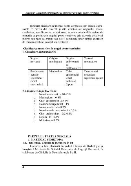
Clasificarea <strong>tumorilor</strong> <strong>de</strong> <strong>unghi</strong> <strong>ponto</strong>-<strong>cerebelos</strong><br />
1. Clasificare histopatologică<br />
Origine<br />
nervoasă<br />
Neurinoame:<br />
-acustic<br />
-trigemin<strong>al</strong><br />
-faci<strong>al</strong><br />
-nervi mixti<br />
Origine<br />
meningi<strong>al</strong>ă<br />
Origine<br />
embrionară<br />
şi<br />
m<strong>al</strong>formative<br />
Meningiom Chist<br />
epi<strong>de</strong>rmoid<br />
Chist<br />
arahnoid<br />
Lipom<br />
2. Clasificare după frecvenţă:<br />
o Neurinom acustic – 80-85%<br />
o Meningiom – 6-8%<br />
o Chist epi<strong>de</strong>rmoid -2,5-3%<br />
o Neurinom trigemin<strong>al</strong> – 1%<br />
o Neurinom faci<strong>al</strong> – 0,7%<br />
o Neurinom <strong>de</strong> nervi micşti – 0,5%<br />
o Chist arahnoidian – 0,2-0,4%<br />
o Lipom - 0,1-0,5%<br />
o Metastaze – 0,2%<br />
Tumori<br />
metastatice<br />
Determinări<br />
secundare<br />
leptomeninge<strong>al</strong>e<br />
PARTEA II : PARTEA SPECIALĂ<br />
1. MATERIAL ŞI METODĂ<br />
1.1. Obiective. Criterii <strong>de</strong> inclu<strong>de</strong>re în lot<br />
Lucrarea a fost efectuată în cadrul Clinicii <strong>de</strong> Radiologie şi<br />
Imagistică Medic<strong>al</strong>ă din Spit<strong>al</strong>ul Universitar <strong>de</strong> Urgenţă Bucureşti, în<br />
colaborare cu Clinicile <strong>de</strong> Neurochirurgie I şi II.