Jahresbericht der Klinik - Klinik für Kardiologie - UniversitätsSpital ...
Jahresbericht der Klinik - Klinik für Kardiologie - UniversitätsSpital ...
Jahresbericht der Klinik - Klinik für Kardiologie - UniversitätsSpital ...

Sie wollen auch ein ePaper? Erhöhen Sie die Reichweite Ihrer Titel.
YUMPU macht aus Druck-PDFs automatisch weboptimierte ePaper, die Google liebt.
Giovanni G. Camici, PhD<br />
Research Coordinator & Group Lea<strong>der</strong><br />
Basic Cardiovascular Research<br />
Vascular Aging, ACS and Stroke<br />
Due to the progressively aging population, aging represents a crucial<br />
research topic and vascular aging is the research focus of the group<br />
headed by Dr. Giovanni G. Camici. Aging is a major risk factor for the<br />
development of cardiovascular diseases, however, its direct effects<br />
on the cardiovascular systems are not clear since it always occurs in<br />
parallel to atherosclerosis and other risk factors.<br />
Mice offer an ideal model to study aging since they do not spontaneously<br />
develop atherosclerosis or age-related risk factors. However,<br />
they still show an age-dependent decay in vascular function [Fig. 2<br />
and 3]. Based on this model, Dr. Simon Stämpfli recently demonstrated<br />
that aging per se – contrary to the common belief – does not<br />
worsen arterial thrombosis, un<strong>der</strong>scoring the importance of controlling<br />
modifiable risk factors.<br />
In this context, several genes implicated in promoting vascular aging<br />
and age-related cardiovascular disease are a major research focus;<br />
amongst these the adaptor protein p66Shc, the MAP Kinase JNK and<br />
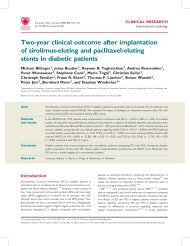
PKC beta. Recently, F. C. Franzeck, D. Hof and R. D. Spescha showed<br />
that some of these genes, namely p66Shc, are similarly upregulated<br />
in patients suffering from acute coronary syndromes [Fig. 1] un<strong>der</strong>lining<br />
that aging genes also play a role in cardiovascular disease.<br />
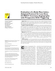
Fig. 2 Giovanni G. Camici (PhD) and Stephan Keller (Chief Technician)<br />
are assessing the morphological alterations of an aged mouse brain<br />
after middle cerebral artery occlusion / reperfusion.<br />
1.5 × 10 −4<br />
p = 0.0249<br />
p = 0.007<br />
ΔCt (p66shc / S18 rRNA)<br />
1.0 × 10 −4<br />
p = 0.367<br />
5.0 × 10 −5 0<br />
Control<br />
CAD<br />
ACS<br />
Fig. 1 p66Shc mRNA expression, relative to expression of 18S rRNA,<br />
in peripheral blood monocytes from angiographically confirmed<br />
CAD-free controls, stable CAD patients and patients with ACS.<br />
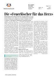
Fig. 3 Cerebral arteries of young and aged mice in the process of<br />
being homogenized prior to protein analysis.