student research day - Case Western Reserve University School of ...
student research day - Case Western Reserve University School of ...
student research day - Case Western Reserve University School of ...
Create successful ePaper yourself
Turn your PDF publications into a flip-book with our unique Google optimized e-Paper software.
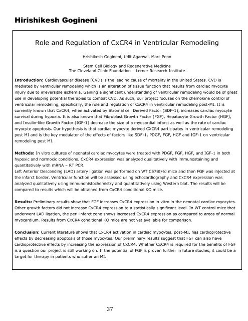
Hirishikesh Gogineni<br />
Role and Regulation <strong>of</strong> CxCR4 in Ventricular Remodeling<br />
Hrishikesh Gogineni, Udit Agarwal, Marc Penn<br />
Stem Cell Biology and Regenerative Medicine<br />
The Cleveland Clinic Foundation – Lerner Research Institute<br />
Introduction: Cardiovascular disease (CVD) is the leading cause <strong>of</strong> mortality in the United States. CVD is<br />
mediated by ventricular remodeling which is an alteration <strong>of</strong> tissue function that results from cardiac myocyte<br />
injury due to irreversible ischemia. Gaining a significant understanding <strong>of</strong> ventricular remodeling would be <strong>of</strong> great<br />
use in developing potential therapies to combat CVD. As such, our project focuses on the chemokine control <strong>of</strong><br />
ventricular remodeling, specifically, the role and regulation <strong>of</strong> CxCR4 in ventricular remodeling post-MI. It is<br />
currently known that CxCR4, when activated by Stromal cell Derived Factor (SDF-1), increases cardiac myocyte<br />
survival during hypoxia. It is also known that Fibroblast Growth Factor (FGF), Hepatocyte Growth Factor (HGF),<br />
and Insulin-like Growth Factor (IGF-1) decrease the size <strong>of</strong> a myocardial infarct as well as the rate <strong>of</strong> cardiac<br />
myocyte apoptosis. Our hypothesis is that cardiac myocyte derived CXCR4 participates in ventricular remodeling<br />
post MI and is the key modulator <strong>of</strong> the effects <strong>of</strong> factors like SDF-1, PDGF, FGF, HGF and IGF-1 on ventricular<br />
remodeling post MI.<br />
Methods: In vitro cultures <strong>of</strong> neonatal cardiac myocytes were treated with PDGF, FGF, HGF, and IGF-1 in both<br />
hypoxic and normoxic conditions. CxCR4 expression was analyzed qualitatively with immunostaining and<br />
quantitatively with mRNA – RT PCR.<br />
Left Anterior Descending (LAD) artery ligation was performed on WT C57BI/6J mice and then FGF was injected at<br />
the infarct border. Ventricular function will be assessed using echocardiography and CxCR4 expression was<br />
analyzed qualitatively using immunohistochemistry and quantitatively using <strong>Western</strong> blot. The results will be<br />
compared to results which will be obtained from CxCR4 conditional KO mice.<br />
Results: Preliminary results show that FGF increases CxCR4 expression in vitro in the neonatal cardiac myocytes.<br />
Other growth factors did not increase CxCR4 expression to a statistically significant level. In WT control mice that<br />
underwent LAD ligation, the peri-infarct zone shows increased CxCR4 expression as compared to areas <strong>of</strong> normal<br />
myocardium. Results from CxCR4 conditional KO mice are not yet available for comparison.<br />
Conclusion: Current literature shows that CxCR4 activation in cardiac myocytes, post-MI, has cardioprotective<br />
effects by decreasing apoptosis <strong>of</strong> those myocytes. Our preliminary results suggest that FGF can also have<br />
cardioprotective effects by increasing the expression <strong>of</strong> CxCR4. Whether CxCR4 is required for the benefits <strong>of</strong> FGF<br />
is a question our project is still working on. If the potential <strong>of</strong> FGF is proven further in future studies, it could be a<br />
target for therapy in patients who suffer an MI.<br />
37


