student research day - Case Western Reserve University School of ...
student research day - Case Western Reserve University School of ...
student research day - Case Western Reserve University School of ...
You also want an ePaper? Increase the reach of your titles
YUMPU automatically turns print PDFs into web optimized ePapers that Google loves.
GEORGE ANESI<br />
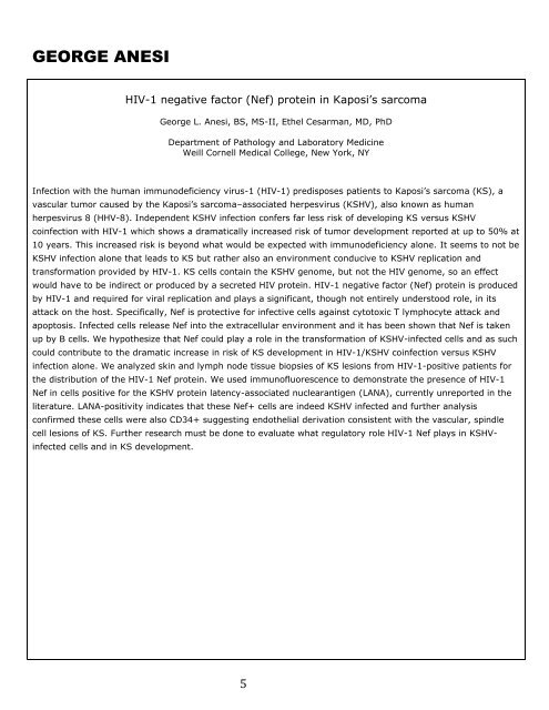
HIV-1 negative factor (Nef) protein in Kaposi’s sarcoma<br />
George L. Anesi, BS, MS-II, Ethel Cesarman, MD, PhD<br />
Department <strong>of</strong> Pathology and Laboratory Medicine<br />
Weill Cornell Medical College, New York, NY<br />
Infection with the human immunodeficiency virus-1 (HIV-1) predisposes patients to Kaposi’s sarcoma (KS), a<br />
vascular tumor caused by the Kaposi’s sarcoma–associated herpesvirus (KSHV), also known as human<br />
herpesvirus 8 (HHV-8). Independent KSHV infection confers far less risk <strong>of</strong> developing KS versus KSHV<br />
coinfection with HIV-1 which shows a dramatically increased risk <strong>of</strong> tumor development reported at up to 50% at<br />
10 years. This increased risk is beyond what would be expected with immunodeficiency alone. It seems to not be<br />
KSHV infection alone that leads to KS but rather also an environment conducive to KSHV replication and<br />
transformation provided by HIV-1. KS cells contain the KSHV genome, but not the HIV genome, so an effect<br />
would have to be indirect or produced by a secreted HIV protein. HIV-1 negative factor (Nef) protein is produced<br />
by HIV-1 and required for viral replication and plays a significant, though not entirely understood role, in its<br />
attack on the host. Specifically, Nef is protective for infective cells against cytotoxic T lymphocyte attack and<br />
apoptosis. Infected cells release Nef into the extracellular environment and it has been shown that Nef is taken<br />
up by B cells. We hypothesize that Nef could play a role in the transformation <strong>of</strong> KSHV-infected cells and as such<br />
could contribute to the dramatic increase in risk <strong>of</strong> KS development in HIV-1/KSHV coinfection versus KSHV<br />
infection alone. We analyzed skin and lymph node tissue biopsies <strong>of</strong> KS lesions from HIV-1-positive patients for<br />
the distribution <strong>of</strong> the HIV-1 Nef protein. We used immun<strong>of</strong>luorescence to demonstrate the presence <strong>of</strong> HIV-1<br />
Nef in cells positive for the KSHV protein latency-associated nuclearantigen (LANA), currently unreported in the<br />
literature. LANA-positivity indicates that these Nef+ cells are indeed KSHV infected and further analysis<br />
confirmed these cells were also CD34+ suggesting endothelial derivation consistent with the vascular, spindle<br />
cell lesions <strong>of</strong> KS. Further <strong>research</strong> must be done to evaluate what regulatory role HIV-1 Nef plays in KSHV-<br />
infected cells and in KS development.<br />
5


