student research day - Case Western Reserve University School of ...
student research day - Case Western Reserve University School of ...
student research day - Case Western Reserve University School of ...
You also want an ePaper? Increase the reach of your titles
YUMPU automatically turns print PDFs into web optimized ePapers that Google loves.
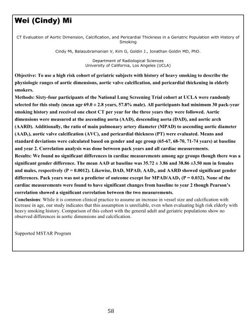
Wei (Cindy) Mi<br />
CT Evaluation <strong>of</strong> Aortic Dimension, Calcification, and Pericardial Thickness in a Geriatric Population with History <strong>of</strong><br />
Smoking<br />
Cindy Mi, Balasubramanian V, Kim G, Goldin J., Jonathan Goldin MD, PhD.<br />
Department <strong>of</strong> Radiological Sciences<br />
<strong>University</strong> <strong>of</strong> California, Los Angeles (UCLA)<br />
Objective: To use a high risk cohort <strong>of</strong> geriatric subjects with history <strong>of</strong> heavy smoking to describe the<br />
physiologic ranges <strong>of</strong> aortic dimensions, aortic valve calcification, and pericardial thickening in elderly<br />
smokers.<br />
Methods: Sixty-four participants <strong>of</strong> the National Lung Screening Trial cohort at UCLA were randomly<br />
selected for this study (mean age 69.0 ± 2.8 years, 57.8% male). All participants had minimum 30 pack-year<br />
smoking history and received one chest CT per year for the three years they were followed. Aortic<br />
dimensions were measured at the ascending aorta (AAD), descending aorta (DAD), and aortic arch<br />
(AARD). Additionally, the ratio <strong>of</strong> main pulmonary artery diameter (MPAD) to ascending aortic diameter<br />
(AAD1), aortic valve calcification (AVC), and pericardial thickness (PT) were evaluated. Means and<br />
standard deviations were calculated based on gender and age group (65-67, 68-70, 71-74 years) at baseline<br />
and year 2. Correlation analysis was done between pack years and all cardiac measurements.<br />
Results: We found no significant differences in cardiac measurements among age groups though there was a<br />
significant gender difference. The mean AAD at baseline was 35.72 ± 3.86 and 38.86 ±3.50 mm in females<br />
and males, respectively (P = 0.0012). Likewise, DAD, MPAD, AAD1, and AARD showed significant gender<br />
differences. Pack years was not a predictor <strong>of</strong> outcome except for MPAD/AAD1 (P = 0.032). None <strong>of</strong> the<br />
cardiac measurements were found to have significant changes from baseline to year 2 though Pearson’s<br />
correlation showed a significant correlation between the two measurements.<br />
Conclusions: While it is common clinical practice to assume an increase in vessel size and calcification with<br />
increase in age, our study indicates that this assumption is unreliable, even when evaluating high risk elderly with<br />
heavy smoking history. Comparison <strong>of</strong> this cohort with the general adult and geriatric populations show no<br />
observed differences in aortic dimensions and calcification.<br />
Supported MSTAR Program<br />
58


