student research day - Case Western Reserve University School of ...
student research day - Case Western Reserve University School of ...
student research day - Case Western Reserve University School of ...
Create successful ePaper yourself
Turn your PDF publications into a flip-book with our unique Google optimized e-Paper software.
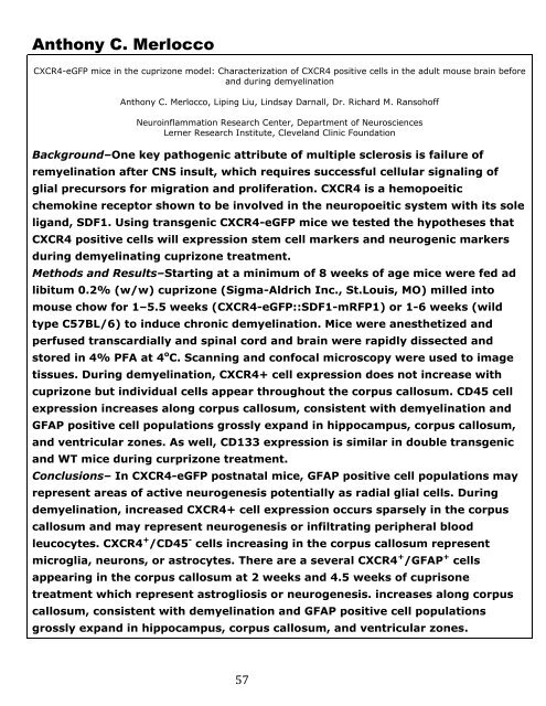
Anthony C. Merlocco<br />
CXCR4-eGFP mice in the cuprizone model: Characterization <strong>of</strong> CXCR4 positive cells in the adult mouse brain before<br />
and during demyelination<br />
Anthony C. Merlocco, Liping Liu, Lindsay Darnall, Dr. Richard M. Ransoh<strong>of</strong>f<br />
Neuroinflammation Research Center, Department <strong>of</strong> Neurosciences<br />
Lerner Research Institute, Cleveland Clinic Foundation<br />
Background–One key pathogenic attribute <strong>of</strong> multiple sclerosis is failure <strong>of</strong><br />
remyelination after CNS insult, which requires successful cellular signaling <strong>of</strong><br />
glial precursors for migration and proliferation. CXCR4 is a hemopoeitic<br />
chemokine receptor shown to be involved in the neuropoeitic system with its sole<br />
ligand, SDF1. Using transgenic CXCR4-eGFP mice we tested the hypotheses that<br />
CXCR4 positive cells will expression stem cell markers and neurogenic markers<br />
during demyelinating cuprizone treatment.<br />
Methods and Results–Starting at a minimum <strong>of</strong> 8 weeks <strong>of</strong> age mice were fed ad<br />
libitum 0.2% (w/w) cuprizone (Sigma-Aldrich Inc., St.Louis, MO) milled into<br />
mouse chow for 1–5.5 weeks (CXCR4-eGFP::SDF1-mRFP1) or 1-6 weeks (wild<br />
type C57BL/6) to induce chronic demyelination. Mice were anesthetized and<br />
perfused transcardially and spinal cord and brain were rapidly dissected and<br />
stored in 4% PFA at 4 o C. Scanning and confocal microscopy were used to image<br />
tissues. During demyelination, CXCR4+ cell expression does not increase with<br />
cuprizone but individual cells appear throughout the corpus callosum. CD45 cell<br />
expression increases along corpus callosum, consistent with demyelination and<br />
GFAP positive cell populations grossly expand in hippocampus, corpus callosum,<br />
and ventricular zones. As well, CD133 expression is similar in double transgenic<br />
and WT mice during curprizone treatment.<br />
Conclusions– In CXCR4-eGFP postnatal mice, GFAP positive cell populations may<br />
represent areas <strong>of</strong> active neurogenesis potentially as radial glial cells. During<br />
demyelination, increased CXCR4+ cell expression occurs sparsely in the corpus<br />
callosum and may represent neurogenesis or infiltrating peripheral blood<br />
leucocytes. CXCR4 + /CD45 - cells increasing in the corpus callosum represent<br />
microglia, neurons, or astrocytes. There are a several CXCR4 + /GFAP + cells<br />
appearing in the corpus callosum at 2 weeks and 4.5 weeks <strong>of</strong> cuprisone<br />
treatment which represent astrogliosis or neurogenesis. increases along corpus<br />
callosum, consistent with demyelination and GFAP positive cell populations<br />
grossly expand in hippocampus, corpus callosum, and ventricular zones.<br />
57


