student research day - Case Western Reserve University School of ...
student research day - Case Western Reserve University School of ...
student research day - Case Western Reserve University School of ...
You also want an ePaper? Increase the reach of your titles
YUMPU automatically turns print PDFs into web optimized ePapers that Google loves.
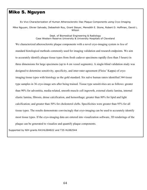
Mike S. Nguyen<br />
Ex Vivo Characterization <strong>of</strong> Human Atherosclerotic Iliac Plaque Components using Cryo-Imaging<br />
Mike Nguyen, Olivier Salvado, Debashish Roy, Grant Steyer, Meredith E. Stone, Robert D. H<strong>of</strong>fman, David L.<br />
Wilson<br />
Dept. <strong>of</strong> Biomedical Engineering & Radiology<br />
<strong>Case</strong> <strong>Western</strong> <strong>Reserve</strong> <strong>University</strong> & <strong>University</strong> Hospitals <strong>of</strong> Cleveland<br />
We characterized atherosclerotic plaque components with a novel cryo-imaging system in lieu <strong>of</strong><br />
standard histological methods commonly used for imaging validation and <strong>research</strong> endpoints. We aim<br />
to accurately identify plaque tissue types from fresh cadaver specimens rapidly (less than 5 hours) in<br />
three dimensions for large specimens (up to 4 cm vessel segments). A single-blind validation study was<br />
designed to determine sensitivity, specificity, and inter-rater agreement (Fleiss’ Kappa) <strong>of</strong> cryo-<br />
imaging tissue types with histology as the gold standard. Six naïve human raters identified 344 tissue<br />
type samples in 36 cryo-image sets after being trained. Tissue type sensitivities are as follows: greater<br />
than 90% for adventitia, media-related, smooth muscle cell ingrowth, external elastic lamina, internal<br />
elastic lamina, fibrosis, dense calcification, and hemorrhage; greater than 80% for lipid and light<br />
calcification; and greater than 50% for cholesterol clefts. Specificities were greater than 95% for all<br />
tissue types. The results demonstrate convincingly that cryo-imaging can be used to accurately identify<br />
most tissue types. If the cryo-imaging data are entered into visualization s<strong>of</strong>tware, 3D renderings <strong>of</strong> the<br />
plaque can be generated to visualize and quantify plaque components.<br />
Supported by NIH grants R41HL084822 and T35 HL082544<br />
64


