student research day - Case Western Reserve University School of ...
student research day - Case Western Reserve University School of ...
student research day - Case Western Reserve University School of ...
You also want an ePaper? Increase the reach of your titles
YUMPU automatically turns print PDFs into web optimized ePapers that Google loves.
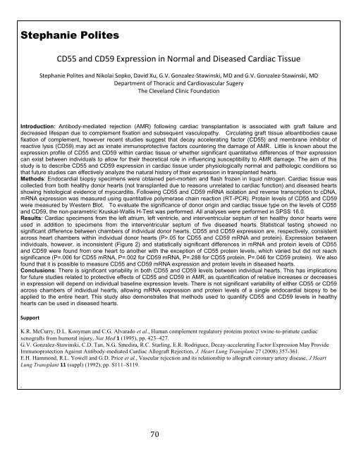
Stephanie Polites<br />
CD55 and CD59 Expression in Normal and Diseased Cardiac Tissue<br />
Stephanie Polites and Nikolai Sopko, David Xu, G.V. Gonzalez-Stawinski, MD and G.V. Gonzalez-Stawinski, MD<br />
Department <strong>of</strong> Thoracic and Cardiovascular Sugery<br />
The Cleveland Clinic Foundation<br />
Introduction: Antibody-mediated rejection (AMR) following cardiac transplantation is associated with graft failure and<br />
decreased lifespan due to complement fixation and subsequent vasculopathy. Circulating graft tissue alloantibodies cause<br />
fixation <strong>of</strong> complement, however recent studies suggest that decay accelerating factor (CD55) and membrane inhibitor <strong>of</strong><br />
reactive lysis (CD59) may act as innate immunoprotective factors countering the damage <strong>of</strong> AMR. Little is known about the<br />
expression pr<strong>of</strong>ile <strong>of</strong> CD55 and CD59 within cardiac tissue or whether significant quantitative differences <strong>of</strong> their expression<br />
can exist between individuals to allow for their theoretical role in influencing susceptibility to AMR damage. The aim <strong>of</strong> this<br />
study is to describe CD55 and CD59 expression in cardiac tissue under physiologically normal and pathologic conditions so<br />
that future studies can effectively analyze the natural history <strong>of</strong> their expression in transplanted hearts.<br />
Methods: Endocardial biopsy specimens were obtained peri-mortem and flash frozen in liquid nitrogen. Cardiac tissue was<br />
collected from both healthy donor hearts (not transplanted due to reasons unrelated to cardiac function) and diseased hearts<br />
showing histological evidence <strong>of</strong> myocarditis. Following CD55 and CD59 mRNA isolation and reverse transcription to cDNA,<br />
mRNA expression was measured using quantitative polymerase chain reaction (RT-PCR). Protein levels <strong>of</strong> CD55 and CD59<br />
were measured by <strong>Western</strong> Blot. To evaluate the significance <strong>of</strong> donor origin and cardiac tissue type on the levels <strong>of</strong> CD55<br />
and CD59, the non-parametric Kruskal-Wallis H-Test was performed. All analyses were performed in SPSS 16.0.<br />
Results: Cardiac specimens from the left atrium, left ventricle, and interventricular septum <strong>of</strong> ten healthy donor hearts were<br />
used in addition to specimens from the interventricular septum <strong>of</strong> five diseased hearts. Statistical testing showed no<br />
significant difference between chambers <strong>of</strong> individual donor hearts. CD55 and CD59 expression are, respectively, consistent<br />
across heart chambers within individual donor hearts (P>.05 for CD55 and CD59 mRNA and protein). Expression between<br />
individuals, however, is inconsistent (Figure 2) and statistically significant differences in mRNA and protein levels <strong>of</strong> CD55<br />
and CD59 were found from one heart to another with the exception <strong>of</strong> CD55 protein levels, which varied but did not reach<br />
significance (P=.006 for CD55 mRNA, P=.002 for CD59 mRNA, P=.288 for CD55 protein, P=.046 for CD59 protein). We also<br />
found that it is possible to measure CD55 and CD59 mRNA expression and protein levels in diseased hearts.<br />
Conclusions: There is significant variability in both CD55 and CD59 levels between individual hearts. This has implications<br />
for future studies related to protective effects <strong>of</strong> CD55 and CD59 in AMR, as quantification <strong>of</strong> relative increases or decreases<br />
in expression will depend on individual baseline expression levels. There is not significant variability <strong>of</strong> either CD55 or CD59<br />
across chambers <strong>of</strong> individual hearts, allowing mRNA expression and protein levels <strong>of</strong> a single endocardial biopsy to be<br />
applied to the entire heart. This study also demonstrates that methods used to quantify CD55 and CD59 levels in healthy<br />
hearts can be used in diseased hearts.<br />
Support<br />
K.R. McCurry, D.L. Kooyman and C.G. Alvarado et al., Human complement regulatory proteins protect swine-to-primate cardiac<br />
xenografts from humoral injury, Nat Med 1 (1995), pp. 423–427.<br />
G.V. Gonzalez-Stawinski, C.D. Tan, N.G. Smedira, R.C. Starling, E.R. Rodriguez, Decay-accelerating Factor Expression May Provide<br />
Immunoprotection Against Antibody-mediated Cardiac Allograft Rejection, J. Heart Lung Transplant 27 (2008) 357-361.<br />
E.H. Hammond, R.L. Yowell and G.D. Price et al., Vascular rejection and its relationship to allograft coronary artery disease, J Heart<br />
Lung Transplant 11 (suppl) (1992), pp. S111–S119.<br />
.<br />
70


