student research day - Case Western Reserve University School of ...
student research day - Case Western Reserve University School of ...
student research day - Case Western Reserve University School of ...

You also want an ePaper? Increase the reach of your titles
YUMPU automatically turns print PDFs into web optimized ePapers that Google loves.
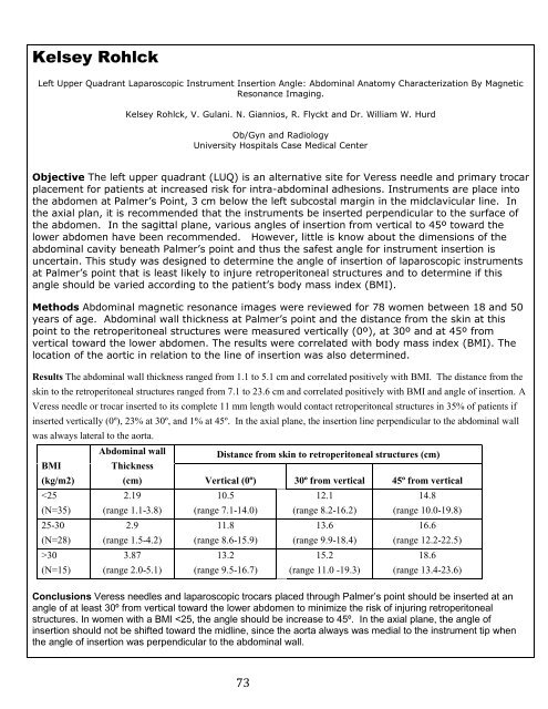
Kelsey Rohlck<br />
Left Upper Quadrant Laparoscopic Instrument Insertion Angle: Abdominal Anatomy Characterization By Magnetic<br />
Resonance Imaging.<br />
Kelsey Rohlck, V. Gulani. N. Giannios, R. Flyckt and Dr. William W. Hurd<br />
Ob/Gyn and Radiology<br />
<strong>University</strong> Hospitals <strong>Case</strong> Medical Center<br />
Objective The left upper quadrant (LUQ) is an alternative site for Veress needle and primary trocar<br />
placement for patients at increased risk for intra-abdominal adhesions. Instruments are place into<br />
the abdomen at Palmer’s Point, 3 cm below the left subcostal margin in the midclavicular line. In<br />
the axial plan, it is recommended that the instruments be inserted perpendicular to the surface <strong>of</strong><br />
the abdomen. In the sagittal plane, various angles <strong>of</strong> insertion from vertical to 45º toward the<br />
lower abdomen have been recommended. However, little is know about the dimensions <strong>of</strong> the<br />
abdominal cavity beneath Palmer’s point and thus the safest angle for instrument insertion is<br />
uncertain. This study was designed to determine the angle <strong>of</strong> insertion <strong>of</strong> laparoscopic instruments<br />
at Palmer’s point that is least likely to injure retroperitoneal structures and to determine if this<br />
angle should be varied according to the patient’s body mass index (BMI).<br />
Methods Abdominal magnetic resonance images were reviewed for 78 women between 18 and 50<br />
years <strong>of</strong> age. Abdominal wall thickness at Palmer’s point and the distance from the skin at this<br />
point to the retroperitoneal structures were measured vertically (0º), at 30º and at 45º from<br />
vertical toward the lower abdomen. The results were correlated with body mass index (BMI). The<br />
location <strong>of</strong> the aortic in relation to the line <strong>of</strong> insertion was also determined.<br />
Results The abdominal wall thickness ranged from 1.1 to 5.1 cm and correlated positively with BMI. The distance from the<br />
skin to the retroperitoneal structures ranged from 7.1 to 23.6 cm and correlated positively with BMI and angle <strong>of</strong> insertion. A<br />
Veress needle or trocar inserted to its complete 11 mm length would contact retroperitoneal structures in 35% <strong>of</strong> patients if<br />
inserted vertically (0º), 23% at 30º, and 1% at 45º. In the axial plane, the insertion line perpendicular to the abdominal wall<br />
was always lateral to the aorta.<br />
BMI<br />
(kg/m2)<br />
30<br />
(N=15)<br />
Abdominal wall<br />
Thickness<br />
(cm)<br />
2.19<br />
(range 1.1-3.8)<br />
2.9<br />
(range 1.5-4.2)<br />
3.87<br />
(range 2.0-5.1)<br />
Distance from skin to retroperitoneal structures (cm)<br />
Vertical (0º) 30º from vertical 45º from vertical<br />
10.5<br />
(range 7.1-14.0)<br />
11.8<br />
(range 8.6-15.9)<br />
13.2<br />
(range 9.5-16.7)<br />
73<br />
12.1<br />
(range 8.2-16.2)<br />
13.6<br />
(range 9.9-18.4)<br />
15.2<br />
(range 11.0 -19.3)<br />
14.8<br />
(range 10.0-19.8)<br />
16.6<br />
(range 12.2-22.5)<br />
18.6<br />
(range 13.4-23.6)<br />
Conclusions Veress needles and laparoscopic trocars placed through Palmer’s point should be inserted at an<br />
angle <strong>of</strong> at least 30º from vertical toward the lower abdomen to minimize the risk <strong>of</strong> injuring retroperitoneal<br />
structures. In women with a BMI