

European Human Genetics Conference 2007 June 16 – 19, 2007 ...
European Human Genetics Conference 2007 June 16 – 19, 2007 ...
European Human Genetics Conference 2007 June 16 – 19, 2007 ...

Create successful ePaper yourself
Turn your PDF publications into a flip-book with our unique Google optimized e-Paper software.
Clinical genetics<br />
plicated: two cousins - brother and sister with symptoms of psychomotoric<br />
retardation and one child with Down syndrome from the other<br />
cousin‘s family. The dysmorphic features were seen from the birth of<br />
this girl. Facial dysmorphism is characterised by the triangular high<br />
forehead with expressed sagital suture, hypertrichosis, wide spaced<br />
eyes, ptosis, down-slanting palpebral fissures, strabismus, broad nasal<br />
root, very short nose with anteverted nares, short grooved philtrum,<br />
triangular mouth, thin upper lip, everted lower lip, high narrow palate,<br />
micrognathia, malformed ears with preauricular sinus on one of sides.<br />
The characteristic skin lesions are typical for chromosomal mosaicism:<br />
they involve streaked, whorled and mottled areas of hypopigmentation<br />
on trunk and limbs. The psychomotor development of our patient is<br />
with severe features of delay: she sat alone only at two years. Clinical<br />
follow-up showed these clinical findings: CT scan showed corpus<br />
callosum agenesis and hydrocephaly, X-ray: abnormal feet position<br />
- equinovarus bilaterally.<br />
Cytogenetic analysis of peripheral blood lymphocytes revealed a mosaic<br />
karyotype 47,XX, +mar/46,XX in girl with dysmorphism of phenotype.<br />
Chromosome analysis was performed from GTG banded metaphases.<br />
The resolution level was 400-500 bands. The exact nature of<br />
the marker chromosome could not be identified in our patient. Parental<br />
karyotypes were normal.<br />
P0097. Disproportional high frequency of CLCN1 mutations<br />
among patients with myotonic dystrophy type 2.<br />
T. Suominen 1 , B. Schoser 2 , O. Raheem 3 , S. Auvinen 4 , R. Krahe 5 , H. Lochmüller<br />
2 , W. Kress 6 , B. Udd 7 ;<br />
1 Neurogenetics, University of Tampere, Tampere, Finland, 2 Friedrich-Baur-Institute,<br />
Ludwig-Maximilians-University Munich, Germany, 3 Department of Pathology,<br />
Pirkanmaa Hospital District, Center for Laboratory Medicine, Tampere,<br />
Finland, 4 Department of Neurology, Central Finland Central Hospital, Jyväskylä,<br />
Finland, 5 Department of Molecular <strong>Genetics</strong>, University of Texas M. D. Anderson<br />
Cancer Center, Houston, TX, United States, 6 Institute of <strong>Human</strong> <strong>Genetics</strong>,<br />
University of Wuerzburg, Germany, 7 Department of Neurology, Tampere University<br />
Hospital, Tampere, Finland.<br />
Background: Myotonic dystrophy type 2 (DM2) is a multiorgan disease<br />
caused by (CCTG)n repeat expansion mutation in ZNF9 gene. Clinical<br />
core features are myotonia, muscle weakness and cataracts. The<br />
phenotype is highly variable ranging from mild to severe forms, which<br />
makes clinical classification difficult. DM2 mutation causes aberrant<br />
splicing of different genes including CLCN1. Mutations in ClC1 chloride<br />
channel gene (CLCN1) cause recessive and rarely dominant myotonia<br />
congenita (MC), characterized by myotonia and muscle hypertrophy.<br />
Objective: to clarify whether co-segregation of frequent recessive<br />
CLC1 mutations may have a modifier effect on the DM2 phenotype.<br />
Methods: CLCN1 mutations R894X, F413C and A531V were analysed<br />
in 200 Finnish and German DM2 patients and 200 controls by TaqMan<br />
Sequence Detection System (ABI) using specific primers for PCR and<br />
fluorescent oligonucleotide probes.<br />
Results: CLCN1 mutations R894X, F413C and A531V in DM2 patients<br />
and controls.<br />
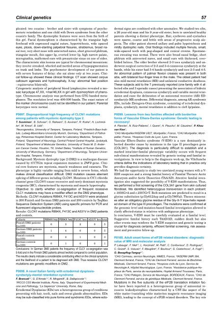
Cohorts R894X hoz R894X hez F413C hez A531V hez total<br />
Finnish DM2 patients<br />
(n=100)<br />
0 3 2 0 5<br />
German DM2 patients<br />
(n=100)<br />
1 4 0 0 5<br />
Finnish controls<br />
(n=100)<br />
0 2 1 0 3<br />
German controls<br />
(n=100)<br />
0 1 0 0 1<br />
Conclusions: In German DM2 patients the frequency of CLC1 co-segregation was<br />
5-fold and in the Finnish DM2 patients almost twofold compared to control population.<br />
The results clearly indicate a considerable contributing effect on the clinical symptoms<br />
and the likelihood of a patient to be diagnosed with DM2. Thus recessive CLCN1<br />
mutations are genetic modifiers in DM2.<br />
P0098. A novel Italian family with ectodermal dysplasiasyndactyly-mental<br />
retardation syndrome<br />
F. Brancati1,2 , G. D’Amato1,2 , R. Mingarelli1 , B. Dallapiccola1,2 ;<br />
1 2 IRCCS CSS Mendel Institute, Rome, Italy, Department of Experimental Medicine<br />
and Pathology, “La Sapienza” University, Rome, Italy.<br />
Ectodermal Dysplasias (EDs) are a heterogeneous group of conditions<br />
presenting with hair, tooth, nails and sweat glands abnormalities. EDs<br />
may be sub-classified into pure forms and syndromic EDs, where ecto-<br />
dermal signs are combined with other anomalies. We studied two sibs,<br />
a 26 year-old man and his 9 year-old sister, born to unrelated healthy<br />
parents showing a distinct phenotype. Hair, eyebrows and eyelashes<br />
were sparse, coarse and brittle with areas of progressive scalp alopecia.<br />
The older patient showed axillary and pubic hypotrichosis and<br />
mildly dystrophic nails. Oral findings included multiple frenula, small,<br />
wide-spaced teeth with peg-shaped and conical crowns. Spontaneous<br />
sweating was normal. There were also broad nasal bridge, short<br />
philtrum with anteverted nares, and small ears with thickened, overfolded<br />
helices. The older brother showed 2-3 toes syndactyly and underwent<br />
surgical correction of 2-3 and 3-4 cutaneous syndactyly at the<br />
hands, while his sister had 3-4 hands syndactyly and toes 2-3 and 4-5.<br />
An abnormal pattern of palmar flexion creases was present in both<br />
sibs, with bilateral four-finger lines in the male. The oldest patient had<br />
also mild mental retardation (MR) and unilateral conductive deafness.<br />
These subjects add to the 7 previously reported (one family with 4 affected<br />
sibs and 3 sporadic cases) presenting the association of hidrotic<br />
ectodermal dysplasia, cutaneous syndactyly and variable mental retardation<br />
and ease the delineation of an autosomal recessive (AR) EDsyndactyly-MR<br />
syndrome. Differential diagnosis, among AR syndromic<br />
EDs, include Zlotogora-Orun syndrome, consisting of ectodermal dysplasia,<br />
syndactyly, mental retardation in addition to cleft lip/palate.<br />
P0099. Lessons from two families affected with borderline<br />
forms of Vascular Elhers-Danlos syndrome: Genetic testing is<br />
necessary!<br />
A. Plancke1 , N. Ruiz-Pallares1 , I. Quere2 , H. Plauchu3 , M. Claustres1 , P. Khau<br />
van Kien1 ;<br />
1 2 CHU Montpellier/INSERM U827, Montpellier, France, CHU Montpellier, Montpellier,<br />
France, 3Hospices Civils de Lyon, Lyon, France.<br />
Vascular Elhers-Danlos syndrome (V-EDS) is a rare dominantly inherited<br />
disorder cause by mutations in the type III procollagen gene<br />
(COL3A1). The diagnosis is particularly difficult to establish and a<br />
marked intra/inter-familial phenotypic variability occurs with frequent<br />
sub-clinical manifestations revealed by genetic testing and familial investigations.<br />
In view to help to the diagnosis work-up, the Villefranche<br />
criteria define the indications of laboratory testing that in practice only<br />
provides diagnosis certainty.<br />
We had the opportunity to study two unrelated young women with a V-<br />
EDS suspicion and a strong familial history of Familial Thoracic Aortic<br />
Aneurysm and/or Aortic Dissection (TAA/AD). Attentive familial investigation<br />
also suggested the possibility of V-EDS manifestations. Thus,<br />
we performed a first screening of the COL3A1 gene from skin cultured<br />
fibroblast. We identified heterozygous transversion in each probant:<br />
c.1835G>A and c.2357G>T. Both mutations are predicted to result in a<br />
glycine substitution (p.Gly612Asp and p.Gly786Val). Theses missenses<br />
alter an obligatory glycine residue of the Gly-X-Y tripe-helix repeated<br />
domain of the type III procollagen. The mutations were confirmed at<br />
the genomic level and mutation-based familial investigation allowed to<br />
clarify affected-status in relatives with ambiguous phenotype.<br />
In conclusion, V-EDS must be carefully evaluated at a familial level.<br />
Suggestive familial history such TAAD/AD, sudden death but also<br />
other events may reinforce the V-EDS suspicion and genetic testing is<br />
crucial for diagnosis certainty, efficient familial screening, risk assessment<br />
and preventive follow-up.<br />
P0100. Adult onset forms of eIF2B related disorders: diagnostic<br />
value of MRI and molecular analysis<br />
P. Labauge 1 , F. Niel 2,3 , L. Horzinski 2 , M. Petit 3 , C. Confavreux 4 , D. Rodriguez 5 ,<br />
C. Goizet 6 , S. Vukusic 4 , F. Maugière 4 , F. Bouhour 4 , G. Casterlnovo 1 , A. Fogli 2,3 ,<br />
O. Boespflug-Tanguy 2,7 ;<br />
1 CHU Carmeau, service Neurologie, NIMES, France, 2 INSERM UMR 384,<br />
Clermont ferrand, France, 3 CHU de Clermont Ferrand, service de Biochimie<br />
Médicale, Clermont ferrand, France, 4 Hospices civils de Lyon, Service de<br />
Neurologie A, Hôpital Neurologique, Lyon, France, 5 Assistance publique-Hopitaux<br />
de Paris, service de neuropédiatrie, Hopital Armand Trousseau, Paris,<br />
France, 6 CHU Pellegrin, Service de Neurologie, BORDEAUX, France, 7 CHU de<br />
Clermont Ferrand, service de génétique Médicale, Clermont ferrand, France.<br />
Mutations in the five subunits of the eIF2B translation initiation factor<br />
have been reported in a heterogeneous group of autosomal recessive<br />
leukodystrophies characterized by a CSF-like aspect of the<br />
white matter (vanishing white matter)on magnetic resonance imaging<br />
(MRI), leading to the concept of eIF2B related disorders. The key role